Ozempic, Wegovy and Mounjaro makes food taste sweeter and saltier, and that may quiet cravings

New research being presented at the Annual Meeting of the European Association for the Study of Diabetes (EASD) in Vienna, Austria (September 15-19) shows that some individuals who are taking Ozempic, Wegovy or Mounjaro find that foods taste sweeter or saltier than before.

About one in five of those participating in the real-word study, published in the journal Diabetes, Obesity and Metabolism, perceived sweetness more intensely and a similar number were more sensitive to salt – and these changes were linked to a reduction in appetite.

“Incretin-based therapies such as Ozempic, Wegovy and Mounjaro are widely used for weight management but their effect on taste perception has been unclear,” says Othmar Moser, of University of Bayreuth, Bayreuth, Germany, who led the research.

“If changes in taste are linked to greater appetite control and weight loss, this could help clinicians better select therapies, provide more tailored dietary advice and improve long-term treatment outcomes for patients.”

To find out more, Professor Moser and colleagues from the Medical University of Vienna surveyed hundreds of individuals with overweight and obesity who were taking Ozempic, Wegovy or Mounjaro for weight loss about their sense of taste and appetite.

Of the 411 participants (69.6% female), 148 were on Ozempic, 217 were on Wegovy and 46 were taking Mounjaro.

Median duration of treatment was similar for the three groups (Ozempic: 43 weeks; Wegovy: 40 weeks; and Mounjaro: 47 weeks), with all of the participants receiving treatment for at least three consecutive months. The average BMI before starting treatment was 34.7 kg/m2 (Ozempic), 35.6 kg/m2 (Wegovy) and 36.2 kg/m2 (Mounjaro).

The participants, who were recruited online, were asked if their sense of taste (perception of sweetness, saltiness, sourness and bitterness) had changed since starting treatment.

They were also asked about changes to appetite, satiety and food cravings, as well changes to lifestyle factors, such as smoking, and for self-reported data on height and weight before and during treatment.

Reductions in BMI, adjusted for duration of treatment, dose, baseline BMI, age and sex, were 17.4% with Ozempic, 17.6% with Wegovy and 15.5% with Mounjaro.

Around a fifth of the participants said that food tasted sweeter (21.3%) or saltier (22.6%) than before. Their perception of bitterness and sourness did not change.

Some 26.7% of participants in the Wegovy group reported that food tasted saltier than before, compared with 16.2% in the Ozempic group and 15.2% in the Mounjaro group. Increases in sweetness were reported at similar frequencies in all groups (Wegovy 19.4%, Ozempic 21.6%, Mounjaro 21.7%).

More than half of the participants (58.4%) reported they were less hungry in general, i.e. their appetite had decreased (Ozempic: 62.1%, Wegovy: 54.4%, Mounjaro: 56.5%).

Almost two-thirds of the participants (63.5%) reported increased satiety i.e. they felt full sooner Ozempic: 58.8%, Wegovy: 66.8%, Mounjaro: 63.1%). Food cravings were also reduced, with 41.3% of Mounjaro users reporting a strong reduction in cravings, i.e. their cravings were much less intense that before, compared with 34.1% of those taking Wegovy and 29.7% of those taking Ozempic.

Further analysis revealed links between changes to sense of taste and appetite and satiety.

Participants who reported that food tasted sweeter since starting incretin-based therapy were twice as likely to report increased satiety, compared with participants who said their perception of sweetness had not changed.

Those with an increase in the perception of sweetness were also 67% more likely to report a reduction in appetite and 85% more likely to report a reduction in cravings, compared with those whose perception of sweetness was unchanged.

Similarly, participants who said food tasted saltier than before were about twice as likely (2.17 times) to also reported increased satiety, compared with those whose perception of saltiness was unchanged.

Professor Moser says: “These drugs act not only in the gut and brain areas that control hunger but also on taste bud cells and brain regions that process taste and reward. This means they can subtly change how strong flavours, like sweetness or saltiness, are perceived. This, in turn, may affect appetite.”

However, there was no link between changes in taste perception and reduction in BMI. The researchers speculate this is because sense of taste is just one of many factors involved in weight loss.

Professor Moser explains: “Shifts in taste may affect how satisfying or appealing food feels in the moment, which influences appetite control. However, weight loss depends on many other factors – like metabolism, long-term eating patterns, and activity – so changes to taste alone may not be enough to directly drive body weight reduction.”

The study’s limitations include inability to prove causation, the self-reporting of data and the possibility that the participants weren’t representative of the patient group as a whole.

Professor Moser concludes: “Drugs like Wegovy, Ozempic and Mounjaro may alter sense of taste, making foods seem sweeter or saltier and helping people feel full sooner and less hungry. For clinical practice, this suggests that monitoring patients’ taste changes could provide useful clues about treatment response, even though taste alone does not directly drive weight loss.

“For example, tracking changes in taste could help gauge whether the treatment is working beyond weight loss.

“It could also perhaps be used to tailor dietary advice, for example by helping patients find alternatives to foods with flavours that have become overwhelming or less appealing.”

Why so many young kids with ADHD are getting the wrong treatment

Young children with attention deficit/hyperactivity disorder often receive medication just after being diagnosed, which contravenes treatment guidelines endorsed by the American Academy of Pediatrics, a Stanford Medicine-led study has found.

The finding, published on Aug. 29 in JAMA Network Open, highlights a gap in medical care for 4- and 5-year-olds with ADHD. Treatment guidelines recommend that these young children and their families try six months of behavior therapy before starting ADHD medication.

But pediatricians often prescribe medication immediately upon diagnosis, according to an analysis of medical records from nearly 10,000 young children with ADHD who received care in eight pediatric health networks in the United States.

“We found that many young children are being prescribed medications very soon after their diagnosis of ADHD is documented,” said the study’s lead author, Yair Bannett, MD, assistant professor of pediatrics. “That’s concerning, because we know starting ADHD treatment with a behavioral approach is beneficial; it has a big positive effect on the child as well as on the family.”

In addition, stimulant medications prescribed for the condition cause more side effects in young patients than they do in older children, Bannett said. Before age 6, children’s bodies don’t fully metabolize the drugs.

“We don’t have concerns about the toxicity of the medications for 4- and 5-year-olds, but we do know that there is a high likelihood of treatment failure, because many families decide the side effects outweigh the benefits,” he said. Stimulant medication can make young children more irritable, emotional and aggressive.

ADHD is a developmental disorder characterized by hyperactivity, difficulty paying attention and impulsive behavior.

“It’s important to catch it early because we know these kids are at higher risk for having academic problems and not completing school,” Bannett said. Early identification and effective treatment for ADHD improve children’s academic performance. Research has shown that good treatment also helps prepare individuals with ADHD for many aspects of adulthood, such as maintaining employment, having successful relationships and avoiding trouble with the law.

Complementary treatments

Behavioral therapy and medication, the two mainstays of ADHD treatment, have different purposes.

“Behavioral treatment works on the child’s surroundings: the parents’ actions and the routine the child has,” Bannett said. The therapy helps parents and kids build skills and establish habits compatible with how the child’s brain works.

The evidence-based behavioral treatment recommended by the American Academy of Pediatrics is called parent training in behavior management. The training helps parents build strong, positive relationships with their children; offers guidance in rewarding a child’s good behaviors and ignoring negative behaviors; and recommends tools that help kids with ADHD, such as making visual schedules to help them stay organized.

In contrast, medication relieves ADHD symptoms such as hyperactivity and inattentiveness, with effects that wear off as the body breaks down each dose of the drug.

Both approaches are needed for most kids with ADHD to do well. But previous studies of preschoolers diagnosed at age 4 or 5 show that it’s best to start with six months of behavioral treatment before prescribing any medication.

Rapid prescriptions

The researchers analyzed data from electronic health records for children seen at primary care practices affiliated with eight U.S. academic medical centers. They began with 712,478 records from children who were 3, 4 or 5 years old and were seen by their primary care physician at least twice, over a period of at least six months, between 2016 and 2023.

From these records, the scientists identified 9,708 children who received an ADHD diagnosis, representing 1.4% of the children in the initial sample. They found that 42.2% of these children — more than 4,000 kids — were prescribed medication within a month of their ADHD diagnosis. Only 14.1% of children with ADHD first received medication more than six months after diagnosis. The researchers did not have access to data on referrals to behavioral therapy, but since young children are supposed to try the therapy alone for six months before receiving medication, any who were prescribed medication sooner were likely not being treated according to academy guidelines. A smaller study of recommendations for behavior therapy, published in 2021, found only 11% of families got the therapy in line with guidelines.

Children who were initially given a formal diagnosis of ADHD were more likely to get medication within the first 30 days than those whose medical charts initially noted some ADHD symptoms, with a diagnosis at a later time. But even among preschoolers who did not initially meet full criteria for the condition, 22.9% received medication within 30 days.

Barriers to behavioral treatment?

Because the study was based on an analysis of electronic medical records, the researchers could not ask why physicians made the treatment decisions they did. However, Bannett’s team had informal conversations with physicians, outside the scope of the study, in which they asked why they prescribed medication.

“One important point that always comes up is access to behavioral treatment,” Bannett said. Some locales have few or no therapists who offer the treatment, or patients’ insurance may not cover it. “Doctors tell us, ‘We don’t have anywhere to send these families for behavioral management training, so, weighing the benefits and risks, we think it’s better to give medication than not to offer any treatment at all.'”

Bannett said he hopes to educate primary care pediatricians on how to bridge this gap. For example, free or low-cost online resources are available for parents who want to learn principles of the behavioral approach.

And while the study focused on the youngest ADHD patients, behavioral management therapy also helps older children with the diagnosis.

“For kids 6 and above, the recommendation is both treatments, because behavioral therapy teaches the child and family long-term skills that will help them in life,” Bannett said. “Medication will not do that, so we never think of medication as the only solution for ADHD.”

Researchers contributed to the study from the Children’s Hospital of Philadelphia, the Perelman School of Medicine at the University of Pennsylvania, Nationwide Children’s Hospital, The Ohio State University College of Medicine, Cincinnati Children’s Hospital Medical Center, University of Cincinnati College of Medicine, Texas Children’s Hospital, Baylor College of Medicine, Ann and Robert H. Lurie Children’s Hospital of Chicago, Children’s Hospital of Philadelphia, the University of Colorado, and Nemours Children’s Hospital. `

This work was supported by the Stanford Medicine Maternal and Child Health Research Institute; the National Institute of Mental Health (grant K23MH128455); and the National Heart, Lung, and Blood Institute (grant K23HL157615). The study was conducted using PEDSnet, A Pediatric Clinical Research Network. PEDSnet was developed with funding from the Patient-Centered Outcomes Research Institute.

Scientists test an anti-aging cream that actually works

Against the backdrop of high market demand for effective anti-ageing cosmetics, a team of Chinese researchers assessed the clinical effectiveness of a 0.1 % pterostilbene-containing skincare emulsion against a control emulsion over 28 days with 31 participants.

The study employed a double-blind, split-face design, comparing the left and right sides of the face and using advanced instruments along with subject self-assessments. The set of instruments used, together with the findings, is reported in the team’s published article in the Journal of Dermatologic Science and Cosmetic Technology.

“Our results indicated that the pterostilbene emulsion remarkably improved skin elasticity, firmness, and reduced wrinkles, such as forehead, undereye, and Crow’s feet wrinkles, shares co-author Zhiyuan Chen, Founder of Guangzhou Luanying Cosmetics Co., Ltd. “It also increased the thickness of the epidermis layer, enhanced collagen and elastic fibers, and minimized skin pores.”

Compared to the control emulsion, the pterostilbene emulsion brought about statistically significant improvements, and all subjects expressed higher satisfaction with the pterostilbene emulsion. These results collectively demonstrated the superior anti-aging efficacy of the pterostilbene emulsion through multiple mechanisms.

According to corresponding author Xueping Chen, the findings advance our understanding of pterostilbene’s role in skincare by providing evidence of its potent anti-ageing effects and supporting its use as an active ingredient in cosmetics. “It also offers a new perspective on natural compounds’ applications in the skincare industry,” says Chen. “The significant improvements in various skin parameters and the subjects’ positive feedback highlight the effectiveness of pterostilbene, which may change the way we approach anti-ageing skincare formulations.”

The authors propose a longer study period to fully understand the impact of pterostilbene.

Daily eye drops could make reading glasses obsolete

Everybody develops presbyopia as they age – a difficulty in focusing on near objects and text – and often have to resort to reading glasses. However, the solution might be as simple as using special eye drops two or three times a day.

A retrospective study of 766 patients presented on September 14 at the 43rd Congress of the European Society of Cataract and Refractive Surgeons (ESCRS) [1], found that the majority could read an extra two, three or more lines on the eye chart used for testing near visual acuity (the Jaeger chart) after using specially formulated eye drops. This improvement was sustained for up to two years.

Dr Giovanna Benozzi, director of the Center for Advanced Research for Presbyopia, in Buenos Aires, Argentina, said: “We conducted this research due to the significant unmet medical need in presbyopia management. Current solutions such as reading glasses or surgical interventions have limitations, including inconvenience, social discomfort, and potential risks or complications. There is a group of presbyopia patients who have limited options besides spectacles, and who are not candidates for surgery; these are our primary focus of interest. We sought to provide robust clinical evidence supporting an innovative pharmacological solution to offer patients a non-invasive, convenient and effective alternative.”

The eye drops, developed by Dr Benozzi’s father, the late Dr Jorge Benozzi of the same centre, contain a combination of two active agents: pilocarpine, a drug that constricts the pupils and contracts the ciliary muscle, which is a muscle controlling the eye’s accommodation for seeing objects at varying distances, and diclofenac, a non-steroidal anti-inflammatory drug (NSAID) that reduces inflammation and the discomfort that pilocarpine often causes.

Patients administered the eye drops twice a day, usually on waking and again approximately six hours later, with an optional third dose if symptoms recurred or additional visual comfort was needed. The group of patients (373 women and 393 men, with an average age of 55) were divided into three groups to receive one of three eye drop formulations. Each formulation had a fixed dose of diclofenac but concentrations of pilocarpine were 1%, 2% and 3%.

The researchers assessed the improvement in how well patients could read the Jaeger chart without reading glasses (uncorrected near visual acuity) one hour after the first administration of the drops, and they followed up the patients for two years.

Dr Benozzi told the Congress: “Our most significant result showed rapid and sustained improvements in near vision for all three concentrations. One hour after having the first drops, patients had an average improvement of 3.45 Jaeger lines. The treatment also improved focus at all distances.

“Impressively, 99% of 148 patients in the 1% pilocarpine group reached optimal near vision and were able to read two or more extra lines. Approximately 83% of all patients maintained good functional near vision at 12 months. Importantly, no significant adverse events like increased intraocular pressure or retinal detachment were observed.”

In the 2% group, 69% of 248 patients were able to read three or more extra lines on the Jaeger chart, and in the 3% group, 84% of 370 patients could read three or more extra lines.

The improvement in the patients’ vision was sustained for up to two years, with a median duration of 434 days. Adverse side effects were mild, with the most common being temporary dim vision, which occurred in 32% of cases, irritation when the drops were instilled (3.7%) and headache (3.8%). No patients discontinued the treatment.

Common adverse side effects of pilocarpine can also include eye redness, watery eyes, blurred vision, dim or dark vision, sensitivity to light or problems changing focus between objects, seeing flashes of light or “floaters” in vision, and, in rare cases, detached retinas.

Dr Benozzi continued: “Nearly all patients experienced positive improvements in near visual acuity, although the magnitude of the improvement depended on the status of their vision before treatment at baseline. Our study revealed that optimal pilocarpine concentrations could be individualized depending on the baseline severity of presbyopia as assessed by the initial Jaeger scores. Patients with less severe presbyopia responded best to 1% concentrations, while those with more advanced presbyopia required higher 2% or 3% concentrations to achieve significant visual improvement.”

She concluded: “These results suggest this combination therapy offers a safe, effective, and well-tolerated alternative to traditional presbyopia management. It significantly reduces dependence on reading glasses, providing a convenient, non-invasive option for patients, although these eye drops may not eliminate the need for glasses in all individuals.

“Importantly, this treatment is not intended to replace surgical interventions, but rather to serve as a valuable solution for patients who need safe, effective, and personalised alternatives and seek freedom from the inconvenience of eyewear. Eye care professionals now have an evidence-based pharmacological option that expands the spectrum of presbyopia care beyond glasses and surgery.”

Besides the group of patients in this study, Dr Benozzi has other patients who have received the treatment for more than ten years. Dr Benozzi plans further research to measure improvements in patients’ quality of life, and to explore the underlying physiological mechanisms of the eye drops.

Strengths of the study include the large number of patients included and the long follow-up time. It is the first systematic evaluation comparing three different pilocarpine concentrations in combination with diclofenac. A limitation is that it is a retrospective, single-centre study, which could limit the generalizability of the findings and introduce selection bias.

ESCRS President-Elect, Professor Burkhard Dick, chair of the ophthalmology department at the University Eye Hospital Bochum, Germany, was not involved in the research. He commented: “While surgery for age-related near vision loss has advanced, some patients are not candidates. The single-centre retrospective study by Dr Benozzi suggests that eye drops containing pilocarpine and diclofenac may improve near vision for up to two years, but the limited design means the results may not apply to everyone. Long-term pilocarpine use can sometimes cause side effects such as reduced night vision, dimmer vision in low light, eye strain, irritation and, in rare cases, retinal detachment, while prolonged topical NSAID use may pose corneal risks. Broader, long-term, multi-centre studies are needed to confirm safety and effectiveness before this treatment can be widely recommended.”

Notes

  1. Abstract number: ESCRS25-FP-3944, ‘Dose-dependent efficacy and safety of pilocarpine-diclofenac eye drops for presbyopia: a real-world single-center study,” by Giovanna Benozzi et al. Free paper session on ‘Miscellaneous topics in cataract and refractive surgery’, 16:30-18:00 hrs CEST, Sunday, September 14, https://pag.virtual-meeting.org/escrs/escrs2025/en-GB/pag/presentation/570375
1 in 8 Americans have already tried Ozempic and similar weight loss medications

Nearly 12% of Americans have used GLP-1 drugs for weight loss, including about one-fifth of women aged 50 to 64, according to a new RAND report.

Surveying a nationally representative sample of 8,793 Americans, researchers found that 11.8% have used GLP-1 agonists and 14% say they are interested in using the drugs. Meanwhile, 74% say they do not plan to take the medications.

Among those who have used GLP-1 drugs, about half report they have experienced nausea and about one-third experienced diarrhea — the two most-common side effects associated with the medications.

The report is the most-recent and largest survey to date that estimates how many Americans have used the drugs that have revolutionized weight loss care. The report outlines use of GLP-1 drugs by both age and sex.

The use of Ozempic and other GLP-1 medications has increased sharply in recent years since studies demonstrated the drugs can help people significantly aid weight loss. Since 2020, the number of prescriptions for the drugs has more than tripled.

RAND researchers surveyed participants of the RAND American Life Panel to ask about use of GLP-1 drugs and their experiences with side effects. The survey was performed during April and May of 2025.

The results show that women tend to use GLP-1 drugs at higher rates than men, although there is considerable variability within age groups.

Use of GLP-1 drugs is most common among those between the ages of 50 and 64, with the highest rate of use found among women in the age group. Among those 65 and older, use of GLP-1 drugs is somewhat higher for men than for women. Conversely, among those between the ages of 30 and 49, women are more than twice as likely to have used a GLP-1 than their male peers.

The RAND American Life Panel is a probability sample-based survey panel of about 11,000 active, regularly interviewed respondents aged 12 and older. The panel was developed by RAND in 2006.

The report, “New Weight Loss Drugs: GLP-1 Agonist Use and Side Effects in the United States,” is available at www.rand.org. Authors of the report are Robert Bozick, Shannon Donofry and Katherine M. Rancaño.

The publication is the first in what is planned to be a series of brief reports that highlights topline descriptive findings from surveys run on the RAND American Life Panel.

The RAND Education and Labor division is dedicated to improving education and expanding economic opportunities for all through research and analysis.

Sleepless nights may raise dementia risk by 40%, Mayo Clinic reveals
  • Chronic insomnia — trouble sleeping at least three days a week for three months or more — could speed up brain aging.
  • People with chronic insomnia were 40% more likely to develop dementia or mild cognitive issues than people without insomnia.
  • Insomnia with perceived reduced sleep was associated with lower cognition comparable to being four years older.
  • Better sleep isn’t just beauty rest — it might protect your brain health.

People with chronic insomnia may experience faster declines in memory and thinking skills as they age — along with brain changes that can be seen on imaging scans — than people who do not have chronic insomnia, according to a study published in the September 10, 2025, issue of Neurology, the medical journal of the American Academy of Neurology.

The study found that people with chronic insomnia — trouble sleeping at least three days a week for three months or more — had a 40% higher risk of developing mild cognitive impairment or dementia than those without insomnia, which is the equivalent of 3.5 additional years of aging. The study does not prove that insomnia causes brain aging, it only shows an association.

“Insomnia doesn’t just affect how you feel the next day — it may also impact your brain health over time,” said study author Diego Z. Carvalho, MD, of the Mayo Clinic in Rochester, Minnesota, and a member of the American Academy of Neurology. “We saw faster decline in thinking skills and changes in the brain that suggest chronic insomnia could be an early warning sign or even a contributor to future cognitive problems.”

The study tracked a group of cognitively healthy older adults — 2,750 people with an average age of 70 — for an average of 5.6 years. Of participants, 16% had chronic insomnia.

At the start of the study, participants were asked whether they had been sleeping more or less than usual during the past two weeks. They also took yearly thinking and memory tests, and some had brain scans to look for white matter hyperintensities — areas where small vessel disease may have damaged brain tissue — and for amyloid plaques, a protein that can build up and is linked to Alzheimer’s disease.

During the study, 14% of the people with chronic insomnia developed mild cognitive impairment or dementia, compared to 10% of those who did not have insomnia.

After accounting for factors like age, high blood pressure, use of sleep medications and a diagnosis of sleep apnea, they found that people with insomnia were 40% more likely to develop mild cognitive impairment or dementia than those without insomnia. They also had faster declines on tests measuring different thinking skills.

Among those with insomnia, researchers determined the type: those getting less sleep than usual in the past two weeks; or those getting more sleep than usual in the past two weeks.

People who reported getting less sleep than usual were more likely to have lower cognitive test scores at the beginning of the study, comparable to being four years older. They also had more white matter hyperintensities and amyloid plaques. For amyloid, the effect was similar to what is seen in people with the APOE ε4 gene, a known genetic risk factor.

People who reported getting more sleep than usual, on the other hand, were more likely to have fewer white matter hyperintensities at the start of the study.

Some groups were especially vulnerable. Participants who carry the APOE ε4 gene — linked to higher Alzheimer’s risk — showed steeper declines in memory and thinking skills.

“Our results suggest that insomnia may affect the brain in different ways, involving not only amyloid plaques, but also small vessels supplying blood to the brain,” Carvalho said. “This reinforces the importance of treating chronic insomnia — not just to improve sleep quality but potentially to protect brain health as we age. Our results also add to a growing body of evidence that sleep isn’t just about rest — it’s also about brain resilience.”

A limitation of the study was that insomnia diagnoses came from medical records, which do not capture undiagnosed cases or reflect how severe symptoms are.

The study was supported by the National Institutes of Health, GHR Foundation, Mayo Foundation for Medical Education and Research, and a grant from Sleep Number Corporation to the Mayo Clinic.

Fatty liver breakthrough: A safe, cheap vitamin shows promise

Approximately 30% of the global population is affected by metabolic-associated fatty liver disease (MASLD), a condition that previously lacked targeted treatments. In a groundbreaking discovery, researchers have identified a genetic factor that exacerbates the disease, and remarkably, the FDA-approved drug that most effectively targets this factor is vitamin B3.

A collaborative research team led by Professor Jang Hyun Choi from the Department of Life Sciences at UNIST, in partnership with Professor Hwayoung Yun from the College of Pharmacy and Research Institute for Drug Development at Pusan National University (PNU), and Professor Neung Hwa Park from Ulsan University Hospital (UUH), has, for the first time globally, elucidated the role of microRNA-93 (miR-93), which is expressed in the liver, as a key genetic regulator in the development and progression of MASLD.

MiR-93 is a specialized RNA molecule expressed in hepatocytes that functions to suppress the expression of specific target genes. The team observed abnormally elevated levels of miR-93 in both patients with fatty liver disease and animal models. Through molecular analysis, they demonstrated that miR-93 promotes lipid accumulation, inflammation, and fibrosis by inhibiting the expression of SIRT1, a gene involved in lipid metabolism within liver cells.

In experiments utilizing gene editing techniques to eliminate miR-93 production in mice, researchers observed a marked reduction in hepatic fat accumulation, along with significant improvements in insulin sensitivity and liver function indicators. Conversely, mice with overexpressed miR-93 exhibited worsened hepatic metabolic function.

Furthermore, screening 150 FDA-approved drugs revealed that niacin (vitamin B3) most effectively suppresses miR-93. Mice treated with niacin showed a significant decrease in hepatic miR-93 levels and a notable increase in SIRT1 activity. The activated SIRT1 restored disrupted lipid metabolism pathways, thereby normalizing liver lipid homeostasis.

The research team explained, “This study precisely elucidates the molecular origin of MASLD and demonstrates the potential for repurposing an already approved vitamin compound to modulate this pathway, which has high translational clinical relevance.”

They added, “Given that niacin is a well-established and safe medication used to treat hyperlipidemia, it holds promise as a candidate for combination therapies targeting miRNA pathways in MASLD.”

This research was supported by various including the National Research Foundation of Korea (NRF) and the Korea Research Institute of Bioscience and Biotechnology (KRIBB). The findings were published online in the prestigious biomedical journal, Metabolism: Clinical and Experimental. Participants include Dr. Yo Han Lee and Kieun Park from UNIST, along with Professor Joonho Jeong from Ulsan University Hospital and Jinyoung Lee from Pusan National University, as co-first authors.

Hidden gut molecule found to wreck kidneys

A molecule made by bacteria in the gut can hitch a ride to the kidneys, where it sets off a chain reaction of inflammation, scarring and fibrosis — a serious complication of diabetes and a leading cause of kidney failure — according to a new study from researchers at the University of Illinois Urbana-Champaign and Mie University in Japan.

After finding high levels of corisin — a small peptide produced by Staphylococcus bacteria in the gut — in the blood of patients with diabetic kidney fibrosis, the researchers used computer simulations and tissue and mouse experiments to track how corisin affects the kidneys, how it gets there from the gut, and a possible method of countering it with antibody treatment.

“Our earlier studies showed corisin can damage cells and worsen tissue scarring and fibrosis in other organs, so we suspected it might be a hidden driver of kidney fibrosis,” said Illinois animal sciences professor Isaac Cann, who led the study with Mie University immunology professor Dr. Esteban Gabazza. Cann and Gabazza are affiliates of the Carl R. Woese Institute for Genomic Biology at Illinois. “Our new findings suggest corisin is indeed a hidden culprit behind progressive kidney damage in diabetes, and that blocking it could offer a new way to protect kidney health in patients.”

The researchers published their findings in the journal Nature Communications.

Diabetic kidney fibrosis is a major cause of kidney failure worldwide, yet the key drivers of it have remained a mystery, and no treatments can stop the process, said Dr. Taro Yasuma of Mie University, a medical doctor and the first author of the manuscript.

“Many people with longstanding diabetes eventually develop kidney fibrosis, and once it progresses, there are limited options beyond dialysis or kidney transplantation. Current treatments mainly focus on controlling blood sugar and blood pressure, but there’s no cure that stops or reverses the scarring or fibrotic process,” Yasuma said.

The researchers began by screening the blood and urine of patients with diabetic kidney disease. They found that patients had significantly more corisin than their healthy counterparts, and that the amount of corisin in the blood correlated with the extent of kidney damage.

Upon seeing the same results in mice with kidney fibrosis, the researchers tracked what corisin was doing in the kidneys of the mice. They found that corisin speeds up aging in kidney cells, setting off a chain reaction from inflammation to cell death to a buildup of scar tissue, eventually resulting in the loss of kidney function and worsening fibrosis.

But how was corisin getting from the gut to the kidneys? Cann and Gabazza’s groups collaborated with U. of I. chemical and biomolecular engineering professor Diwakar Shukla’s group to produce computer simulations and laboratory experiments to follow corisin’s journey from the gut to the bloodstream. They found that corisin can attach to albumin, one of the most common proteins in blood, and ride it through the bloodstream. When it reaches the kidneys, corisin detaches from the albumin to attack the delicate structures that filter blood and urine.

To confirm that corisin was the main culprit behind the kidney damage, the researchers gave the mice antibodies against corisin. They saw a dramatic reduction in the speed of kidney damage.

“When we treated the mice with an antibody that neutralizes corisin, it slowed the aging of kidney cells and greatly reduced kidney scarring,” said Gabazza, who also is an adjunct professor of animal sciences at Illinois. “While no such antibody is currently approved for use in humans, our findings suggest it could be developed into a new treatment.”

Next, the researchers plan to test anticorisin treatments in more advanced animal models, such as pigs, to explore how they could be adapted for safe use in humans. The U. of I. and Mie University have a joint invention disclosure on corisin antibodies.

“Our work suggests that blocking corisin, either with antibodies or other targeted therapies, could slow down or prevent kidney scarring in diabetes and thus enhance the quality of life for patients,” Cann said.

This study was supported by the Japan Science and Technology Agency, the Japan Society for the Promotion of Science, the Takeda Science Foundation, the

Japan Association for Diabetes Education and Care, the Eli Lilly Japan Innovation Research Grant, the Daiwa Security Foundation and the Charles and Margaret Levin Family Foundation. Cann is also a professor of microbiology and nutritional sciences and a member of the Center for East Asian and Pacific Studies at Illinois.

One daily habit could save you from chronic back pain

A major study has investigated the relationship between walking and the risk of developing chronic lower back problems. The findings could save the healthcare system significant amounts of money while also alleviating many people’s back pain – if we just follow the simple advice provided.

The results are clear: People who walk a lot have less back pain than people who do not walk much – and the volume is what matters most, not the intensity.. It is better to walk a lot than to walk fast.

“People who walk more than 100 minutes every day have a 23 per cent lower risk of lower back problems than those who walk 78 minutes or less,” said Rayane Haddadj.

He is a PhD candidate at the Department of Public Health and Nursing at the Norwegian University of Science and Technology (NTNU), and is part of a research group that specifically studies musculoskeletal disorders.

The results of the new study were published in the JAMA Network Open journal. The article has already received a lot of attention.

Even leisurely strolls are beneficial

It probably comes as no surprise that physical activity is good for your back, but until now we have not actually known whether the amount of low-intensity walking we do also helps.

“Intensity also plays a role in the risk of long-term back problems, but not as much as the daily amount of walking,” emphasized Haddadj.

A total of 11,194 people participated in the study, which is part of the Trøndelag Health Study (The HUNT Study). What makes this study unique is that the volume and intensity of daily walking were measured using two sensors that participants wore on their thigh and back for up to a week.

The results may be important in relation to preventing chronic back problems. Until now, there has been little research on the prevention of these types of musculoskeletal problems. It is well known that physical activity can prevent a wide range of illnesses and ailments. This study is important because it confirms that physical activity, and especially daily walking, can help prevent long-term lower back problems.

Back pain is a very common ailment

“The findings highlight the importance of finding time to be physically active – to prevent both chronic back problems and a number of other diseases. Over time, this could lead to major savings for society,” said Paul Jarle Mork, a professor at NTNU’s Department of Public Health and Nursing.

Back and neck problems cost society several billion kroner every year. Musculoskeletal disorders are likely the largest expense within the Norwegian healthcare system.

Back pain is one of the most common health problems in Norway. Depending on what you include, between 60 and 80 per cent of us will experience back problems at some point in our lives. At any given time, around one in five Norwegians has back trouble.

The causes are many and complex, but the solution might be as simple as putting on your shoes and going for a walk – each and every day.

Blood test spots hidden mesothelioma that scans can’t see

People with operable diffuse pleural mesothelioma may benefit from immunotherapy before and after surgery, based on results of a clinical trial exploring the sequence of treatment and the role of surgery for this difficult to treat cancer.

Mesothelioma is a rare cancer that affects the tissue that lines many organs of the body. Approximately 30,000 cases are diagnosed every year worldwide, most of them in the pleura, or lining of the lungs. It occurs most often in people who have been exposed to asbestos.

“Mesothelioma is a difficult tumor to treat,” said the study’s lead author Joshua Reuss, MD, a thoracic medical oncologist with Georgetown’s Lombardi Comprehensive Cancer Center. “Our study demonstrated the feasibility and safety of using immunotherapy before surgery for patients who have tumors that can potentially be removed surgically.

“Immunotherapy is making substantial contributions to extending the lives of patients with lung cancer and many other solid tumors. This is an important step in identifying mesothelioma patients who could benefit from immunotherapy in the perioperative period, meaning right before or after their surgery and in choosing patients who are actually candidates for that surgery,” said Reuss, who is also an attending physician at MedStar Georgetown University Hospital.

Reuss designed the clinical trial during fellowship training at the Johns Hopkins Kimmel Cancer Center, the primary site where the study was conducted. He presented the results of the phase II study, Neoadjuvant Nivolumab or Nivolumab plus Ipililumab in Resectable Diffuse Pleural Mesothelioma, at the 2025 World Conference on Lung Cancer in Barcelona, Spain on September 8 and is lead author of the study published concurrently in the journal Nature Medicine (DOI 10.1038/s41591-025-03958-3).

Phase II clinical trials are designed to assess whether it is possible to deliver innovative treatments to specific patient populations, and whether the potential benefits of the therapy outweigh any adverse effects that patients experience.

“When looking at patient outcomes to date, the issue of whether any mesothelioma is truly resectable is controversial,” said Reuss. “Several major studies have not shown improvement in survival when surgery is incorporated into systemic therapy for mesothelioma. This study incorporates immunotherapy into the treatment of patients who might benefit from surgery.

“Since they occur in the tissue that lines the lungs, mesotheliomas don’t grow and spread like other cancers.” Reuss said. “They don’t typically form solid masses or nodules. These tumors are more fluid, or diffuse throughout the lining of the lung. That makes it more difficult to use our usual methods to determine how extensive a tumor is or to measure whether a treatment is effective by standard imaging assessments.”

In this study, the clinical team worked closely with scientists in the laboratory to test a novel approach studying circulating tumor DNA (ctDNA) in their patient’s blood. Tumors frequently shed cancer DNA into the blood stream. Oncologists can test the blood to detect the presence of this ctDNA, but their role in clinical decision-making is an evolving area of interest. This is particularly challenging in mesothelioma, a tumor type that has a low number of cancer mutations that can be detected by traditional ctDNA techniques.

“Imaging doesn’t always capture what’s happening with mesothelioma, especially during treatment,” said the study’s senior author, Valsamo Anagnostou, MD, PhD, the Alex Grass professor of oncology and co-director of the upper aerodigestive cancers program at Johns Hopkins. “By using an ultra-sensitive genome-wide ctDNA sequencing method, we were able to detect microscopic signs of cancer that imaging missed and predict which patients were most likely to benefit from treatment or experience relapse.”

“This approach may give us a baseline to monitor the efficacy of that treatment,” Reuss said. “If the ctDNA decreases or disappears, it is a good indication that the therapy is working, If not, it indicates a change in therapy may be warranted.” Reuss added that further validation of this methodology is required before it can routinely be incorporated into clinical practice.

“These analyses contribute to our understanding of which patients with mesothelioma may be candidates for surgery,” Reuss said. “Up until now, ctDNA assessments have not been part of the clinical landscape in the management of diffuse pleural mesothelioma, but our analyses suggest this may be nearing a change in the future.”

Phase II clinical trials are not designed to measure the clinical efficacy of treatment options but both arms of this trial showed improvements in the time from treatment to when the tumors began to grow again and overall length of survival.

Reuss cautions against drawing conclusions about that data, but notes that the results do provide positive signals about the potential value of neoadjuvant immunotherapy for mesothelioma patients with tumors that can be surgically removed and point the way to future studies.

“This is a small study,” he said, “and it does not tell us whether neoadjuvant immunotherapy will improve outcomes for these patients, but it does open windows of opportunity. We need to take what we learned and do further studies, dig deeper so that we can develop better therapies for patients with mesothelioma.”

The study was conducted across multiple academic cancer centers. The trial was sponsored by Bristol Myers Squibb. The research was supported in part by the Department of Defense Congressionally Directed Medical Research Programs grant CA190755, the Johns Hopkins Kimmel Cancer Center NCI Support Grant NCI CCSG P30 CA006973, the US Food and Drug Administration grant U01FD005942-FDA, National Institutes of Health grant CA1211113, the Bloomberg~Kimmel Institute for Cancer Immunotherapy, the ECOG-ACRIN Thoracic Malignancies Integrated Translational Science Center Grant UG1CA233259, the Robyn Adler Fellowship Award, the Commonwealth Foundation, the Mark Foundation for Cancer Research, and the Florence Lomax Eley Fund.

Reuss reports receives research funding through Georgetown University from Genentech/Roche, Verastem, Nuvalent, Arcus, Revolution Medicines, Regeneron, Amgen, DualityBio, and AstraZeneca, and serves in a consultant/advisory role for AstraZeneca, Bristol Myers Squibb, Daiichi Sankyo, Seagen, Gilead, Janssen, Novocure, Regeneron, Summit Therapeutics, Pfizer, Lilly, Natera, Merck, EMD Serono, Roche Diagnostics, and OncoHost. Anagnostou reports receiving research funding from Astra Zeneca and Personal Genome Diagnostics, Bristol-Myers Squibb, and Delfi Diagnostics, is an advisor to Astra Zeneca and Neogenomics and receives honoraria from Foundation Medicine, Guardant Health, Roche and Personal Genome Diagnostics. Other author disclosures are included in the manuscript.

Additional authors include Paul K. Lee, Reza J. Mehran, Chen Hu, Suqi Ke, Amna Jamali, Mimi Najjar, Noushin Niknafs, Jaime Wehr, Ezgi Oner, Qiong Meng, Gavin Pereira, Samira Hosseini-Nami, Mark Sausen, Marianna Zahurak, Richard J. Battafarano, Russell K. Hales, Joseph Friedberg, Boris Sepesi, Julie S. Deutsch, Tricia Cottrell, Janis Taube, Peter B. Illei, Kellie N. Smith, Drew M. Pardoll, Anne S. Tsao, Julie R. Brahmer, and Patrick M. Forde.

Your morning coffee could secretly be weakening antibiotics

Ingredients of our daily diet – including caffeine – can influence the resistance of bacteria to antibiotics. This has been shown in a new study by a team of researchers at the Universities of Tübingen and Würzburg led by Professor Ana Rita Brochado. They discovered bacteria such as Escherichia coli (E. coli) orchestrate complex regulatory cascades to react to chemical stimuli from their direct environment which can influence the effectiveness of antimicrobial drugs.

In a systematic screening, Brochado’s team investigated how 94 different substances – including antibiotics, prescription drugs, and food ingredients – influence the expression of key gene regulators and transport proteins of the bacterium E. coli, a potential pathogen. Transport proteins function as pores and pumps in the bacterial envelope and control which substances enter or leave the cell. A finely tuned balance of these mechanisms is crucial for the survival of bacteria.

Researchers describe phenomenon as an ‘antagonistic interaction’

“Our data show that several substances can subtly but systematically influence gene regulation in bacteria,” says PhD student Christoph Binsfeld, first author of the study. The findings suggest even everyday substances without a direct antimicrobial effect – e.g. caffeinated drinks – can impact certain gene regulators that control transport proteins, thereby changing what enters and leaves the bacterium. “Caffeine triggers a cascade of events starting with the gene regulator Rob and culminating in the change of several transport proteins in E. coli – which in turn leads to a reduced uptake of antibiotics such as ciprofloxacin,” explains Ana Rita Brochado. This results in caffeine weakening the effect of this antibiotic. The researchers describe this phenomenon as an ‘antagonistic interaction.’

“Caffeine triggers a cascade of events starting with the gene regulator Rob and culminating in the change of several transport proteins in E. coli – which in turn leads to a reduced uptake of antibiotics such as ciprofloxacin.” Ana Rita Brochado

This weakening effect of certain antibiotics was not detectable in Salmonella enterica, a pathogen closely related to E. coli. This shows that even in similar bacterial species, the same environmental stimuli can lead to different reactions – possibly due to differences in transport pathways or their contribution to antibiotic uptake. President Prof. Dr. Dr. h.c. (Dōshisha) Karla Pollmann emphasizes: “Such fundamental research into the effect of substances consumed on a daily basis underscores the vital role of science in understanding and resolving real-world problems.”

The study, which has been published in the scientific journal PLOS Biology, makes an important contribution to the understanding of what is called ‘low-level’ antibiotic resistance, which is not due to classic resistance genes, but to regulation and environmental adaptation. This could have implications for future therapeutic approaches, including what is taken during treatment and in what amount, and whether another drug or food ingredient – should be given greater consideration.

Don’t toss cannabis leaves. Scientists just found rare compounds inside

Analytical chemists from Stellenbosch University (SU) have provided the first evidence of a rare class of phenolics, called flavoalkaloids, in Cannabis leaves.

Phenolic compounds, especially flavonoids, are well-known and sought after in the pharmaceutical industry because of their antioxidant, anti-inflammatory, and anti-carcinogenic properties.

The researchers identified 79 phenolic compounds in three strains of Cannabis grown commercially in South Africa, of which 25 were reported for the first time in Cannabis. Sixteen of these compounds were tentatively identified as flavoalkaloids. Interestingly, the flavoalkaloids were mainly found in the leaves of only one of the strains. The results were published in the Journal of Chromatography A recently.

Dr Magriet Muller, an analytical chemist in the LC-MS laboratory of the Central Analytical Facility (CAF) at Stellenbosch University and first author on the paper, says the analysis of plant phenolics is challenging due to their low concentration and extreme structural diversity.

“Most plants contain highly complex mixtures of phenolic compounds, and while flavonoids occur widely in the plant kingdom, the flavoalkaloids are very rare in nature,” she explains.

“We know that Cannabis is extremely complex – it contains more than 750 metabolites – but we did not expect such high variation in phenolic profiles between only three strains, nor to detect so many compounds for the first time in the species. Especially the first evidence of flavoalkaloids in Cannabis was very exciting.”

For her postgraduate studies in SU’s Department of Chemistry and Polymer Science, she developed powerful analytical methods combining comprehensive two-dimensional liquid chromatography and high-resolution mass spectrometry for the detailed characterisation of phenolic compounds.

“We were looking for a new application for the methods that I developed, after successfully testing them on rooibos tea, grapes and wine. I then decided to apply the methods to Cannabis because I knew it was a complex sample, and that Cannabis phenolics have not been well characterised,” she explains.

According to Prof. André de Villiers, her study leader and main author on the paper, he was blown away by the chromatographic results that Muller obtained: “The excellent performance of two-dimensional liquid chromatography allowed separation of the flavoalkaloids from the much more abundant flavonoids, which is why we were able to detect these rare compounds for the first time in Cannabis.” He leads the analytical chemistry research group in SU’s Department of Chemistry and Polymer Science.

Prof. De Villiers says it is obvious there is still much to gain from studying Cannabis, as the bulk of research in this field to date has been focused on the pharmacological properties of the mood-effecting cannabinoids.

“Our analysis again highlights the medicinal potential of Cannabis plant material, currently regarded as waste. Cannabis exhibits a rich and unique non-cannabinoid phenolic profile, which could be relevant from a biomedical research perspective,” he concludes.

Smog in the brain: Dirty air speeds Alzheimer’s decline

Exposure to high concentrations of air pollution may worsen Alzheimer’s disease (AD) by accelerating the buildup of toxic proteins in the brain and speeding up cognitive decline. For the first time, post-mortem tissue from people with AD revealed that those who lived in areas with higher concentrations of fine particulate matter in the air even just one year had more severe accumulation of amyloid plaques and tau tangles — hallmarks of Alzheimer’s pathology — compared to those with less exposure. These individuals also experienced faster cognitive and functional decline, including memory loss, impaired judgment, and difficulty with personal care, according to research published today (September 8) in JAMA Neurologyfrom the Perelman School of Medicine at the University of Pennsylvania.

“This study shows that air pollution doesn’t just increase the risk of dementia — it actually makes Alzheimer’s disease worse,” said Edward Lee, MD, PhD, co-director of Penn’s Institute on Aging. “As researchers continue to search for new treatments, it’s important to uncover all of the factors that contribute to the disease, including the influence of the environment in which they live.”

Health risks from tiny air particles

Air pollution is made up of fine particulate matter, or the tiny, inhalable particles, ranging from 10 micrometers to less than 2.5 micrometers wide, about half the width of a single strand of spider web. It can come from wildfire smoke, car exhaust, construction site debris, or combustion from factories. Particulate matter 2.5 micrometers and smaller (PM2.5) is so small that when inhaled, the particles can be absorbed into the blood stream and cause health concerns. Previous research has linked air pollution containing PM2.5 with dementia, loss of cognitive function, and accelerated cognitive decline.

The researchers examined brain samples from over 600 autopsies from the Penn Medicine Brain Bank. Using data from satellites and local air quality monitors, the researchers modeled the amount of PM2.5 in the air based on where each person lived. They found that for every increase of 1 microgram per cubic meter of PM2.5, the risk for worse Alzheimer’s disease amyloid and tau buildup increased by 19 percent.

Further, when they examined the clinical records of these individuals, researchers found that those who lived in areas with high concentrations of PM2.5 with advanced pathology also had greater cognitive impairment and more rapid onset of symptoms, including memory loss, difficulty with speech, and diminished judgement, compared to people who lived in areas with lower concentrations of air pollution.

While this study focused on exposures to PM2.5 based on geographic location, researchers acknowledgethat they could not account for individual-specific exposures to air pollution, such as exposure to second-hand smoke in the home, or working with potentially dangerous chemicals.

“In the United States, air pollution is at the lowest levels in decades, but even just a year living in an area with high levels of pollution can have a big impact on a person’s risk for developing Alzheimer’s disease,” said Lee. “It underscores the value of environmental justice efforts that focus on reducing air pollution to improve public health.”

This research is funded by the National Institutes of Health and the National Institute of Environmental Health Sciences (P30AG072979, P01AG066597, U19AG062418, P01AG084497, and P30ES013508).

How orangutans thrive in feast and famine without gaining weight

Humans could learn a thing or two from orangutans when it comes to maintaining a balanced, protein-filled diet.

Great apes native to the rainforests of Indonesia and Malaysia, orangutans are marvels of adaptation to the vagaries of food supply in the wild, according to an international team of researchers led by a Rutgers University-New Brunswick scientist. The critically endangered primates outshine modern humans in avoiding obesity through their balanced choices of food and exercise, the scientists found.

The researchers reported their findings, based on 15 years of firsthand observations of wild orangutans in the jungles of Borneo, in Science Advances.

“These findings show how wild Bornean orangutans adapt to changes in their environment by adjusting their nutrient intake, behavior and energy use,” said Erin Vogel, the Henry Rutgers Term Chair Professor in the Department of Anthropology in the School of Arts and Sciences, who led the study. “The work highlights the importance of understanding natural dietary patterns and their impact on health, both for orangutans and humans.”

Orangutans are one of the closest living relatives to humans, sharing a common ancestor, Vogel said. This evolutionary relationship means that orangutans and humans have similar physiological and metabolic processes, dietary needs and behavioral adaptations. Studying orangutans can provide insights into the evolutionary adaptations that might also be relevant to humans, she said.

Humans also exhibit metabolic flexibility, Vogel said, but modern diets high in processed foods can disrupt this balance, leading to metabolic disorders such as diabetes.

While orangutans reduce physical activity during low fruit periods to conserve energy, Vogel said, humans, especially those with sedentary lifestyles, may not adjust their energy expenditure to match their caloric intake, leading to weight gain and associated health issues.

“Understanding these adaptations can help us learn more about how humans can manage their diets and health,” Vogel said. “It also highlights the importance of conserving orangutan habitats to ensure their survival.”

The research was conducted at the Tuanan Orangutan Research Station in the Mawas Conservation Area in Central Kalimantan, Indonesia, on the island of Borneo. The conservation area, a peat swamp forest, protects about 764,000 acres, an area roughly the size of Rhode Island. Peat forests are richly biodiverse, ancient ecosystems with landscapes dominated by waterlogged trees that grow on layers of dead leaves and plant material.

Understanding the dietary strategies of orangutans can inform better nutritional practices for humans, said Vogel, who also is director of the Center for Human Evolutionary Studies at Rutgers.

“In essence, the research on orangutans underscores the importance of dietary balance and metabolic flexibility, which are crucial for maintaining health in both orangutans and humans,” Vogel said. “It suggests that modern dietary habits, characterized by high consumption of processed foods rich in sugars and fats, can lead to metabolic imbalances and health issues.”

In earlier studies, Vogel and an international team of colleagues established the patterns by which orangutans fed. Orangutans prefer to eat fruit because it is rich in carbohydrates, but when fruit is scarce, they switch to eating more leaves, bark and other foods that can provide more protein but fewer sugary carbohydrates. In times of high fruit availability, orangutans still consume protein but get most of their energy from carbohydrates and fats in the fruit.

“We wanted to find out how their bodies handle these changes,” Vogel said. “We tested how the availability of fruit affects their diet and how their bodies adapt to avoid energy imbalance. We looked at how they switch between different types of fuel – like fats and proteins – when preferred food availability changes.”

To conduct the study, Vogel, research colleagues, students and a staff that mostly included field technicians indigenous to the island of Borneo collected data for more than a decade on what the orangutans ate daily and analyzed their urine to see how their bodies responded to any nutritional changes. This required staying in close proximity to the ape in the equatorial, humid jungle from dawn until night.

The scientists made a number of key findings:

  • Orangutans avoid obesity as part of a response to the significant fluctuations – in both magnitude and duration – in fruit availability in their natural habitat. Unlike humans in Western culture, who have constant access to high-calorie foods, orangutans experience periods of both abundance and scarcity. The periods of scarcity and resulting low caloric intake, similar to humans’ intermittent fasting, may help maintain their health by reducing oxidative stress.
  • During periods of fruit scarcity, orangutans exhibit metabolic flexibility, switching to using stored body fat and muscle protein for energy. This allows them to survive when food is scarce.
  • During periods of fruit scarcity, orangutans exhibit behavioral adaptability, relying on reduced physical activity as well as stored energy and muscles to conserve energy. They rest more, go to sleep earlier, travel less and spend less time with other orangutans. This flexibility enables them to use body fat and protein for fuel when needed. They rebuild fat reserves and muscle when fruit availability is high.
  • The orangutan diet also prioritizes a consistent level of protein, which contrasts with a modern Western diet, which often can be rich in low-cost, energy-dense, protein-poor foods. Those choices contribute to obesity and metabolic diseases in humans.

This research builds on a report published earlier this year in The American Journal of Biological Anthropology, led by doctoral student Will Aguado, as the first author. This study found that orangutans at Tuanan get most of their protein from the leaves and seeds of just one out of nearly 200 species in the diet — a vine called Bowringia callicarpa. The protein in this plant fuels orangutans through seasons of fruit scarcity and likely allows orangutans at Tuanan to persist and for their population to grow.

Other scientists on the study from Rutgers included Malcolm Watford, a professor in the Department of Nutritional Sciences, Rutgers School of Environmental and Biological Sciences; and former Rutgers doctoral student Rebecca Brittain, Tatang Mitra-Setia and Sri Suci Utami from Universitas Nasional in Indonesia, graduate students William Aguado, Astri Zulfa and Alysse Moldawer, all with the Department of Anthropology in the School of Arts and Sciences. Former graduate student Timothy Bransford, who also contributed to the study, is now at Eckerd College, St. Petersburg, Fla.

Researchers from the following institutions also contributed to the study: The Max Planck Institute of Animal Behavior and the University of Konstanz in Germany; Yale University; Jagiellonian University in Krakow, Poland; the University of Cincinnati; the University of Colorado; Eckerd College in St. Petersburg, Fla.; Universitas Nasional in Jakarta, Indonesia; National Research and Innovation Agency in Cibinong-Bogor, Indonesia; University of Zurich in Switzerland; Hunter College of the City University of New York; and the University of Sydney in Australia.

Heart attacks may actually be infectious

A pioneering study by researchers from Finland and the UK has demonstrated for the first time that myocardial infarction may be an infectious disease. This discovery challenges the conventional understanding of the pathogenesis of myocardial infarction and opens new avenues for treatment, diagnostics, and even vaccine development.

According to the recently published research, an infection may trigger myocardial infarction. Using a range of advanced methodologies, the research found that, in coronary artery disease, atherosclerotic plaques containing cholesterol may harbor a gelatinous, asymptomatic biofilm formed by bacteria over years or even decades. Dormant bacteria within the biofilm remain shielded from both the patient’s immune system and antibiotics because they cannot penetrate the biofilm matrix.

A viral infection or another external trigger may activate the biofilm, leading to the proliferation of bacteria and an inflammatory response. The inflammation can cause a rupture in the fibrous cap of the plaque, resulting in thrombus formation and ultimately myocardial infarction.

Professor Pekka Karhunen, who led the study, notes that until now, it was assumed that events leading to coronary artery disease were only initiated by oxidized low-density lipoprotein (LDL), which the body recognizes as a foreign structure.

“Bacterial involvement in coronary artery disease has long been suspected, but direct and convincing evidence has been lacking. Our study demonstrated the presence of genetic material — DNA — from several oral bacteria inside atherosclerotic plaques,” Karhunen explains.

The findings were validated by developing an antibody targeted at the discovered bacteria, which unexpectedly revealed biofilm structures in arterial tissue. Bacteria released from the biofilm were observed in cases of myocardial infarction. The body’s immune system had responded to these bacteria, triggering inflammation which ruptured the cholesterol-laden plaque.

The observations pave the way for the development of novel diagnostic and therapeutic strategies for myocardial infarction. Furthermore, they advance the possibility of preventing coronary artery disease and myocardial infarction by vaccination.

The study was conducted by Tampere and Oulu Universities, Finnish Institute for Health and Welfare and the University of Oxford. Tissue samples were obtained from individuals who had died from sudden cardiac death, as well as from patients with atherosclerosis who were undergoing surgery to cleanse carotid and peripheral arteries.

The research is part of an extensive EU-funded cardiovascular research project involving 11 countries. Significant funding was also provided by the Finnish Foundation for Cardiovascular Research and Jane and Aatos Erkko Foundation.

The research article “Viridans Streptococcal Biofilm Evades Immune Detection and Contributes to Inflammation and Rupture of Atherosclerotic Plaques” was published in the Journal of the American Heart Association.

This common sugar builds stronger cancer-killing T cells

For cancer, and infection-fighting T cells, glucose offers far more than a simple sugar rush.

A new discovery by Van Andel Institute scientists reveals that glucose, an essential cellular fuel that powers immune cells, also aids in T cells’ internal communication and boosts their cancer-fighting properties. The findings may help optimize T cells’ ability to combat cancer and other diseases.

A study describing the work published on September 2 in Cell Metabolism.

“Immune cells are highly influenced by their environment” said Joseph Longo, Ph.D., the study’s first author and a postdoctoral fellow in the lab of Russell Jones, Ph.D. “We knew that T cells need access to glucose to function, but we didn’t know exactly why. It was previously thought that T cells mainly break down glucose for energy, but our new work shows that T cells use glucose as a building block for other molecules that are necessary to support T cells’ anti-cancer properties.”

The findings reveal that T cells allocate significant portions of glucose to build large molecules called glycosphingolipids (GSLs). These sugar-fat compounds are essential for T cell growth and making proteins that T cells use to combat cancer.

GSLs help form fat-rich structures on T cell surfaces called lipid rafts, which bring together cell signaling proteins that instruct the T cell to kill cancer cells. Without GSLs, these signals are weaker, making T cells less effective at destroying tumors.

“Both T cells and cancer cells leverage different nutrients to support varying aspects of their function,” Jones said. “The more we know about these different fuel sources, the better we can support T cells’ innate cancer-fighting abilities while also developing ways to possibly make cancer cells more vulnerable to immune attack.”

Other authors include Lisa M. DeCamp, Brandon M. Oswald, Ph.D., Robert Teis, Alfredo Reyes-Oliveras, Ph.D., Michael S. Dahabieh, Ph.D., Abigail E. Ellis, Michael P. Vincent, Ph.D., Hannah Damico, M.B., Kristin L. Gallik, Ph.D., Nicole M. Foy, Shelby E. Compton, Ph.D., Colt D. Capan, M.S., Kelsey S. Williams, Ph.D., Corinne R. Esquibel, Ph.D., Zachary B. Madaj, M.S., Hyoungjoo Lee, Ph.D., Connie Krawczyk, Ph.D., Brian B. Haab, Ph.D., and Ryan D. Sheldon, Ph.D., of VAI; and Dominic G. Roy, Ph.D., of Université de Montréal.

Research reported in this publication was supported by the National Institute of Allergy and Infectious Diseases of the National Institutes of Health under award no. R01AI165722 (Jones). The content is solely the responsibility of the authors and does not necessarily represent the official views of the National Institutes of Health.

Scientists just made CRISPR three times more effective
  • CRISPR gene-editing machinery could transform medicine but is difficult to get into tissues and disease-relevant cells
  • New delivery system loads CRISPR machinery inside spherical nucleic acid (SNA) nanoparticles
  • Particles entered cells three times more effectively, tripled gene-editing efficiency, and decreased toxicity compared to current delivery methods

With the power to rewrite the genetic code underlying countless diseases, CRISPR holds immense promise to revolutionize medicine. But until scientists can deliver its gene-editing machinery safely and efficiently into relevant cells and tissues, that promise will remain out of reach.

Now, Northwestern University chemists have unveiled a new type of nanostructure that dramatically improves CRISPR delivery and potentially extends its scope of utility.

Called lipid nanoparticle spherical nucleic acids (LNP-SNAs), these tiny structures carry the full set of CRISPR editing tools — Cas9 enzymes, guide RNA and a DNA repair template — wrapped in a dense, protective shell of DNA. Not only does this DNA coating shield its cargo, but it also dictates which organs and tissues the LNP-SNAs travel to and makes it easier for them to enter cells.

In lab tests across various human and animal cell types, the LNP-SNAs entered cells up to three times more effectively than the standard lipid particle delivery systems used for COVID-19 vaccines, caused far less toxicity and boosted gene-editing efficiency threefold. The new nanostructures also improved the success rate of precise DNA repairs by more than 60% compared to current methods.

The study was published on Sept. 5 in the Proceedings of the National Academy of Sciences.

The study paves the way for safer, more reliable genetic medicines and underscores the importance of how a nanomaterial’s structure — rather than its ingredients alone — can determine its potency. This principle underlies structural nanomedicine, an emerging field pioneered by Northwestern’s Chad A. Mirkin and his colleagues and pursued by hundreds of researchers around the world.

“CRISPR is an incredibly powerful tool that could correct defects in genes to decrease susceptibility to disease and even eliminate disease itself,” said Mirkin, who led the new study. “But it’s difficult to get CRISPR into the cells and tissues that matter. Reaching and entering the right cells — and the right places within those cells — requires a minor miracle. By using SNAs to deliver the machinery required for gene editing, we aimed to maximize CRISPR’s efficiency and expand the number of cell and tissue types that we can deliver it to.”

A nanotechnology and nanomedicine pioneer, Mirkin is the George B. Rathmann Professor of Chemistry at Northwestern’s Weinberg College of Arts and Sciences; professor of chemical and biological engineering, biomedical engineering and materials science and engineering at the McCormick School of Engineering; professor of medicine at the Feinberg School of Medicine; executive director of the International Institute for Nanotechnology; and a member of the Robert H. Lurie Comprehensive Cancer Center of Northwestern University.

CRISPR needs a ride

When CRISPR machinery reaches its target inside a cell, it can disable genes, fix mutations, add new functions and more. But CRISPR machinery cannot enter cells by itself. It always needs a delivery vehicle.

Currently, scientists typically use viral vectors and lipid nanoparticles (LNPs) to perform this function. Naturally good at sneaking into cells, viruses are efficient, but they can cause the human body to mount an immune response, leading to painful or even dangerous side effects. LNPs, on the other hand, are safer but inefficient. They tend to get stuck in endosomes, or compartments within the cell, where they cannot release their cargo.

“Only a fraction of the CRISPR machinery actually makes it into the cell and even a smaller fraction makes it all the way into the nucleus,” Mirkin said. “Another strategy is to remove cells from the body, inject the CRISPR components and then put the cells back in. As you can imagine, that’s extremely inefficient and impractical.”

A DNA-wrapped taxi

To overcome this barrier, Mirkin’s team turned to SNAs, which are globular — rather than linear — forms of DNA and RNA previously invented in Mirkin’s lab at Northwestern. The spherical genetic material surrounds a nanoparticle core, which can be packed with cargo. Roughly 50 nanometers in diameter, the tiny structures possess a proven ability to enter cells for targeted delivery. Seven SNA-based therapies are already in human clinical trials, including a Phase 2 clinical trial for Merkel cell carcinoma being developed by Flashpoint Therapeutics, a clinical-stage biotechnology startup.

In the new study, Mirkin’s team started with an LNP core carrying the CRISPR machinery inside. Then, they decorated the particle’s surface with a dense layer of short strands of DNA. Because the DNA can interact with a cell’s surface receptors, cells easily absorb SNAs. The DNA also can be engineered with sequences that target specific cell types, making delivery more selective.

“Simple changes to the particle’s structure can dramatically change how well a cell takes it up,” Mirkin said. “The SNA architecture is recognized by almost all cell types, so cells actively take up the SNAs and rapidly internalize them.”

Boosted performance across the board

After successfully synthesizing LNP-SNAs with CRISPR cargo, Mirkin and his team added them to cellular cultures, which included skin cells, white blood cells, human bone marrow stem cells and human kidney cells.

Then, the team observed and measured several key factors: how efficiently the cells internalized the particles, whether the particles were toxic to cells and if the particles successfully delivered a gene. They also analyzed the cells’ DNA to determine if CRISPR had made the desired gene edits. In every category, the system demonstrated its ability to successfully deliver CRISPR machinery and enable complex genetic modifications.

Next, Mirkin plans to further validate the system in multiple in vivo disease models. Because the platform is modular, researchers can adapt it for a wide range of systems and therapeutic applications. Northwestern biotechnology spin-out Flashpoint Therapeutics is commercializing the technology with the goal of rapidly moving it toward clinical trials.

“CRISPR could change the whole field of medicine,” Mirkin said. “But how we design the delivery vehicle is just as important as the genetic tools themselves. By marrying two powerful biotechnologies — CRISPR and SNAs — we have created a strategy that could unlock CRISPR’s full therapeutic potential.”

The study, “A general genome editing strategy using CRISPR lipid nanoparticle spherical nucleic acids,” was supported by the Air Force Office of Scientific Research (award number FA9550-22-1-0300), the National Science Foundation (award number DMR-2428112) and Edgar H. Bachrach through the Bachrach Foundation.

Strange new bacteria found in Amazon sand flies. Could it spread to humans?

A new species of bacteria of the genus Bartonella has been found in the Amazon National Park in the state of Pará, Brazil, in phlebotomine insects, also known as sand flies. This type of insect is generally associated with transmitting leishmaniasis, but according to the researchers, the DNA of the newly discovered microorganism is similar to that of two other Andean species of bacteria, B. bacilliformis and B. ancashensis. These bacteria cause Carrión’s disease (also known as Peruvian wart and Oroya fever) and are both transmitted by phlebotomine sand flies.

There is currently no evidence in Brazil that this new species of bacteria can cause disease. However, since species of the genus Bartonella are responsible for several diseases in other countries, further studies are needed.

The research was conducted by Marcos Rogério André in partnership with Eunice Aparecida Bianchi Galati. Both researchers are affiliated with Brazilian institutions: the Faculty of Agricultural and Veterinary Sciences of São Paulo State University (FCAV-UNESP) in Jaboticabal campus and the School of Public Health of the University of São Paulo (FSP-USP). The study was supported by FAPESP through two projects (22/08543-2 and 22/16085-4).

It was published in the scientific journal Acta Tropica and included the participation of researchers Paulo Vitor Cadina Arantes, Israel de Souza Pinto, Daniel Antônio Braga Lee, Anna Cláudia Baumel Mongruel, and Rosângela Zacarias Machado.

What is the disease?

Bartonellosis is a term that refers to a group of diseases caused by bacteria of the genus Bartonella. These bacteria are transmitted by various vectors. In addition to sand flies, they can be transmitted by fleas and lice.

Symptoms usually include infections that take a long time to clear up in both humans and animals. These bacteria can remain in the body for a long time without being detected and end up harming patients with preexisting immune problems.

“Bartonellosis is a neglected disease. The disease best known to health professionals is cat scratch disease, caused by Bartonella henselae. It’s important to understand the real prevalence of these diseases, especially in isolated regions with low human development indices, where populations don’t have easy access to health services,” explains André.

The objective of the study was to investigate the presence of Bartonella spp. DNA in 297 specimens of female sand flies (Diptera: Psychodidae) collected in the Amazon National Park in the state of Pará. “This park has caves and receives many visitors, so it’s important to study it,” says the researcher.

The phlebotomine sand flies were collected between February 2022 and February 2023. Every month, the researchers collected samples along two trails near the banks of the Uruá and Tracoá rivers, which are located within the conservation unit.

“The discovery of Bartonella species in phlebotomine sand flies here in Brazil may indicate that B. bacilliformis and B. ancashensis, which cause Carrión’s disease or Peruvian wart, can adapt to non-Andean species and be transmitted in areas outside the Andes. This isn’t too much of an extrapolation, as two species that have been identified as vectors of B. bacilliformis, Pintomyia robusta and Pintomyia maranonensis in Peru, are very similar to species found in Brazil, namely Pintomyia serrana and Pintomyia nevesi,” explains Galati.

In recent years, the group has been studying the diversity of bacteria found in this genus and the diseases they cause in both humans and animals. According to the scientists, the sequences found in the Amazon differ from those found in Peru; however, the results corroborate data collected in a previous study.

According to André, this second article by the research group confirms evidence found in previous studies, such as the discovery of new species of Bartonella in Acre, another Brazilian state in the Amazon region. Therefore, they decided to expand the investigation and analyze samples from Pará and other locations.

“We’re detecting a strain here in Brazil that’s never been described and is very similar to two species of the Bartonella genus that cause disease in Andean countries. Despite this similarity, we don’t yet have information on whether it can cause disease with distinct symptoms. That’s why we need to study them further,” the professor points out.

To continue mapping the insects and the bacteria with which they may be infected, the researchers are collecting samples in various biomes.

“The next steps are to continue investigations involving more populations of phlebotomine sand flies and other diptera from different biomes in search of these strains, as well as to look for other strains,” says Galati.

According to the researcher, the next step would be to investigate what animals these insects feed on to find “reservoirs.”

“I have a project funded by FAPESP in which I was able to store many specimens of phlebotomine sand flies from the Atlantic Forest of São Paulo, and the idea is to explore this material in partnership with Professor André,” reveals Galati.

Although the results are preliminary, the project has helped the researchers uncover the possibility of finding disease agents that had not yet been detected.

According to André, since this is a new finding, it would be beneficial for doctors and researchers to collaborate on investigating this group of bacteria in individuals with fever of unknown origin.

“Could people with fever who are often sent home and who have repeated episodes of fever be infected with this pathogen? Could patients with Leishmania also be co-infected with this new species of Bartonella?” asks the professor.

The fat you can’t see may be damaging your heart, even if you exercise

Excessive amounts of visceral fat — the hidden fat surrounding organs — is linked with faster aging of the heart, a new study has found.

Aging is the biggest risk factor for heart disease, but why some people age faster than others isn’t fully understood. The scientists leading the research say that visceral body fat could play an important role in accelerating aging of the heart and blood vessels. This type of fat is known to be harmful to health and this study now links it to faster heart aging.

The study, led by scientists from the Medical Research Council (MRC) Laboratory of Medical Sciences, in London, UK, also found differences between men and women, and discovered that fat around the hips and thighs could potentially slow heart aging in women.

In the study, published in the European Heart Journal, the scientists analyzed data from 21,241 participants in UK Biobank, which includes whole body imaging to map the amount of fat and where it is located in the body.

The UK Biobank data also includes detailed imaging of the heart and blood vessels. Artificial intelligence was used to analyse these images to capture signs of organ aging — such as tissues becoming stiff and inflamed. An individual was given a “heart age” which can be compared to their actual age at the time of the scan.

The researchers found that faster heart aging was linked to having more visceral adipose tissue. Visceral adipose tissue is fat found deep inside the abdomen around organs such as the stomach, intestines, and liver. This type of fat cannot be seen from the outside, and some people can have large amounts of visceral fat despite having a healthy weight.

The researchers found signs on blood tests that visceral fat is linked to increased inflammation in the body – which is a potential cause of premature aging.

They also found differences between the sexes. Male-type fat distribution (fat around the belly, often called ‘apple’ shaped) was particularly predictive of early aging in men.

In contrast, a genetic predisposition to female-type fat (fat on the hips and thighs, often called “pear” shaped) was protective against heart aging in women.

The researchers also found a link between higher oestrogen levels in premenopausal women and a slowing of heart aging, which they suggest could indicate a role for hormones in protecting against heart aging.

Professor Declan O’Regan, who led the research at the MRC Laboratory of Medical Sciences and Imperial College London, and is the British Heart Foundation Professor of Cardiovascular AI, said: “We have known about the apple and pear distinction in body fat, but it hasn’t been clear howit leads to poor health outcomes. Our research shows that “bad” fat, hidden deep around the organs, accelerates aging of the heart. But some types of fat could protect against aging- specifically fat around the hips and thighs in women.”

“We also showed that BMI wasn’t a good way of predicting heart age which underscores the importance of knowing where fat is stored in the body and not just total body weight.”

“The goal of our research is to find ways to increase healthy lifespan. While being active is important, we found that hidden fat could still be harmful even in fit people. In the future we plan to investigate how drug therapies, such as GLP-1 inhibitors (e.g. Ozempic) could improve not just diabetes and obesity, but target the aging effects of hidden visceral fat.”

This study was funded by the Medical Research Council, British Heart Foundation, and the National Institute for Health and Care Research Imperial College Biomedical Research Centre.

Professor Bryan Williams OBE, chief scientific and medical officer at the British Heart Foundation said: “We already know excess visceral fat around the heart and liver can lead to increased blood pressure and high cholesterol, so it is concerning that it could also help to speed up aging of the heart and blood vessels.

“As the pattern of fat distribution typically seen in women’s bodies is linked to oestrogen, that hormone may be key to future therapies developed to tackle heart aging.

“Eating a healthier diet and becoming more active can help to reduce visceral fat levels.”
 

Experts warn: Smartphones before 13 could harm mental health for life

Owning a smartphone before age 13 is associated with poorer mind health and wellbeing in early adulthood, according to a global study of more than 100,000 young people.

Published recently in the peer-reviewed Journal of Human Development and Capabilities, the study found that 18- to 24-year-olds who had received their first smartphone at age 12 or younger were more likely to report suicidal thoughts, aggression, detachment from reality, poorer emotional regulation, and low self-worth.

The data also shows evidence that these effects of smartphone ownership at an early age are in large part associated with early social media access and higher risks of cyberbullying, disrupted sleep, and poor family relationships by adulthood.

A team of experts from Sapien Labs, which hosts the world’s largest database on mental wellbeing, the Global Mind Project — where the data for this research was pooled from — are calling for urgent action to protect the mind health of future generations.

“Our data indicate that early smartphone ownership — and the social media access it often brings — is linked with a profound shift in mind health and wellbeing in early adulthood,” says lead author neuroscientist Dr Tara Thiagarajan, who is the founder and Chief Scientist of Sapien Labs.

“These correlations are mediated through several factors, including social media access, cyberbullying, disrupted sleep, and poor family relationships leading to symptoms in adulthood that are not the traditional mental health symptoms of depression and anxiety and can be missed by studies using standard screeners. These symptoms of increased aggression, detachment from reality and suicidal thoughts can have significant societal consequences as their rates grow in younger generations.

“Based on these findings, and with the age of first smartphones now well under age 13 across the world, we urge policymakers to adopt a precautionary approach, similar to regulations on alcohol and tobacco, by restricting smartphone access for under 13s, mandating digital literacy education and enforcing corporate accountability.”

Since the early 2000s, smartphones have reshaped how young people connect, learn and form identities. But alongside these opportunities come growing concerns over how AI-driven social media algorithms may amplify harmful content and encourage social comparison — while also impacting on other activities such as face-to-face interaction and sleep.

Although many social media platforms set a minimum user age of 13, enforcement is inconsistent. Meanwhile, the average age of first smartphone ownership continues to fall, with many children spending hours a day on their devices.

Currently, it is a mixed picture internationally around the banning on phones in schools, at least. In recent years, several countries have banned or restricted cell phone use in institutions, including France, the Netherlands, Italy, and New Zealand. Results of these moves are limited, however a study commissioned by the Dutch government has found improved focus among students. This month, policymakers in New York have announced it was to become the largest US state yet to ban smartphones in schools, joining locations such as Alabama, Arkansas, Nebraska, North Dakota, Oklahoma and West Virginia which have all passed legislation requiring schools to have policies that at least limit access to smartphones.

Overall, previous studies into screen time, social media and smartphone access and various mental health outcomes have shown negative effects, but also mixed, often conflicting results — making it hard for policymakers, schools, and families to navigate this issue. Possibly this may have to do with the use of screeners that miss the critical associated symptoms.

For this new analysis, the team at Sapien drew data from their Global Mind Project, and then used the Mind Health Quotient (MHQ) — a self-assessment tool that measures social, emotional, cognitive, and physical wellbeing — to generate an overall ‘mind health’ score.

Their results showed:

· The specific symptoms most strongly linked with earlier smartphone ownership include suicidal thoughts, aggression, detachment from reality, and hallucinations.

· Young adults who received their first smartphone before age 13 had lower MHQ scores, with scores progressively declining the younger the age of first ownership. For example, those who owned a smartphone at age 13 scored an average of 30, dropping to just 1 for those who had one at age five.

· Correspondingly, the percentage considered distressed or struggling (with scores indicating they had five or more severe symptoms) rose by 9.5% for females and 7% for males. This pattern was consistent across all regions, cultures and languages, pointing to a critical window of heightened vulnerability.

· That younger ownership is also associated with diminished self-image, self-worth and confidence, and emotional resilience among females, and lower stability and calmness, self-worth and empathy among males.

Further analysis indicated that early access to social media explains about 40% of the association between earlier childhood smartphone ownership and later mind health, with poor family relationships (13%), cyberbullying (10%) and disrupted sleep (12%) also playing significant downstream roles.

The researchers acknowledge the COVID-19 pandemic may have magnified these patterns, but the consistency of these trends across all global regions suggests a broader developmental impact of early smartphone access.

While current evidence does not yet prove direct causation between early smartphone ownership and later mind health and wellbeing, a limitation of the paper, the authors argue that the scale of the potential harm is too great to ignore and justifies a precautionary response.

They recommend four key areas for policymakers to address:

· A requirement of mandatory education on digital literacy and mental health.

· To strengthen the active identification of social media age violations and ensure meaningful consequences for technology companies.

· Restricting access to social media platforms.

· Implementing graduated access restrictions for smartphones.

“Altogether, these policy recommendations aim to safeguard mind health during critical developmental windows,” states Dr Thiagarajan, whose research specialism focuses on the impact of environment on the brain and mind, with an interest in understanding and enabling the productive evolution of the human mind and human systems.

“Their implementation requires substantial political and societal will, effective enforcement, and a multi-stakeholder approach, but successful precedents do exist. For example, in the United States, underage alcohol access and consumption is regulated through a combination of parental, commercial, and corporate accountability.”

Concluding she states: “Our evidence suggests childhood smartphone ownership, an early gateway into AI-powered digital environments, is profoundly diminishing mind health and wellbeing in adulthood with deep consequences for individual agency and societal flourishing.

“I was initially surprised by how strong the results are. However when you give it due consideration, it does begin to make sense that the younger developing mind is more compromised by the online environment given their vulnerability and lack of worldly experience.

“That said, I think it is also important to point out that smartphones and social media are not the only assault to mental health and crisis facing younger adults. It explains some of the overall decline but not all of it. “Now, while more research is needed to unravel the causal mechanisms, waiting for irrefutable proof in the face of these population-level findings unfortunately risks missing the window for timely, preventative action.”

This paper is part of a special cohesive set, entitled ‘The Policy Forum’, in the upcoming publication of Journal of Human Development and Capabilities.

Scientists reveal how breakfast timing may predict how long you live

As we age, what and how much we eat tends to change. However, how meal timing relates to our health remains less understood. Researchers at Mass General Brigham and their collaborators studied changes to meal timing in older adults and discovered people experience gradual shifts in when they eat meals as they age. They also found characteristics that may contribute to meal timing shifts and revealed specific trajectories linked to an earlier death. The results are published in Communications Medicine.

“Our research suggests that changes in when older adults eat, especially the timing of breakfast, could serve as an easy-to-monitor marker of their overall health status. Patients and clinicians can possibly use shifts in mealtime routines as an early warning sign to look into underlying physical and mental health issues,” said lead author Hassan Dashti, PhD, RD, a nutrition scientist and circadian biologist at Massachusetts General Hospital, a founding member of the Mass General Brigham healthcare system. “Also, encouraging older adults in having consistent meal schedules could become part of broader strategies to promoting healthy aging and longevity.”

Dashti and his colleagues — including senior author Altug Didikoglu, MSc, PhD, of the Izmir Institute of Technology in Turkey — examined key aspects of meal timing that are significant for aging populations to determine whether certain patterns might signal, or even influence, health outcomes later in life. The research team analyzed data, including blood samples, from 2,945 community-dwelling adults in the UK aged 42-94 years old who were followed for more than 20 years. They found that as older adults age, they tend to eat breakfast and dinner at later times, while also narrowing the overall time window in which they eat each day.

Later breakfast time was consistently associated with having physical and mental health conditions such as depression, fatigue and oral health problems. Difficulty with meal preparation and worse sleep were also linked with later mealtimes. Notably, later breakfast timing was associated with an increased risk of death during follow-up. Individuals genetically predisposed to characteristics associated with being a “night owl” (preferring later sleep and wake times) tended to eat meals at later times.

“Up until now, we had a limited insight into how the timing of meals evolves later in life and how this shift relates to overall health and longevity,” said Dashti. “Our findings help fill that gap by showing that later meal timing, especially delayed breakfast, is tied to both health challenges and increased mortality risk in older adults. These results add new meaning to the saying that ‘breakfast is the most important meal of the day,’ especially for older individuals.”

Dashti noted that this has important implications as time-restricted eating and intermittent fasting gain popularity, where the health impacts of shifting meal schedules may differ significantly in aging populations from those in younger adults.

Authorship: In addition to Dashti, Mass General Brigham authors include Chloe Liu, Hao Deng and Anushka Sharma.

Funding: This study was supported by the National Institute of Health (R00HL153795).

Scientists watch Parkinson’s protein drill holes in brain cells

A toxic protein forms dynamic pores in the membranes of brain cells – and that may be the key to understanding how Parkinson’s disease develops. This is the conclusion of a new study from Aarhus University, where researchers have developed an advanced method to track molecular attacks in real time.

Parkinson’s disease often begins subtly. A slight tremor in the hand. A bit of stiffness. But over time, brain cells begin to die, and the symptoms worsen. The cause has long remained a mystery – but scientists may now be a step closer to an explanation.

At the center of attention is the protein α-synuclein, which plays a role in cell-to-cell communication in the healthy brain. In Parkinson’s, however, it starts to behave abnormally and clumps into toxic structures.

Until now, most research has focused on the large aggregates known as fibrils, which are visible in brain tissue from patients with Parkinson’s. But a new study focuses on smaller, less understood, and more toxic structures: α-synuclein oligomers. According to the researchers, these are the ones that drill microscopic holes in the membranes of nerve cells.

The study was recently published in the prestigious journal ACS Nano, published by the American Chemical Society.

Tiny revolving doors in the cells

“We are the first to directly observe how these oligomers form pores – and how the pores behave,” says Mette Galsgaard Malle, postdoctoral researcher at both Aarhus University and Harvard University.

The process unfolds in three steps. First, the oligomers attach to the membrane, especially at curved regions. Then they partially insert themselves into the membrane. Finally, they form a pore that allows molecules to pass through and potentially disrupt the cell’s internal balance.

But these are not static holes. The pores constantly open and close like tiny revolving doors.

“This dynamic behavior may help explain why the cells don’t die immediately,” says Bo Volf Brøchner, PhD student and first author of the study. “If the pores remained open, the cells would likely collapse very quickly. But because they open and close, the cell’s own pumps might be able to temporarily compensate.”

Molecular movie in slow motion

This is the first time such pore dynamics have been observed in real time. It was made possible by a newly developed single-vesicle analysis platform that allows researchers to follow interactions between individual proteins and individual vesicles.

Vesicles are small artificial bubbles that mimic cell membranes and serve as simplified models of real cells.

“It’s like watching a molecular movie in slow motion,” explains Mette Galsgaard Malle. “Not only can we see what happens – we can also test how different molecules affect the process. That makes the platform a valuable tool for drug screening.”

Long road to treatment

In fact, the team has already tested nanobodies – small antibody fragments – developed to specifically bind these oligomers. They show promise as highly selective diagnostic tools. However, as a treatment, there is still some way to go.

“The nanobodies did not block the pore formation,” says Bo Volf Brøchner. “But they may still help detect oligomers at very early stages of the disease. That’s crucial, since Parkinson’s is typically diagnosed only after significant neuronal damage has occurred.”

The study also shows that the pores are not formed randomly. They tend to emerge in specific membrane types – especially those resembling the membranes of mitochondria, the cell’s energy factories. This could indicate that the damage begins there.

One step at a time

However, the researchers emphasise that the study was conducted in model systems – not in living cells. The next step will be to replicate the findings in biological tissue, where more complex factors come into play.

“We created a clean experimental setup where we can measure one thing at a time. That’s the strength of this platform,” says Mette Galsgaard Malle. “But now we need to take the next step and investigate what happens in more complex biological systems.”

A 3-minute brainwave test could spot Alzheimer’s years before symptoms

A simple brainwave test developed at the University of Bath has been shown to detect signs of memory impairment linked to Alzheimer’s disease years before clinical diagnosis is typically possible.

Published in the journal Brain Communications the study by academics from the University of Bath and the University of Bristol, reports that Fastball EEG, a three-minute passive test that records electrical activity in the brain while participants view a stream of images, can reliably identify memory problems in people with Mild Cognitive Impairment (MCI) — a condition that can lead to Alzheimer’s. This follows the group’s previous study in 2021 that demonstrated Fastball was sensitive to memory impairment in Alzheimer’s disease.

Crucially, the research team has demonstrated for the first time that the test can be administered in people’s homes, outside of a clinical environment. Researchers say this opens the door to wider screening and monitoring using accessible, low-cost technology.

With the development of the breakthrough Alzheimer’s drugs, donanemab and lecanemab, an early diagnosis is more important than ever before. The drugs are clinically proven to be the most effective in the early stages of Alzheimer’s. Despite this, in England, it is estimated that as many as 1 in 3 people do not currently have a dementia diagnosis, delaying treatments, support and research opportunities to tackle the condition.

The study was led by Dr George Stothart, a cognitive neuroscientist in the Department of Psychology at the University of Bath. He said:

“We’re missing the first 10 to 20 years of Alzheimer’s with current diagnostic tools. Fastball offers a way to change that — detecting memory decline far earlier and more objectively, using a quick and passive test.”

How the test works

Fastball is a passive EEG test that monitors the brain’s automatic responses to images — without requiring participants to follow instructions or recall information. This makes it more objective and accessible than traditional memory tests.

Key findings:

  • Detected early memory issues in people with MCI likely to develop Alzheimer’s.
  • Delivered reliable results in real-world home settings.
  • Showed reduced memory responses even in patients who later progressed to dementia.

Researchers say Fastball could be scaled for use in GP surgeries, memory clinics, or at home — helping deliver earlier, more accurate diagnoses.

Dr Stothart added: “There’s an urgent need for accurate, practical tools to diagnose Alzheimer’s at scale. Fastball is cheap, portable, and works in real-world settings.”

The study was funded by the Academy of Medical Sciences and supported by dementia research charity BRACE.

Chris Wiliams, CEO of BRACE Dementia Research, said: “Fastball is an incredible tool that could offer anyone who, for whatever reason, cannot access a dementia diagnosis in a clinical setting.

BRACE has been supporting the development of Fastball for several years, and we are excited to see what Dr Stothart’s team will achieve over the next few years with ongoing support from the charity.”

Overworked neurons burn out and fuel Parkinson’s disease

Certain brain cells are responsible for coordinating smooth, controlled movements of the body. But when those cells are constantly overactivated for weeks on end, they degenerate and ultimately die. This new observation made by scientists at Gladstone Institutes may help explain what goes awry in the brains of people with Parkinson’s disease.

Researchers have long known that a particular subset of neurons die as Parkinson’s disease progresses, but they aren’t sure why. The new work, published in the scientific journal eLife, shows that in mice, chronic activation of these neurons can directly cause their demise. The scientists hypothesize that in Parkinson’s, neuron overactivation could be triggered by a combination of genetic factors, environmental toxins, and the need to compensate for other neurons that are lost.

“An overarching question in the Parkinson’s research field has been why the cells that are most vulnerable to the disease die,” says Gladstone Investigator Ken Nakamura, MD, PhD, who led the study. “Answering that question could help us understand why the disease occurs and point toward new ways to treat it.”

Too Much Buzz

More than 8 million people worldwide are living with Parkinson’s disease, a degenerative brain disease that causes tremors, slowed movement, stiff muscles, and problems walking and balancing.

Scientists know that a set of neurons that produce dopamine and support voluntary movements die in people with Parkinson’s. Many lines of evidence also suggest that the activity of these cells actually increases with disease, both before and after degeneration begins. But whether this change in activity can directly cause cell death is poorly understood.

In the new study, Nakamura and his colleagues tackled this question by introducing a receptor specifically into dopamine neurons in mice that allowed them to increase the cells’ activity by treating the animals with a drug, clozapin-N-oxide (CNO). Uniquely, the scientists added CNO to the animals’ drinking water, driving chronic activation of the neurons.

“In previous work, we and others have transiently activated these cells with injections of CNO or by other means, but that only led to short bursts of activation,” says Katerina Rademacher, a graduate student in Nakamura’s lab and first author of the study. “By delivering CNO through drinking water, we get a relatively continuous activation of the cells, and we think that’s important in modeling what happens in people with Parkinson’s disease.”

Within a few days of overactivating dopamine neurons, the animals’ typical cycle of daytime and nighttime activities became disrupted. After one week, the researchers could detect degeneration of the long projections (called axons) extending from some dopamine neurons. By one month, the neurons were beginning to die.

Importantly, the changes mostly affected one subset of dopamine neurons — those found in the region of the brain known as the substantia nigra, which is responsible for movement control — while sparing dopamine neurons in brain regions responsible for motivation and emotions. This is the same pattern of cellular degeneration seen in people with Parkinson’s disease.

A Link to Human Disease

To gain insight into why overactivation leads to neuronal degeneration, the researchers studied the molecular changes that occurred in the dopamine neurons before and after the overactivation. They showed that overactivation of the neurons led to changes in calcium levels and in the expression of genes related to dopamine metabolism.

“In response to chronic activation, we think the neurons may try to avoid excessive dopamine — which can be toxic — by decreasing the amount of dopamine they produce,” Rademacher explains. “Over time, the neurons die, eventually leading to insufficient dopamine levels in the brain areas that support movement.”

When the researchers measured the levels of genes in brain samples from patients with early-stage Parkinson’s, they found similar changes; genes related to dopamine metabolism, calcium regulation, and healthy stress responses were turned down.

The research did not reveal why activity of the dopamine neurons might increase with Parkinson’s disease, but Nakamura hypothesizes that there could be multiple causes, including genetic and environmental factors. The overactivity could also be part of a vicious cycle initiated early in disease. As dopamine neurons become overactive, they gradually shut down dopamine production, which worsens movement problems. Remaining neurons work even harder to compensate, ultimately leading to cell exhaustion and death.

“If that’s the case, it raises the exciting possibility that adjusting the activity patterns of vulnerable neurons with drugs or deep brain stimulation could help protect them and slow disease progression,” Nakamura says.

Metformin’s mysterious metal effect could explain its big health benefits

The widely used diabetes drug metformin changes blood metal levels in humans. The Kobe University study is an important step in understanding the drug’s many actions and designing better ones in the future.

Metformin is the most widely prescribed diabetes drug in the world. Apart from lowering blood sugar levels, it is also known to have a broad range of beneficial side effects such as against tumors, inflammations and atherosclerosis. However, although it has been used for more than 60 years now, its mechanism of action is still not clear, hampering the development of even better drugs against these conditions.

Kobe University endocrinologist Wataru Ogawa says: “It is known that diabetes patients experience changes in the blood levels of metals such as copper, iron and zinc. In addition, chemical studies found that metformin has the ability to bind certain metals, such as copper, and recent studies showed that it is this binding ability that might be responsible for some of the drug’s beneficial effects. So, we wanted to know whether metformin actually affects blood metal levels in humans, which had not been clarified.” To do so, Ogawa and his team enlisted about 200 diabetes patients at Kobe University Hospital, half of which took metformin and half of which did not, in a study to analyze their blood serum levels for those metals and various metal deficiency indicators.

In the journal BMJ Open Diabetes Research & Care, the Kobe University team now published the first clinical evidence of altered blood metal levels in patients taking metformin. They showed that drug-taking patients have significantly lower copper and iron levels and heightened zinc levels. Ogawa says: “It is significant that we could show this in humans. Furthermore, since decreases in copper and iron concentrations and an increase in zinc concentration are all considered to be associated with improved glucose tolerance and prevention of complications, these changes may indeed be related to metformin’s action.”

Recently, Japan has approved the use of imeglimin, a new diabetes drug that is a derivative of metformin but that should not be able to bind metals the same way as its parent. “Imeglimin is thought to have a different method of action, and we are already conducting studies to compare the effects the two drugs have,” says Ogawa.

It is not just about understanding the current drugs, however. Ogawa explains the bigger picture, saying: “We need both clinical trials and animal experiments to pinpoint the causal relationship between the drug’s action and its effects. If such studies progress further, they may lead to the development of new drugs for diabetes and its complications by properly adjusting the metal concentrations in the body.”

This research was funded by the Japan Society for the Promotion of Science (grant 24H00638) and the Manpei Suzuki Diabetes Foundation. It was conducted in collaboration with a researcher from the Kagayaki Diabetes and Endocrinology Clinic Sannomiya.

Why Alzheimer’s attacks the brain’s memory hub first

One of the first parts of the brain affected by Alzheimer’s disease is the entorhinal cortex — a region that plays a big role in memory, spatial navigation, and the brain’s internal mapping system.

With support from the Commonwealth of Virginia’s Alzheimer’s and Related Diseases Research Award Fund (ARDRAF), Fralin Biomedical Research Institute at VTC scientists Sharon Swanger and Shannon Farris are working to understand why this area is especially vulnerable.

Swanger studies how brain cells communicate across synapses in disease-susceptible brain circuits, while Farris focuses on how different circuits in the brain’s memory center function at the molecular level. Their overlapping expertise made the collaboration a natural fit.

“We’ve both been studying how circuits differ at the molecular level for a while,” said Swanger, an assistant professor at the research institute. “This new collaborative project brings together my work on synapses and Shannon’s on mitochondria in a way that addresses a big gap in the Alzheimer’s disease field.”

“This kind of state-level support is critical,” Farris said. “It gives researchers in Virginia the chance to ask questions that may eventually make a difference for people living with Alzheimer’s. It’s meaningful to be part of research that could help people facing that journey.”

A key focus of their research is mitochondria — tiny structures inside brain cells that provide the energy needed for a variety of cellular functions in neurons including synaptic transmission. In Alzheimer’s disease, mitochondria stop working properly in the course of the disease.

Farris and Swanger are investigating whether mitochondria in a vulnerable memory-related circuit may become overloaded with calcium, a key signaling chemical for multiple neuronal and synaptic processes. That overload could contribute to the early breakdown of memory circuits.

“The connection between these cells is one of the first to fail in Alzheimer’s,” Farris said. “We found that this synapse has unusually strong calcium signals in nearby mitochondria — so strong we can see them clearly under a light microscope. Those kinds of signals are hard to ignore. It gives us a model where we can really watch what’s happening as things start to go wrong.”

To test their hypothesis, the researchers will study brain tissue from healthy mice and mice with certain aspects of Alzheimer’s pathology. By comparing how mitochondria function and how brain cells communicate across synapses in each group, they hope to find early signs of stress or failure in the entorhinal cortex-hippocampus circuit.

Swanger and Farris are members of the Fralin Biomedical Research Institute’s Center for Neurobiology Research and also faculty in the Department of Biomedical Sciences and Pathobiology of the Virginia-Maryland College of Veterinary Medicine.

Study finds cannabis improves sleep where other drugs fail

Insomnia patients taking cannabis-based medical products reported better quality sleep after up to 18 months of treatment, according to a study published August 27 in the open-access journal PLOS Mental Health by Arushika Aggarwal from Imperial College London, U.K., and colleagues.

About one out of every three people has some trouble getting a good night’s rest, and 10 percent of adults meet the criteria for an insomnia disorder. But current treatments can be difficult to obtain, and the drugs approved for insomnia run the risk of dependence. To understand how cannabis-based medical products might affect insomnia symptoms, the authors of this study analyzed a set of 124 insomnia patients taking medical cannabis products. They examined the patient’s reports of their sleep quality, anxiety/depression, and quality of life changes between one and 18 months of treatment.

The patients reported improved sleep quality that lasted over the 18 months of treatment. They also showed significant improvements in anxiety/depression as well as reporting less pain. About nine percent of the patients reported adverse effects such as fatigue, insomnia, or dry mouth, but none of the side effects were life-threatening. While randomized controlled trials will be needed to prove that the products are safe and effective, the authors suggest that cannabis-based medical products could improve sleep quality in insomnia patients.

Co-author Dr. Simon Erridge, Research Director at Curaleaf Clinic, summarizes: “Over an 18-month period, our study showed that treatment for insomnia with cannabis-based medicinal products was associated with sustained improvements in subjective sleep quality and anxiety symptoms. These findings support the potential role of medical cannabis as a medical option where conventional treatments have proven ineffective, though further randomised trials are needed to confirm long-term efficacy.”

He adds: “Conducting this long-term study provided valuable real-world evidence on patient outcomes that go beyond what we typically see in short-term trials. It was particularly interesting to observe signs of potential tolerance over time, which highlights the importance of continued monitoring and individualized treatment plans.”

The next Ozempic: A 4-in-1 breakthrough for lasting weight loss

Weight loss drugs like Ozempic and Wegovy are used by over 15 million adults in the U.S., or 4.5% of the population. Despite their effectiveness, they have drawbacks. Their effect may not last after discontinuing use, and side effects including osteoporosis and muscle loss have raised concerns about long-term harms. They also induce nausea, which can make it difficult to stay the course of treatment.

Now Tufts researchers led by Krishna Kumar, Robinson Professor of Chemistry, have designed a new, next-generation compound with hopes that it could be more effective with fewer side effects, which they report in a paper in the Journal of the American Chemical Society.

While weight loss drugs currently on the market and in development target one, two, or even three hormone receptors related to glucose metabolism and the desire to eat, the Tufts team has identified a fourth target that could potentially further enhance the control strategy.

“Obesity is linked to over 180 different disease conditions, including cancer, cardiovascular disease, osteoarthritis, liver disease, and type 2 diabetes, and affects over 650 million people worldwide,” said Kumar. “What drives us is the idea that we can design a single drug to treat obesity and simultaneously mitigate the risk of developing a long list of health problems plaguing society.”

How the Drugs Work

After we eat a meal, our gut and brain trigger a hormonal “fuel gauge” that regulates levels of glucose and tells us when we have had enough to eat.

The hormone glucagon-like peptide 1 (GLP-1) is released to help stimulate the production of insulin and the uptake of glucose in muscle and other tissues. With the cells now loaded with fuel, the level of glucose in the blood returns to normal. Ozempic uses GLP-1 with slight modifications to increase its availability in the bloodstream. Its success in controlling blood glucose has prompted the American Diabetes Association to recommend it and other GLP-1-based drugs as the new first line injectable treatments for diabetes, ahead of insulin.

But GLP-1 also acts directly on the brain, making us feel full after having a meal, and it slows down the rate that the stomach contents are emptied into the intestines, creating a more evenly paced release of nutrients and glucose into the bloodstream. That’s why it has also become extremely popular as a weight loss treatment.

It’s still not a perfect drug strategy for weight loss, though. “The biggest problem with GLP-1 drugs is that they have to be injected once a week, and they can induce a very strong feeling of nausea,” said Kumar. “As much as 40% of people using these drugs give up after the first month.”

A second hormone released after eating is glucose-dependent insulinotropic peptide (GIP). It also makes us feel full after a meal. GIP looks a lot like GLP-1, so rather than administer two drugs, researchers created one peptide that incorporates structural elements of both — what’s called in drug development a chimera. That drug, called Mounjaro or Zepbound (the brand names for tirzepatide), has the added benefit of significantly reducing nausea. As a more tolerable treatment, it may overtake Ozempic in the weight loss market.

“And then there is a third hormone, glucagon,” said Kumar. “Paradoxically, it actually increases blood glucose, but at the same time increases the expenditure of energy in cells of the body, raises body temperature, and suppresses appetite.” By adding glucagon to the mix, GLP-1 and GIP end up neutralizing its glucose-enhancing effect, leaving the remaining functionalities of all three hormones working together to enhance weight loss.

Glucagon is also similar in structure to GLP-1 and GIP, so drug developers created a single chimera peptide that incorporates elements of all three hormones, which can be recognized by their three separate receptors. That drug, called retatrudide, is currently in clinical trials that indicate even greater achievable weight loss (up to 24%) compared to the original GLP-1 drugs (6-15%).

Going for the Weight Loss Gold Standard with a Fourth Target

“The goal that people are trying to shoot for is bariatric surgery,” said Kumar. That’s a surgical procedure significantly reducing the size of the stomach, which can achieve long-lasting weight loss up to 30%. “For individuals with persistent obesity and potential deadly associated conditions, it becomes a necessary but invasive treatment.”

Current injectable weight loss drugs still fall short of that gold standard, so the Tufts chemists are focused on a drug redesign that could match the 30% weight loss outcome.

“There is one more hormone we wanted to bring in to complete a weight control quartet,” said Tristan Dinsmore, a graduate student in the Kumar lab and the lead author of the study. “It’s called peptide YY (PYY). This molecule is also secreted by the gut after we eat a meal, and its job is to reduce appetite and slow the process of emptying food from the stomach, but via different mechanisms than either GLP-1 or GIP. It may also be involved in directly ‘burning off’ fat.”

PYY is from a separate and structurally unrelated class of hormones than the first three, so blending its structure into a chimeric peptide that also mimics GLP-1, GIP, and glucagon was not easy. Instead, the Tufts team was able to join two peptide segments end-to-end, creating a new ‘tetra-functional’ clinical candidate.

“One of the limitations of the current drugs is that individual variation, possibly including how people express target receptors or respond to their corresponding hormones, can lead to lesser than desired weight loss outcomes in many patients,” said Martin Beinborn, visiting scholar in the Department of Chemistry. “By hitting four different hormone receptors at the same time, we hope to improve the chances of averaging out such variation toward the goal of achieving greater and more consistent overall effectiveness.”

“A second issue is that patients tend to regain weight after discontinuing currently available GLP-1 related drugs,” said Beinborn, who notes that lifestyle changes should ideally be a complement to medication treatment. This two-pronged approach will not only support reaching and keeping one’s target weight, but may also help preserve bone and muscle mass.

“Recent studies indicate that weight rebound after drug discontinuation is delayed with the newer, more effective GLP-1 mimetics,” he said. “Extending from this observation, one may speculate that multi-chimeras along the lines of the one we discovered could get us closer to the bariatric surgery standard of lasting weight loss.”

Exercise may actually reverse your body’s aging clock

A new research perspective was recently published in Aging, titled “Exercise as a geroprotector: focusing on epigenetic aging.”

In this perspective, led by Takuji Kawamura from Tohoku University, researchers reviewed existing evidence from scientific studies showing that regular exercise, physical activity, and fitness may influence epigenetic aging and potentially reverse it, offering a promising way to extend healthspan and improve long-term health.

Epigenetic aging refers to changes in the body’s DNA that reflect how quickly a person is aging at the molecular level. It is measured using epigenetic clocks, which analyze patterns of DNA methylation, a chemical modification that can affect gene activity. Unlike chronological age, which simply counts the number of years lived, epigenetic aging presents a more accurate picture of how well the body’s cells and tissues are functioning. This process is influenced by various factors, including lifestyle, and has become a powerful tool for studying aging.

This perspective highlights that while general physical activity, such as walking or doing household tasks, offers health benefits, structured exercise routines that are planned, repetitive, and goal-directed appear to have stronger effects on slowing epigenetic aging. Physical fitness, especially high cardiorespiratory capacity, is also closely associated with slower epigenetic aging.

The authors also discuss key findings from both human and animal studies. In mice, structured endurance and resistance training reduced age-related molecular changes in muscle tissue. In humans, multi-week exercise interventions demonstrated reductions in biological age markers in blood and skeletal muscle. One study found that sedentary middle-aged women reduced their epigenetic age by two years after just eight weeks of combined aerobic and strength training. Another study showed that older men with higher oxygen uptake levels, a key measure of cardiovascular fitness, had significantly slower epigenetic aging.

“These findings suggest that maintaining physical fitness delays epigenetic aging in multiple organs and supports the notion that exercise as a geroprotector confers benefits to various organs.”

The research also examines which organs benefit most from exercise. While skeletal muscle has been a central focus, new evidence shows that regular physical training may also slow aging in the heart, liver, fat tissue, and even the gut. In addition, Olympic athletes were found to have slower epigenetic aging than non-athletes, suggesting that long-term, intensive physical activity may have lasting anti-aging effects.

The authors call for further research to understand why some individuals respond more strongly to exercise than others and how different types of training influence aging in various organs. They also point out the importance of developing personalized exercise programs to maximize anti-aging benefits. Overall, the findings support the growing recognition that maintaining physical fitness is not only essential for daily health but may also serve as one of the most effective tools for slowing the body’s internal aging process.

Common heart drug taken by millions found useless, possibly risky

Beta blockers—drugs commonly prescribed for a range of cardiac conditions, including heart attacks—provide no clinical benefit for patients who have had an uncomplicated myocardial infarction with preserved heart function. Beta blockers have been the standard treatment for these patients for 40 years.

This is a breakthrough discovery from the “REBOOT Trial” with senior investigator Valentin Fuster, MD, PhD, President of Mount Sinai Fuster Heart Hospital and General Director of Spain’s Centro Nacional de Investigaciones Cardiovasculares (CNIC). The study results, which could overturn a standard treatment paradigm, were presented on Saturday, August 30, during a “Hot Line” session at the European Society of Cardiology Congress in Madrid, and simultaneously published in The New England Journal of Medicine.

Additionally, a REBOOT substudy, published on August 30 in the European Heart Journal, shows that women treated with beta blockers had a higher risk of death, heart attack, or hospitalization for heart failure compared to women not receiving the drug. Men did not have this increased risk.

“This trial will reshape all international clinical guidelines. It joins other previous landmark trials led by CNIC and Mount Sinai—such as SECURE with the polypill and DapaTAVI, with SLT2 inhibition associated to TAVI—that have already transformed some global approaches to cardiovascular disease,” says Dr. Fuster.

The SECURE trial showed a polypill, a single pill that that combines three medications – which contains aspirin, ramipril, and atorvastatin – reduces cardiovascular events by 33 percent in patients treated with this after a heart attack. The DapaTAVI trial showed both dapagliflozin and the related medication empagliflozin – drugs used to treat diabetes- improves the prognosis of patients with aortic stenosis treated by transcatheter aortic valve implantation.

“REBOOT will change clinical practice worldwide,” says Principal Investigator Borja Ibáñez, MD, CNIC’s Scientific Director, who presented the results. “Currently, more than 80 percent of patients with uncomplicated myocardial infarction are discharged on beta blockers. The REBOOT findings represent one of the most significant advances in heart attack treatment in decades.”

Although generally considered safe, beta blockers can cause side effects such as fatigue, bradycardia (low heart rate), and sexual dysfunction. For more than 40 years, beta blockers have been prescribed as a standard treatment after a heart attack, but their benefit in the context of modern treatments was unproven. The REBOOT trial, is the largest clinical trial on this subject. The international study was coordinated by CNIC in collaboration with the Mario Negri Institute for Pharmacological Research in Milan.

Researchers enrolled 8,505 patients across 109 hospitals in Spain and Italy. Participants were randomly assigned to receive or not receive beta blockers after hospital discharge. All patients otherwise received the current standard of care and were followed for a median of nearly four years. The results showed no significant differences between the two groups in rates of death, recurrent heart attack, or hospitalization for heart failure.

A REBOOT subgroup analysis found that women treated with beta blockers experienced more adverse events. Results show women treated with beta-blockers had a 2.7 percent higher absolute risk of mortality compared to those not treated with beta-blockers during the 3.7 years of follow-up of the study. The elevated risk when treated with beta-blockers was restricted to women with a complete normal cardiac function after a heart attack (left ventricular ejection fraction of 50 percent or higher). Those with a mild deterioration in cardiac function did not have an excess risk of adverse outcomes when treated with beta-blockers.

“After a heart attack, patients are typically prescribed multiple medications, which can make adherence difficult,” explains Dr. Ibáñez. “Beta blockers were added to standard treatment early on because they significantly reduced mortality at the time. Their benefits were linked to reduced cardiac oxygen demand and arrhythmia prevention. But therapies have evolved. Today, occluded coronary arteries are reopened rapidly and systematically, drastically lowering the risk of serious complications such as arrhythmias. In this new context—where the extent of heart damage is smaller—the need for beta blockers is unclear. While we often test new drugs, it’s much less common to rigorously question the continued need for older treatments.”

That was the motivation behind REBOOT.

“The trial was designed to optimize heart attack care based on solid scientific evidence and without commercial interests. These results will help streamline treatment, reduce side effects, and improve quality of life for thousands of patients every year,” Dr. Ibanez adds.

REBOOT was conducted without pharmaceutical industry funding.


Scientists reveal breakthrough blood pressure treatment that works when others fail

A new treatment has been shown to significantly lower blood pressure in people whose levels stay dangerously high, despite taking several existing medicines, according to the results of a Phase III clinical trial led by a UCL Professor.

Globally around 1.3 billion people have high blood pressure (hypertension), and in around half of cases the condition is uncontrolled or treatment resistant. These individuals face a much greater risk of heart attack, stroke, kidney disease, and early death. In the UK the number of people with hypertension is around 14 million.

The international BaxHTN trial, led by Professor Bryan Williams (UCL Institute of Cardiovascular Science) and sponsored by AstraZeneca, assessed the new drug baxdrostat – which is taken as a tablet – with participation from nearly 800 patients across 214 clinics worldwide.

The study was supported by the NIHR Biomedical Research Centre at UCLH.

Results were presented on August 30th at the European Society of Cardiology (ESC) Congress 2025 in Madrid and are being simultaneously published in the New England Journal of Medicine.

The trial results showed that, after 12 weeks, patients taking baxdrostat (1 mg or 2 mg once daily in pill form) saw their blood pressure fall by around 9-10 mmHg more than placebo – a reduction large enough to cut cardiovascular risk. About 4 in 10 patients reached healthy blood pressure levels, compared with fewer than 2 in 10 on placebo.

Principal Investigator, Professor Williams, who is presenting the results at ESC, said: “Achieving a nearly 10 mmHg reduction in systolic blood pressure with baxdrostat in the BaxHTN Phase III trial is exciting, as this level of reduction is linked to substantially lower risk of heart attack, stroke, heart failure and kidney disease.”

How baxdrostat works

Blood pressure is strongly influenced by a hormone called aldosterone, which helps the kidneys regulate salt and water balance.

Some people produce too much aldosterone, causing the body to hold onto salt and water. This aldosterone dysregulation pushes blood pressure up and makes it very difficult to control.

Addressing aldosterone dysregulation has been a key effort in research over many decades, but it has been so far difficult to achieve.

Baxdrostat works by blocking aldosterone production, directly addressing this driver of high blood pressure (hypertension).

Professor Williams, Chair of Medicine at UCL, said: “These findings are an important advance in treatment and in our understanding of the cause of difficult to control blood pressure.

“Around half of people treated for hypertension do not have it controlled, however this is a conservative estimate and the number is likely higher, especially as the target blood pressure we try to reach is now much lower than it was previously.*

“In patients with uncontrolled or resistant hypertension, the addition of baxdrostat 1mg or 2mg once daily to background antihypertensive therapy led to clinically meaningful reductions in systolic blood pressure, which persisted up to 32 weeks with no unanticipated safety findings.

“This suggests that aldosterone is playing an important role in causing difficult to control blood pressure in millions of patients and offers hope for more effective treatment in the future.”

Historically higher income Western countries were reported to have far higher levels of hypertension; however, largely due to changing diets (adding less salt to food), the numbers of people living with the condition is now far higher in Eastern and lower income countries. More than half of those affected live in Asia, including 226 million people in China and 199 million in India**.

Professor Williams added: “The results suggest that this drug could potentially help up to half a billion people globally – and as many as 10 million people in the UK alone, especially at the new target level for optimal blood pressure control.”

*The ESC 2024 hypertension guidelines recommended a target blood pressure of less than 130/80 mmHg. Prior to 2024 the target had been 140/90 mmHg.

** Figures from Blood Pressure UK

Cells “vomit” waste in a hidden healing shortcut that could also fuel cancer

When injured, cells have well-regulated responses to promote healing. These include a long-studied self-destruction process that cleans up dead and damaged cells as well as a more recently identified phenomenon that helps older cells revert to what appears to be a younger state to help grow back healthy tissue.

Now, a new study in mice led by researchers at Washington University School of Medicine in St. Louis and the Baylor College of Medicine reveals a previously unknown cellular purging process that may help injured cells revert to a stem cell-like state more rapidly. The investigators dubbed this newly discovered response cathartocytosis, taking from Greek root words that mean cellular cleansing.

Published online in the journal Cell Reports, the study used a mouse model of stomach injury to provide new insights into how cells heal, or fail to heal, in response to damage, such as from an infection or inflammatory disease.

“After an injury, the cell’s job is to repair that injury. But the cell’s mature cellular machinery for doing its normal job gets in the way,” said first author Jeffrey W. Brown, MD, PhD, an assistant professor of medicine in the Division of Gastroenterology at WashU Medicine. “So, this cellular cleanse is a quick way of getting rid of that machinery so it can rapidly become a small, primitive cell capable of proliferating and repairing the injury. We identified this process in the GI tract, but we suspect it is relevant in other tissues as well.”

Brown likened the process to a “vomiting” or jettisoning of waste that essentially adds a shortcut, helping the cell declutter and focus on regrowing healthy tissues faster than it would be able to if it could only perform a gradual, controlled degradation of waste.

As with many shortcuts, this one has potential downsides: According to the investigators, cathartocytosis is fast but messy, which may help shed light on how injury responses can go wrong, especially in the setting of chronic injury. For example, ongoing cathartocytosis in response to an infection is a sign of chronic inflammation and recurring cell damage that is a breeding ground for cancer. In fact, the festering mess of ejected cellular waste that results from all that cathartocytosis may also be a way to identify or track cancer, according to the researchers.

A novel cellular process

The researchers identified cathartocytosis within an important regenerative injury response called paligenosis, which was first described in 2018 by the current study’s senior author, Jason C. Mills, MD, PhD. Now at the Baylor College of Medicine, Mills began this work while he was a faculty member in the Division of Gastroenterology at WashU Medicine and Brown was a postdoctoral researcher in his lab.

In paligenosis, injured cells shift away from their normal roles and undergo a reprogramming process to an immature state, behaving like rapidly dividing stem cells, as happens during development. Originally, the researchers assumed the decluttering of cellular machinery in preparation for this reprogramming happens entirely inside cellular compartments called lysosomes, where waste is digested in a slow and contained process.

From the start, though, the researchers noticed debris outside the cells. They initially dismissed this as unimportant, but the more external waste they saw in their early studies, the more Brown began to suspect that something deliberate was going on. He utilized a model of mouse stomach injury that triggered the reprogramming of mature cells to a stem cell state all at once, making it obvious that the “vomiting” response — now happening in all the stomach cells simultaneously — was a feature of paligenosis, not a bug. In other words, the vomiting process was not just an accidental spill here and there but a newly identified, standard way cells behaved in response to injury.

Although they discovered cathartocytosis happening during paligenosis, the researchers said cells could potentially use cathartocytosis to jettison waste in other, more worrisome situations, like giving mature cells that ability to start to act like cancer cells.

The downside to downsizing

While the newly discovered cathartocytosis process may help injured cells proceed through paligenosis and regenerate healthy tissue more rapidly, the tradeoff comes in the form of additional waste products that could fuel inflammatory states, making chronic injuries harder to resolve and correlating with increased risk of cancer development.

“In these gastric cells, paligenosis — reversion to a stem cell state for healing — is a risky process, especially now that we’ve identified the potentially inflammatory downsizing of cathartocytosis within it,” Mills said. “These cells in the stomach are long-lived, and aging cells acquire mutations. If many older mutated cells revert to stem cell states in an effort to repair an injury — and injuries also often fuel inflammation, such as during an infection — there’s an increased risk of acquiring, perpetuating and expanding harmful mutations that lead to cancer as those stem cells multiply.”

More research is needed, but the authors suspect that cathartocytosis could play a role in perpetuating injury and inflammation in Helicobacter pylori infections in the gut. H. pylori is a type of bacteria known to infect and damage the stomach, causing ulcers and increasing the risk of stomach cancer.

The findings also could point to new treatment strategies for stomach cancer and perhaps other GI cancers. Brown and WashU Medicine collaborator Koushik K. Das, MD, an associate professor of medicine, have developed an antibody that binds to parts of the cellular waste ejected during cathartocytosis, providing a way to detect when this process may be happening, especially in large quantities. In this way, cathartocytosis might be used as a marker of precancerous states that could allow for early detection and treatment.

“If we have a better understanding of this process, we could develop ways to help encourage the healing response and perhaps, in the context of chronic injury, block the damaged cells undergoing chronic cathartocytosis from contributing to cancer formation,” Brown said.

This work was supported by the National Institutes of Health (NIH), grant numbers K08DK132496, R21AI156236, P30DK052574, P30DK056338, R01DK105129, R01CA239645, F31DK136205, K99GM159354 and F31CA236506; the Department of Defense, grant number W81XWH-20-1-0630; the American Gastroenterological Association, grant numbers AGA2021-5101 and AGA2024-13-01; and a Philip and Sima Needleman Student Fellowship in Regenerative Medicine. The content is solely the responsibility of the authors and does not necessarily represent the official views of the NIH.

New AI model predicts which genetic mutations truly drive disease

When genetic testing reveals a rare DNA mutation, doctors and patients are frequently left in the dark about what it actually means. Now, researchers at the Icahn School of Medicine at Mount Sinai have developed a powerful new way to determine whether a patient with a mutation is likely to actually develop disease, a concept known in genetics as penetrance.

The team set out to solve this problem using artificial intelligence (AI) and routine lab tests like cholesterol, blood counts, and kidney function. Details of the findings were reported in the August 28 online issue of Science. Their new method combines machine learning with electronic health records to offer a more accurate, data-driven view of genetic risk.

Traditional genetic studies often rely on a simple yes/no diagnosis to classify patients. But many diseases, like high blood pressure, diabetes, or cancer, don’t fit neatly into binary categories. The Mount Sinai researchers trained AI models to quantify disease on a spectrum, offering more nuanced insight into how disease risk plays out in real life.

“We wanted to move beyond black-and-white answers that often leave patients and providers uncertain about what a genetic test result actually means,” says Ron Do, PhD, senior study author and the Charles Bronfman Professor in Personalized Medicine at the Icahn School of Medicine at Mount Sinai. “By using artificial intelligence and real-world lab data, such as cholesterol levels or blood counts that are already part of most medical records, we can now better estimate how likely disease will develop in an individual with a specific genetic variant. It’s a much more nuanced, scalable, and accessible way to support precision medicine, especially when dealing with rare or ambiguous findings.”

Using more than 1 million electronic health records, the researchers built AI models for 10 common diseases. They then applied these models to people known to have rare genetic variants, generating a score between 0 and 1 that reflects the likelihood of developing the disease.

A higher score, closer to 1, suggests a variant may be more likely to contribute to disease, while a lower score indicates minimal or no risk. The team calculated “ML penetrance” scores for more than 1,600 genetic variants.

Some of the results were surprising, say the investigators. Variants previously labeled as “uncertain” showed clear disease signals, while others thought to cause disease had little effect in real-world data.

“While our AI model is not meant to replace clinical judgment, it can potentially serve as an important guide, especially when test results are unclear. Doctors could in the future use the ML penetrance score to decide whether patients should receive earlier screenings or take preventive steps, or to avoid unnecessary worry or intervention if the variant is low-risk,” says lead study author Iain S. Forrest, MD, PhD, in the lab of Dr. Do at the Icahn School of Medicine at Mount Sinai. “If a patient has a rare variant associated with Lynch syndrome, for instance, and it scores high, that could trigger earlier cancer screening, but if the risk appears low, jumping to conclusions or overtreatment might be avoided.”

The team is now working to expand the model to include more diseases, a wider range of genetic changes, and more diverse populations. They also plan to track how well these predictions hold up over time, whether people with high-risk variants actually go on to develop disease, and whether early action can make a difference.

Ultimately, our study points to a potential future where AI and routine clinical data work hand in hand to provide more personalized, actionable insights for patients and families navigating genetic test results,” says Dr. Do. “Our hope is that this becomes a scalable way to support better decisions, clearer communication, and more confidence in what genetic information really means.”

The paper is titled “Machine learning-based penetrance of genetic variants.”

The study’s authors, as listed in the journal, are Iain S. Forrest, Ha My T. Vy, Ghislain Rocheleau, Daniel M. Jordan, Ben O. Petrazzini, Girish N. Nadkarni, Judy H. Cho, Mythily Ganapathi, Kuan-Lin Huang, Wendy K. Chung, and Ron Do.

This work was supported in part by the following grants: National Institute of General Medical Sciences of the National Institutes of Health (NIH) (T32-GM007280); the National Institute of General Medical Sciences of the NIH (R35-GM124836); the National Institute of Diabetes and Digestive and Kidney Diseases (U24-DK062429); the National Human Genome Research Institute of the NIH (R01-HG010365); the National Institute of General Medical Sciences of the NIH (R35-GM138113); and the National Institute of Diabetes and Digestive and Kidney Diseases (U24-DK062429).

* Mount Sinai Health System member hospitals: The Mount Sinai Hospital; Mount Sinai Brooklyn; Mount Sinai Morningside; Mount Sinai Queens; Mount Sinai South Nassau; Mount Sinai West; and New York Eye and Ear Infirmary of Mount Sinai

This simple diet could help protect memory, even with Alzheimer’s genes

Researchers found dietary changes may help improve cognitive health and stave off dementia.

A new study led by investigators from Mass General Brigham, Harvard T.H. Chan School of Public Health, and the Broad Institute of MIT and Harvard suggests that a Mediterranean-style diet may help reduce dementia risk. The study, published in Nature Medicine, found that people at the highest genetic risk for Alzheimer’s disease benefited more from following a Mediterranean-style diet, showing a greater reduction in dementia risk compared to those at lower genetic risk.

“One reason we wanted to study the Mediterranean diet is because it is the only dietary pattern that has been causally linked to cognitive benefits in a randomized trial,” said study first author Yuxi Liu, PhD, a research fellow in the Department of Medicine at Brigham and Women’s Hospital, a founding member of the Mass General Brigham healthcare system, and a postdoctoral fellow at the Harvard Chan School and Broad. “We wanted to see whether this benefit might be different in people with varying genetic backgrounds, and to examine the role of blood metabolites, the small molecules that reflect how the body processes food and carries out normal functions.”

Over the last few decades, researchers have learned more about the genetic and metabolic basis of Alzheimer’s disease and related dementias. These are among the most common causes of cognitive decline in older adults. Alzheimer’s disease is known to have a strong genetic component, with heritability estimated at up to 80%.

One gene in particular, apolipoprotein E (APOE), has emerged as the strongest genetic risk factor for sporadic Alzheimer’s disease — the more common type develops later in life and is not directly inherited in a predictable pattern. People who carry one copy of the APOE4 variant have a 3-to-4-fold higher risk of developing Alzheimer’s. People with two copies of the APOE4 variant (called APOE4 homozygous) have a 12-fold higher risk of Alzheimer’s than those without.

To explore how the Mediterranean diet may reduce dementia risk and influence blood metabolites linked to cognitive health, the team analyzed data from 4,215 women in the Nurses’ Health Study, following participants from 1989 to 2023 (average age 57 at baseline). To validate their findings, the researchers analyzed similar data from 1,490 men in the Health Professionals Follow-Up Study, followed from 1993 to 2023.

Researchers evaluated long-term dietary patterns using food frequency questionnaires and examined participants’ blood samples for a broad range of metabolites. Genetic data were used to assess each participant’s inherited risk for Alzheimer’s disease. Participants were then followed over time for new cases of dementia. A subset of 1,037 women underwent regular telephone-based cognitive testing.

They found that the people following a more Mediterranean-style diet had a lower risk of developing dementia and showed slower cognitive decline. The protective effect of the diet was strongest in the high-risk group with two copies of the APOE4 gene variant, suggesting that diet may help offset genetic risk.

“These findings suggest that dietary strategies, specifically the Mediterranean diet, could help reduce the risk of cognitive decline and stave off dementia by broadly influencing key metabolic pathways,” Liu said. “This recommendation applies broadly, but it may be even more important for individuals at a higher genetic risk, such as those carrying two copies of the APOE4 genetic variant.”

A study limitation was that the cohort consisted of well-educated individuals of European ancestry. More research is needed in diverse populations.

In addition, although the study reveals important associations, genetics and metabolomics are not yet part of most clinical risk prediction models for Alzheimer’s disease. People often don’t know their APOE genetics. More work is needed to translate these findings into routine medical practice.

“In future research, we hope to explore whether targeting specific metabolites through diet or other interventions could provide a more personalized approach to reducing dementia risk,” Liu said.

Authorship: In addition to Liu, Mass General Brigham authors include Chirag M. Vyas, Cheng Peng, Danyue Dong, Yuhan Li, Oana A. Zeleznik, Jae H. Kang, Molin Wang, Frank B. Hu, Olivia I. Okereke, A. Heather Eliassen, Meir J. Stampfer, and Dong D. Wang. Additional authors include Xiao Gu, Yanping Li, Fenglei Wang, Yu Zhang, Yin Zhang, Walter C. Willett, and Peter Kraft.

Funding: This study was funded in part by the National Institutes of Health (R00DK119412, R01NR019992, R01AG077489, RF1AG083764, U54AG089325, P30DK046200, UM1CA186107, P01CA087969, R01HL034594, R01HL088521, R01HL060712, U01CA167552, R01HL035464).

Cannabis for coping? Why it may trigger paranoia

New research from the Institute of Psychiatry, Psychology & Neuroscience (IoPPN) at King’s College London, in partnership with the University of Bath, has found that the reasons why a person chooses to use cannabis can increase their risk of developing paranoia.

The use and potency of cannabis is increasing worldwide, and dependence and cannabis-induced psychosis are also greatly increasing as a result, especially in North America. Two new research papers, both using data from Cannabis & Me — the largest survey of its kind — have identified key risk factors associated with the more severe forms of paranoia in cannabis users.

The first study, published on August 26 in the BMJ Mental Health, explored the relationship between why people first started using cannabis, and how this affected their subsequent use.

3389 former and current cannabis users aged 18 and over responded to a survey examining their reasons for first and continued use, their weekly consumption of cannabis in THC units, and their mental health.

Researchers established several key findings. Respondents who first started using cannabis to self-medicate an illness, including physical pain, anxiety, depression, or because they were experiencing minor psychotic symptoms, all demonstrated higher paranoia scores.

This was in contrast to those respondents who tried cannabis for fun or curiosity, or with their friends, who reported the lowest average paranoia and anxiety scores.

Dr Edoardo Spinazzola, a Research Assistant at King’s IoPPN and the study’s first author said, “Our study provides vital evidence on how the reason someone first starts using cannabis can dramatically impact their long-term health.

“This research suggests that using cannabis as a mean to self-medicate physical or mental discomfort can have a negative impact on the levels of paranoia, anxiety, and depression. Most of these subgroups had average scores of depression and anxiety which were above the threshold for referral to counseling.”

Respondents were also asked to provide data on the frequency and strength of the cannabis they were using so that researchers could track their average weekly consumption of Tetrahydrocannabinol (THC) — the principle psychoactive component of cannabis.

The researchers found that the average respondent consumed 206 units of THC a week. This might equate to roughly 10-17 ‘joints’ per week, if the user was consuming an expected 20 per cent THC content that is standard for the most common types of cannabis available in London.

However, respondents who started using cannabis to help with their anxiety, depression, or in cases where they started due to others in their household who were already using cannabis, reported on average 248, 254.7, and 286.9 average weekly THC units respectively.

Professor Tom Freeman, Director of the Addiction and Mental Health Group at the University of Bath and one of the study’s authors said, “A key finding of our study is that people who first used cannabis to manage anxiety or depression, or because a family member was using it, showed higher levels of cannabis use overall.

“In future, standard THC units could be used in a similar way to alcohol units — for example, to help people to track their cannabis consumption and better manage its effects on their health.”

In a separate study, published in Psychological Medicine, researchers explored the relationship between childhood trauma, paranoia and cannabis use.

Researchers used the same data set from the Cannabis & Me survey, with just over half of respondents (52 per cent) reporting experience of some form of trauma.

Analysis established that respondents who had been exposed to trauma as children reported higher average levels of paranoia compared to those who hadn’t, with physical and emotional abuse emerging as the strongest predictors.

Researchers also explored the relationship between childhood trauma and weekly THC consumption. Respondents who reported experience of sexual abuse had a markedly higher weekly intake of THC, closely followed by those who reported experiencing emotional and physical abuse.

Finally, the researchers confirmed that the strong association between childhood trauma and paranoia is further exacerbated by cannabis use, but is affected by the different types of trauma experienced. Respondents who said they had experienced emotional abuse or household discord1 were strongly associated with increased THC consumption and paranoia scores. Respondents reporting bullying, physical abuse, sexual abuse, physical neglect and emotional neglect on the other hand did not show the same effects.

Dr Giulia Trotta, a Consultant Psychiatrist and Researcher at King’s IoPPN and the study’s first author said, “This comprehensive study is the first to explore the interplay between childhood trauma, paranoia, and cannabis use among cannabis users from the general population.

“We have not only established a clear association between trauma and future paranoia, but also that cannabis use can further exacerbate the effects of this, depending on what form the trauma takes.

“Our findings will have clear implications for clinical practice as they highlight the importance of early screening for trauma exposure in individuals presenting with paranoia.”

Professor Marta Di Forti, Professor of Drug use, Genetics and Psychosis at King’s IoPPN, Clinical Lead at the South London and Maudsley NHS Foundation Trust’s Cannabis Clinic for Patients with Psychosis, and the senior author on both studies said, “There is extensive national and internation debate about the legality and safety of cannabis use.

“My experience in clinic tells me that there are groups of people who start to use cannabis as a means of coping with physical and emotional pain. My research has confirmed that this is not without significant further risk to their health and wellbeing, and policy makers across the world should be mindful of the impact that legalisation , without adequate public education and health support, could have on both the individual, as well as on healthcare systems more broadly.”

Cannabis & Me was possible thanks to funding from the Medical Research Council (MRC).

The surprising reason x-rays can push arthritis patients toward surgery

Routine x-rays aren’t recommended to diagnose the condition. Instead, GPs can make a diagnosis based on symptoms and medical history.

Yet nearly half of new patients with knee osteoarthritis who visit a GP in Australia are referred for imaging. Osteoarthritis imaging costs the health system A$104.7 million each year.

Our new study shows using x-rays to diagnose knee osteoarthritis can affect how a person thinks about their knee pain – and can prompt them to consider potentially unnecessary knee replacement surgery.

What happens when you get osteoarthritis?

Osteoarthritis arises from joint changes and the joint working extra hard to repair itself. It affects the entire joint, including the bones, cartilage, ligaments and muscles.

It is most common in older adults, people with a high body weight and those with a history of knee injury.

Many people with knee osteoarthritis experience persistent pain and have difficulties with everyday activities such as walking and climbing stairs.

How is it treated?

In 2021–22, more than 53,000 Australians had knee replacement surgery for osteoarthritis.

Hospital services for osteoarthritis, primarily driven by joint replacement surgery, cost $3.7 billion in 2020–21.

While joint replacement surgery is often viewed as inevitable for osteoarthritis, it should only be considered for those with severe symptoms who have already tried appropriate non-surgical treatments. Surgery carries the risk of serious adverse events, such as blood clot or infection, and not everyone makes a full recovery.

Most people with knee osteoarthritis can manage it effectively with:

  • education and self-management
  • exercise and physical activity
  • weight management (if necessary)
  • medicines for pain relief (such as paracetamol and non-steroidal anti-inflammatory drugs).

Debunking a common misconception

A common misconception is that osteoarthritis is caused by “wear and tear”.

However, research shows the extent of structural changes seen in a joint on an x-ray does not reflect the level of pain or disability a person experiences, nor does it predict how symptoms will change.

Some people with minimal joint changes have very bad symptoms, while others with more joint changes have only mild symptoms. This is why routine x-rays aren’t recommended for diagnosing knee osteoarthritis or guiding treatment decisions.

Instead, guidelines recommend a “clinical diagnosis” based on a person’s age (being 45 years or over) and symptoms: experiencing joint pain with activity and, in the morning, having no joint-stiffness or stiffness that lasts less than 30 minutes.

Despite this, many health professionals in Australia continue to use x-rays to diagnose knee osteoarthritis. And many people with osteoarthritis still expect or want them.

What did our study investigate?

Our study aimed to find out if using x-rays to diagnose knee osteoarthritis affects a person’s beliefs about osteoarthritis management, compared to a getting a clinical diagnosis without x-rays.

We recruited 617 people from across Australia and randomly assigned them to watch one of three videos. Each video showed a hypothetical consultation with a general practitioner about knee pain.

One group received a clinical diagnosis of knee osteoarthritis based on age and symptoms, without being sent for an x-ray.

The other two groups had x-rays to determine their diagnosis (the doctor showed one group their x-ray images and not the other).

After watching their assigned video, participants completed a survey about their beliefs about osteoarthritis management.

What did we find?

People who received an x-ray-based diagnosis and were shown their x-ray images had a 36% higher perceived need for knee replacement surgery than those who received a clinical diagnosis (without x-ray).

They also believed exercise and physical activity could be more harmful to their joint, were more worried about their condition worsening, and were more fearful of movement.

Interestingly, people were slightly more satisfied with an x-ray-based diagnosis than a clinical diagnosis.

This may reflect the common misconception that osteoarthritis is caused by “wear and tear” and an assumption that the “damage” inside the joint needs to be seen to guide treatment.

What does this mean for people with osteoarthritis?

Our findings show why it’s important to avoid unnecessary x-rays when diagnosing knee osteoarthritis.

While changing clinical practice can be challenging, reducing unnecessary x-rays could help ease patient anxiety, prevent unnecessary concern about joint damage, and reduce demand for costly and potentially unnecessary joint replacement surgery.

It could also help reduce exposure to medical radiation and lower health-care costs.

Previous research in osteoarthritis, as well as back and shoulder pain, similarly shows that when health professionals focus on joint “wear and tear” it can make patients more anxious about their condition and concerned about damaging their joints.

If you have knee osteoarthritis, know that routine x-rays aren’t needed for diagnosis or to determine the best treatment for you. Getting an x-ray can make you more concerned and more open to surgery. But there are a range of non-surgical options that could reduce pain, improve mobility and are less invasive.

Written by:

  • Belinda Lawford
    Senior Research Fellow in Physiotherapy, The University of Melbourne
  • Kim Bennell
    Professor of Physiotherapy, The University of Melbourne
  • Rana Hinman
    Professor in Physiotherapy, The University of Melbourne
  • Travis Haber
    Postdoctoral Research Fellow in Physiotherapy, The University of Melbourne 
Scientists crack indole’s toughest bond with copper, unlocking new medicines

Indole, a molecule made up of a six-membered benzene ring fused to a five-membered ring containing nitrogen, forms the core structure of many biologically active compounds. Derivatives of indole, where hydrogen atoms are replaced by various chemical groups, are naturally produced by plants, fungi, and even the human body.

Due to their properties, indoles have gained attention as a backbone for synthesizing a wide variety of drugs. Since 2015, the U.S. Food and Drug Administration has approved 14 indole drugs to treat conditions, such as migraines, infections, and hypertension. Chemists have developed many strategies to attach different chemical groups to indoles. Some approaches introduce new groups directly onto the ring, while others involve temporary structural changes through intermediates. However, modifying specific positions on the indole ring, such as the C5 carbon, remains a challenge due to its low reactivity.

In a recent study, researchers at Chiba University, Japan, reported a method for selectively attaching an alkyl group to the C5 position of indole using a relatively inexpensive copper-based catalyst, which produced the desired product in yields of up to 91%. This method offers a more affordable and scalable approach for modifying indoles, which could be especially valuable in drug development.

The study, led by Associate Professor Shingo Harada, included Mr. Tomohiro Isono, B.Pharm., Ms. Mai Yanagawa, M.Pharm., and Professor Tetsuhiro Nemoto from the Graduate School of Pharmaceutical Sciences at Chiba University, and was published online in the journal Chemical Science.

“We developed a direct, regioselective C5-H functionalization reaction of indoles under copper catalysis. The resulting compounds contain structural features commonly found in natural indole alkaloids and drug molecules, highlighting the usefulness of this approach for making biologically important compounds,” says Dr. Harada.

The reaction uses carbenes, highly reactive carbon species that can form new carbon-carbon bonds. In an earlier study, the team used rhodium-based carbenes to attach groups at the C4 position of indole, guided by unsaturated enone groups placed at the 3-position. In this study, they used a similar strategy but altered the reaction conditions to target the C5 position instead.

They tested the reaction using a model compound, N-benzyl indole with an enone group, together with dimethyl α-diazomalonates as the carbene source and different combinations of rhodium, copper, and silver salts as catalysts. Initially, the desired C5-functionalized product formed only in small amounts, with yields up to 18%. However, when they used a combination of copper and silver salts (Cu(OAc)2·H2O and AgSbF6), the yield rose to 62%. Upon carrying out further optimizations, such as adjusting the solvent volume and increasing the concentration, they improved the yield to 77%.

The reaction proved to be highly versatile, working with a wide range of indoles. When the enone group was replaced at the 3-position with a benzoyl group, the yield increased to 91%. Successful reactions were also observed with indoles bearing other substituents, such as methoxybenzyl, allyl, and phenyl groups, opening the door to the synthesis of structurally diverse molecules.

To uncover the reaction mechanism, the team carried out quantum chemical calculations, which suggested that the carbene does not react directly at C5. Instead, it first forms a bond at the C4 position, creating a strained three-membered ring. This intermediate then rearranges, shifting the new bond to the C5 position. The copper catalyst plays a critical role in making this pathway possible by stabilizing the intermediate and lowering the energy barrier for the rearrangement.

This copper-catalyzed strategy offers a reliable and cost-effective approach for modifying indoles at the C5 position, producing compounds that closely resemble biologically active indole-based agents. Dr. Harada highlights the method’s potential for drug discovery by stating, “While it may not cause a significant shift right away, it could foster steady progress in drug discovery, leading to a small yet beneficial long-term impact.”

The team is continuing its research, exploring other metal-carbene reactions to develop more selective and efficient strategies for constructing indole-based molecules that might one day contribute to the treatment of specific diseases.

The hidden DNA organizer linking fertility and cancer

A research team at Kyoto University has discovered STAG3-cohesin, a new mitotic cohesin complex that helps establish the unique DNA architecture of spermaotogonial stem cells (SSCs), the stem cells that give rise to sperm. This “DNA organizer” is crucial for sperm production in mice: without STAG3, SSCs cannot differentiate properly, leading to a fertility problem. In humans, the researchers found that STAG3 is highly expressed in immune B cells and in B-cell lymphomas (a type of blood cancer), and blocking it slowed the growth of these cells. This discovery might open the door to new strategies for treating infertility and certain cancers.

This research is led by Prof. Mitinori Saitou, Director/Principal Investigator at the Institute for the Advanced Study of Human Biology (WPI-ASHBi), Kyoto University (also Professor at the Graduate School of Medicine), Dr. Masahiro Nagano (then Assistant Professor at the Graduate School of Medicine, currently Research Fellow at ASHBi and Postdoctoral Researcher at the Massachusetts Institute of Technology), and Dr. Bo Hu (then Ph.D. student, currently Research Fellow at ASHBi). The results of this study will be published online in Nature Structural & Molecular Biology at 10:00 am GMT (6:00 pm Japan Standard Time) on August 25, 2025.

Background

Our bodies contain many different types of cells, yet they all contain the same DNA. What makes each cell type unique is how this DNA is modified, packaged, folded, and organized. Think of DNA as a very long piece of string. Inside every nucleus, about two meters of this DNA string must be folded and stored in a space smaller than the width of a human hair. This folding is highly organized, with special boundaries called insulation that separate different regions of DNA and control which genes are turned on or off. Ring-shaped protein complexes called cohesins serve as the key players that create these boundaries. Cohesin complexes were previously thought to exist in two main forms: mitotic cohesins (contain STAG1 or STAG2 together with RAD21) and meiotic cohesins (contain STAG3 together with REC8 or RAD21L).

Germ cells are unique because they pass DNA to the next generation, and they undergo major changes in DNA folding during development. These cells undergo massive reorganization of their DNA packaging during development. Notably, SSCs have a unique way of organizing their DNA with unusually weak boundaries, but scientists do not yet understand how this happens.

Key findings

Because cohesin complexes contribute to DNA boundaries, and SSCs are mitotically dividing cells before entering meiosis, the research team decided to map where different cohesin proteins were located in SSCs cultured in vitro, and which proteins were present at each site. They found that RAD21, which normally partners with STAG1 or STAG2 in dividing cells, was instead partnering with STAG3. This protein was previously thought to function only during meiosis. Using immunoprecipitation-mass spectrometry (a technique that identifies which proteins stick together), they confirmed that RAD21 and STAG3 form a complex, revealing a new type of cohesin, which they referred to as STAG3-cohesin.

To find out what this new complex does, the researchers created two types of genetically modified SSCs in vitro: one set completely lacked STAG3, while the other contained only STAG3 (without STAG1 or STAG2). They discovered that STAG3-cohesin is responsible for the unusually weak DNA boundaries in SSCs. Most importantly, in mice missing STAG3, the SSCs could not progress from their stem-cell state to the next stage of sperm development in an efficient manner. This led to a fertility problem, showing that STAG3-cohesin does more than organize DNA and is critical for proper germ cell development.

As STAG3 functions in mitotically dividing cells, the team then investigated whether it might also function in other human cell types. By analyzing large datasets of all human cell types, they found that STAG3 is highly expressed in immune B cells and in B-cell lymphomas, a type of blood cancer. Interestingly, blocking STAG3 caused these lymphoma cells to grow much more slowly in laboratory studies, suggesting that STAG3 could be explored as a possible target for future cancer research.

Outlook

This study has revealed STAG3-cohesin as a new type of DNA-organizing protein complex that works very differently from previously known complexes. Because of its unique properties, further research on this complex is expected to advance our understanding of how gene activity is controlled through DNA organization. One of the most striking discoveries was that simply changing STAG3 levels could alter the proportion of stem cells in the testis. This suggests a novel mechanism that regulates the SSC state at the boundary between normal cell division and the start of meiosis.

Beyond germ cells, the discovery that blocking STAG3 slows the growth of B-cell cancers points to a possible role for STAG3 in future cancer research. Although more research is needed to uncover the precise mechanisms, these findings offer new insights that could advance stem cell biology, reproductive medicine, and cancer treatment.

Glossary

  • Spermatogonial stem cells (SSCs): The stem cells in the testis that self-renew and also differentiate to give rise to sperm.
  • Mitosis: The process by which a cell produces identical copies of itself, resulting in daughter cells with the same genetic information.
  • Meiosis: A specialized form of division unique to germ cells, through which sperm or eggs are generated.
  • Insulation: The “boundaries” within the 3D structure of DNA. They prevent enhancers (DNA elements that help turn genes on) from influencing genes across the boundary, effectively dividing the genome into separate functional regions.
  • B cells: Immune cells that play a central role in antibody production within the immune system.
  • Cohesin complex: A ring-shaped protein complex that holds chromatids together and helps organize DNA into loops essential for gene regulation and mitosis.
Rats walk again after breakthrough spinal cord repair with 3D printing

For the first time, a research team at the University of Minnesota Twin Cities demonstrated a groundbreaking process that combines 3D printing, stem cell biology, and lab-grown tissues for spinal cord injury recovery.

The study was recently published in Advanced Healthcare Materials, a peer-reviewed scientific journal.

According to the National Spinal Cord Injury Statistical Center, more than 300,000 people in the United States suffer from spinal cord injuries, yet there is no way to completely reverse the damage and paralysis from the injury. A major challenge is the death of nerve cells and the inability for nerve fibers to regrow across the injury site. This new research tackles this problem head-on.

The method involves creating a unique 3D-printed framework for lab-grown organs, called an organoid scaffold, with microscopic channels. These channels are then populated with regionally specific spinal neural progenitor cells (sNPCs), which are cells derived from human adult stem cells that have the capacity to divide and differentiate into specific types of mature cells.

“We use the 3D printed channels of the scaffold to direct the growth of the stem cells, which ensures the new nerve fibers grow in the desired way,” said Guebum Han, a former University of Minnesota mechanical engineering postdoctoral researcher and first author on the paper who currently works at Intel Corporation. “This method creates a relay system that when placed in the spinal cord bypasses the damaged area.”

In their study, the researchers transplanted these scaffolds into rats with spinal cords that were completely severed. The cells successfully differentiated into neurons and extended their nerve fibers in both directions — rostral (toward the head) and caudal (toward the tail) — to form new connections with the host’s existing nerve circuits.

The new nerve cells integrated seamlessly into the host spinal cord tissue over time, leading to significant functional recovery in the rats.

“Regenerative medicine has brought about a new era in spinal cord injury research,” said Ann Parr, professor of neurosurgery at the University of Minnesota. “Our laboratory is excited to explore the future potential of our ‘mini spinal cords’ for clinical translation.”

While the research is in its beginning stages, it offers a new avenue of hope for those with spinal cord injuries. The team hopes to scale up production and continue developing this combination of technologies for future clinical applications.

In addition to Han and Parr, the team included Hyunjun Kim and Michael McAlpine from the University of Minnesota Department of Mechanical Engineering; Nicolas S. Lavoie, Nandadevi Patil and Olivia G. Korenfeld from the University of Minnesota Department of Neurosurgery; Manuel Esguerra from the University of Minnesota Department of Neuroscience; and Daeha Joung from the Department of Physics at Virginia Commonwealth University.

This work was funded by the National Institutes of Health, the State of Minnesota Spinal Cord Injury and Traumatic Brain Injury Research Grant Program and the Spinal Cord Society.

Read the full paper entitled, “3D-Printed Scaffolds Promote Enhanced Spinal Organoid Formation for Use in Spinal Cord Injury” on the Advanced Healthcare Materials website.

Artificial sweeteners could soon taste just like sugar

Some artificial sweeteners, such as saccharin and acesulfame K, have a bitter aftertaste that affects consumer acceptance of reduced-calorie foods and beverages. New research in FEBS Open Bio reveals the potential of compounds that inhibit bitter taste receptors to make artificial sweeteners more palatable.

Saccharin and acesulfame K are detected by two types of bitter taste receptors from the taste receptor type 2 (TAS2R) family: TAS2R31 and TAS2R43. When investigators measured the inhibitory effects of various compounds against TAS2R31, they found that menthols reduced the responses of TAS2R31-expressing cells to saccharin. Additionally, another compound called (R)-(-)-carvone (which gives spearmint leaves their sweetish minty smell) showed a strong inhibitory effect on TAS2R31 and TAS2R43 after the use of saccharin and acesulfame K.

Unlike menthol, (R)-(-)-carvone did not have a notable cooling sensation. As cooling sensation is often not desirable in food flavoring, (R)-(-)-carvone is a promising candidate for lessening the unpleasant aftertaste of artificial sweeteners.

“The bitter taste inhibitors identified in this study have potential applications in food products, suggesting their utility in enhancing the palatability of foods containing artificial sweeteners,” said corresponding author Takumi Misaka, PhD, of the University of Tokyo.

Eating meat may protect against cancer, landmark research shows

Eating animal-sourced protein foods is not linked to a higher risk of death and may even offer protective benefits against cancer-related mortality, new research finds.

The study, published in Applied Physiology, Nutrition, and Metabolism, analyzed data from nearly 16,000 adults aged 19 and older using the National Health and Nutrition Examination Survey (NHAMES III).

Researchers examined how much animal and plant protein people typically consume and whether those patterns were associated with their risk of dying from heart disease, cancer or any cause.

They found no increased risk of death associated with higher intake of animal protein. In fact, the data showed a modest but significant reduction in cancer-related mortality among those who ate more animal protein.

“There’s a lot of confusion around protein – how much to eat, what kind and what it means for long-term health. This study adds clarity, which is important for anyone trying to make informed, evidence-based decisions about what they eat,” explains Stuart Phillips, Professor and Chair of the Department of Kinesiology at McMaster University, who supervised the research.

To ensure reliable results, the team employed advanced statistical methods, including the National Cancer Institute (NCI) method and multivariate Markov Chain Monte Carlo (MCMC) modelling, to estimate long-term dietary intake and minimize measurement error.

“It was imperative that our analysis used the most rigorous, gold standard methods to assess usual intake and mortality risk. These methods allowed us to account for fluctuations in daily protein intake and provide a more accurate picture of long-term eating habits,” says Phillips.

The researchers found no associations between total protein, animal protein or plant protein and risk of death from any cause, cardiovascular disease, or cancer. When both plant and animal protein were included in the analysis, the results remained consistent, suggesting that plant protein has a minimal impact on cancer mortality, while animal protein may offer a small protective effect.

Observational studies like this one cannot prove cause and effect; however, they are valuable for identifying patterns and associations in large populations. Combined with decades of clinical trial evidence, the findings support the inclusion of animal proteins as part of a healthy dietary pattern.

“When both observational data like this and clinical research are considered, it’s clear both animal and plant protein foods promote health and longevity,” says lead researcher Yanni Papanikolaou, MPH, president, Nutritional Strategies.

This research was funded by the National Cattlemen’s Beef Association (NCBA), a contractor to the Beef Checkoff. NCBA was not involved in the study design, data collection and analysis or publication of the findings.

Why irregular sleep puts heart failure patients in danger

People recovering from heart failure should consider improving the regularity of their sleep, a study led by Oregon Health & Science University suggests.

The research team found that even moderately irregular sleep doubles the risk of having another clinical event within six months, according to a study published on August 21 in the journal JACC Advances. A clinical event could be another visit to the emergency room, hospitalization or even death.

“Going to bed and waking up at consistent times is important for overall health,” said lead author Brooke Shafer, Ph.D., a research assistant professor in the Sleep, Chronobiology and Health Laboratory in the OHSU School of Nursing. “Our study suggests that consistency in sleep timing may be especially important for adults with heart failure.”

Researchers enrolled 32 patients who had been hospitalized for acutely decompensated heart failure at OHSU Hospital and Hillsboro Medical Center from September 2022 through October 2023. For one week following hospital discharge, participants used sleep diaries to record the time they fell asleep at night, woke up in the morning and the timing of naps they took during the day.

The participants were then categorized as regular sleepers or moderately irregular sleepers, based on their sleep patterns.

The study found:

  • Following discharge from the hospital, 21 participants experienced a clinical event over the course of six months.
  • Of that group, 13 were classified as moderately irregular sleepers compared with eight classified as having a regular sleep schedule.
  • Statistically, the irregular sleepers had more than double the risk of an event across the six-month time span.

The increased risk of a clinical event for moderately irregular sleepers remained even when accounting for possible contributing factors like sleep disorders and other underlying medical conditions. The research team says the study is among the first to examine the impact of sleep regularity in the context of heart failure, and the findings add to a growing body of evidence suggesting the importance of maintaining a regular sleep schedule.

“Improving sleep regularity may be a low-cost therapeutic approach to mitigate adverse events in adults with heart failure,” the authors conclude.

Shafer said the results strengthen the connection between sleep regularity and cardiovascular health.

“When we’re asleep and in a resting state, our blood pressure and heart rate decrease compared with daytime levels,” she said. “But variability in sleep timing may disrupt mechanisms involved in the regulation of the cardiovascular system. Irregular sleep may contribute to adverse outcomes, especially for people already affected by heart failure.”

The next step would be to scale up the research to a larger cohort of participants and see whether improving sleep regularity lowers the risk of another clinical event, she said.

In addition to Shafer, co-authors include Shirin Hiatt, M.P.H., RN, Sophia Kogan, B.S.N., RN, Nathan Dieckmann, Ph.D., Christopher Chien, M.D., Quin Denfeld, Ph.D., RN, and Andrew McHill, Ph.D., all of OHSU; and Christopher Lee, Ph.D., RN, of Boston College.

The study was supported by the National Heart, Lung, and Blood Institute; the Eunice Kennedy Shriver National Institute of Child Health & Human Development; and the National Institute of Nursing Research, all of the National Institutes of Health, awards T32HL083808, K12AR084221 and R01NR019054, respectively; and the OHSU School of Nursing. The content is solely the responsibility of the authors and does not necessarily represent the official views of the NIH.

Your brain works overtime at night to burn fat and prevent sugar crashes

The brain controls the release of glucose in a wide range of stressful circumstances, including fasting and low blood sugar levels.

However, less attention has been paid to its role in day-to-day situations.

In a study published in Molecular Metabolism, University of Michigan researchers have shown that a specific population of neurons in the hypothalamus help the brain maintain blood glucose levels under routine circumstances.

Over the past five decades, researchers have shown that dysfunction of the nervous system can lead to fluctuations in blood glucose levels, especially in patients with diabetes.

Some of these neurons are in the ventromedial nucleus of the hypothalamus, a region of the brain that controls hunger, fear, temperature regulation and sexual activity.

“Most studies have shown that this region is involved in raising blood sugar during emergencies,” said Alison Affinati, M.D., Ph.D., assistant professor of internal medicine and member of Caswell Diabetes Institute.

“We wanted to understand whether it is also important in controlling blood sugar during day-to-day activities because that’s when diabetes develops.”

The group focused on VMHCckbr neurons, which contain a protein called the cholecystokinin b receptor.

They used mouse models in which these neurons were inactivated.

By monitoring the blood glucose levels, the researchers found that VMHCckbr neurons play an important role in maintaining glucose during normal activities, including the early part of the fasting period between the last meal of the day and waking up in the morning.

“In the first four hours after you go to bed, these neurons ensure that you have enough glucose so that you don’t become hypoglycemic overnight,” Affinati said.

To do so, the neurons direct the body to burn fat through a process called lipolysis.

“In the first four hours after you go to bed, these neurons ensure that you have enough glucose so that you don’t become hypoglycemic overnight.”

-Alison Affinati, M.D., Ph.D.

The fats are broken down to produce glycerol, which is used to make sugar.

When the group activated the VMHCckbr neurons in mice, the animals had increased glycerol levels in their bodies.

These findings could explain what happens in patients with prediabetes, since they show an increase in lipolysis during the night.

The researchers believe that in these patients, the VMHCckbr neurons could be overactive, contributing to higher blood sugar.

These nerve cells, however, only controlled lipolysis, which raises the possibility that other cells might be controlling glucose levels through different mechanisms.

“Our studies show that the control of glucose is not an on-or-off switch as previously thought,” Affinati said.

“Different populations of neurons work together, and everything gets turned on in an emergency. However, under routine conditions, it allows for subtle changes.”

The team is working to understand how all the neurons in the ventromedial nucleus co-ordinate their functions to regulate sugar levels during different conditions, including fasting, feeding and stress.

They are also interested in understanding how the brain and nervous system together affect the body’s control of sugar, especially in the liver and pancreas.

The work was carried out by a team of U-M researchers at the Caswell Diabetes Institute who focus on the neuronal control of metabolism — the roles played by the brain and nervous system in metabolic control and disease.

Additional authors: Jiaao Su, Abdullah Hashsham, Nandan Kodur, Carla Burton, Amanda Mancuso, Anjan Singer, Jennifer Wloszek, Abigail J. Tomlinson, Warren T. Yacawych, Jonathan N. Flak, Kenneth T. Lewis, Lily R. Oles, Hiroyuki Mori, Nadejda Bozadjieva-Kramer, Adina F. Turcu, Ormond A. MacDougald and Martin G. Myers.

Funding/disclosures: Research support was provided by the Michigan Diabetes Research Center (NIH grant P30 DK020572), the Mouse Metabolic Phenotyping Center — Live (U2CDK135066) Physiology Phenotyping Core, the Michigan Nutrition and Obesity Center Adipose Tissue Core (P30 DK089503); Department of Veterans Affairs (IK2BX005715); the Warren Alpert Foundation; Endocrine Fellows Foundation; Marilyn H. Vincent Foundation and Novo Nordisk. This work was also supported in part by NIH grant K08 DK1297226.

Hidden venom divide in Australia’s deadliest snake raises urgent treatment questions

A University of Queensland study suggests the antivenom given to people bitten by Eastern Brown Snakes may not be as effective as it could be, prompting a review of hospital cases.

Professor Bryan Fry from UQ’s School of the Environment led a team which assessed the blood-clotting toxins in venoms from every Australian brown snake species.

“We discovered not all brown-snake venoms are the same – meaning that lifesaving antivenom may need an urgent upgrade,” Professor Fry said.

“Some venoms formed a rock-solid clot in blood, while others spun up a rapid but flimsy web of clots that shredded almost instantly.

“Both venoms can kill but they do it in completely different ways.”

The team used a process called thromboelastography, which assesses blood coagulation, and showed Eastern Brown Snakes (Pseudonaja textilis) from southern Australia have a ‘taipan-like’ venom that builds a strong, stable blood clot.

Venom from northern populations of Eastern Brown Snake, as well as all other brown snake species, triggered fragile blood clots, but lightning fast.

“Our data shows the effect on blood of an Eastern Brown Snake bite in northern areas and a bite in southern Australia are chalk and cheese,” Professor Fry said.

“Currently Australia’s brown-snake antivenom is produced using a pool of venom of unstated geographic origin.

“If it doesn’t have both northern and southern Eastern Brown Snake venom, coverage could be patchy and the antivenom efficacy could vary widely.

“Clinical reports have all brown snake bite cases together regardless of species or location so any differences for the southern population versus all other brown snakes could be obscured.

“Our next step is to go back through hundreds of hospital charts to ascertain if there is a difference, which we can do because the southern strong-clot lineage lives where no other brown snake occurs.

“We can re-code every reported bite by geography and tease apart the clotting patterns between the strong and weak clotting types of brown snakes.

“We will also urgently test the available human and veterinary antivenoms to see if the differences in venom biochemistry are mirrored by variations in antivenom efficacy.

“While existing antivenoms have saved lives, with new information we can move to precision toxicology, matching the right antivenom to the right snake, and ultimately, to the right patient.”

Professor Fry’s team is also sequencing the venom genes to pinpoint the mutations responsible for the differences in northern and southern Eastern Brown Snakes.

“We showed the geographic difference in venom effect overlays with a genetic divide within the Eastern Brown Snake,” he said.

“Our research demonstrates how diet steers venom evolution, because the southern populations consume more reptiles than the northern populations which eat more mammals.

“By appreciating both the evolutionary fine-tuning and the clinical outcomes of these venoms we can better tailor our medical responses.”

The research paper has been published in Toxins.

Tiny protein dismantles the toxic clumps behind Alzheimer’s

Scientists at St. Jude Children’s Research Hospital demonstrated for the first time that the protein midkine plays a preventative role against Alzheimer’s disease. Midkine is known to accumulate in Alzheimer’s disease patients. Now, researchers have connected it with amyloid beta, a protein that accumulates in the brain, causing assemblies that are a hallmark of Alzheimer’s.

In work published on August 21 in Nature Structural & Molecular Biology, the researchers revealed that midkine prevents amyloid beta from sticking together, and, consequently, Alzheimer’s disease models lacking midkine show more amyloid beta accumulation. The findings lay the groundwork to better understand the disease-preventing mechanism of midkine and subsequent drug discovery pathways.

Midkine blocks Alzheimer’s amyloid assembly growth

Midkine is a small, multifunctional growth factor protein found abundantly during embryonic development but also involved in normal cell growth. Its role in cell growth means that midkine is often overexpressed in cancer, making it a valuable biomarker. However, beyond some preliminary studies showing its increase in Alzheimer’s, midkine’s link to the neurodegenerative disease has been poorly understood.

Corresponding author Junmin Peng, PhD, Departments of Structural Biology and Developmental Neurobiology, and his team utilized fluorescence assays, circular dichroism, electron microscopy and nuclear magnetic resonance with disease models that replicate amyloid beta accumulation to investigate the role of midkine in Alzheimer’s thoroughly. They found that midkine and amyloid beta have a similar pattern at the protein level.

“We know that correlation is not causative, so we wanted to demonstrate convincingly that real interactions are occurring between the two proteins,” Peng explained.

The researchers used a fluorescent sensor for amyloid beta assemblies, called thioflavin T, to show that the assemblies were broken up in the presence of midkine. Modeling of those data revealed that midkine inhibits amyloid beta elongation and secondary nucleation, two specific phases during assembly formation. Nuclear magnetic resonance confirmed this finding.

“Once the amyloid beta assemblies grow, the signal becomes weaker and broader until it disappears because the technique can only analyze small molecules,” said Peng. “But when we add in midkine, the signal returns, showing that it inhibits the large assemblies.”

Additionally, the researchers used Alzheimer’s disease mouse models that have increased amyloid beta and demonstrated that removing the midkine gene resulted in even higher levels of amyloid beta assemblies. These results point to the protective role the protein has against Alzheimer’s disease.

The researchers have opened a potential avenue for drug discovery by identifying the apparent protective role of midkine. “We want to continue to understand how this protein binds to amyloid beta so we can design small molecules to do the same thing,” said Peng. “With this work, we hope to provide strategies for future treatment.”

Authors and funding

The study’s other co-corresponding authors are Yang Yang, Van Andel Institute, and Ping-Chung Chen, St. Jude. The study’s first authors are Masihuz Zaman, Shu Yang and Ya Huang, St. Jude. The study’s other authors are Geidy Serrano and Thomas Beach, Banner Sun Health Research Institute; Gang Yu, University of Texas Southwestern Medical Center; and Jay Yarbro, Yanhong Hao, Zhen Wang, Danting Liu, Kiara Harper, Hadeer Soliman, Alex Helphill, Sarah Harvey, Shondra Pruett-Miller, Valerie Stewart, Ajay Singh Tanwar, Ravi Kalathur, Christy Grace, Martin Turk, Sagar Chittori, Yun Jiao, Zhiping Wu, Anthony High, and Xusheng Wang, St. Jude.

The study was supported by the National Institutes of Health (R01AG053987, RF1AG064909, RF1AG068581, U19AG069701, P30CA021765, U24NS072026, P30AG019610, P30AG072980), the Arizona Department of Health Services, the Arizona Biomedical Research Commission, the Michael J. Fox Foundation for Parkinson’s Research and the American Lebanese Syrian Associated Charities (ALSAC), the fundraising and awareness organization of St. Jude.

Mysterious “little red dots” could reveal how the first black holes formed

Astronomers at the Center for Astrophysics | Harvard & Smithsonian have proposed a new explanation for some of the universe’s most puzzling early galaxies, nicknamed “little red dots.”

In the study, published in The Astrophysical Journal Letters, Authors Fabio Pacucci and Abraham (Avi) Loeb suggest that these galaxies are the result of very slowly spinning dark matter halos, an extremely rare cosmic structure.

These faint, compact objects, discovered in deep space images from the James Webb Space Telescope (JWST), have challenged scientists’ understanding of how galaxies and black holes formed in the early universe.

Their paper, “Cosmic Outliers: Low-Spin Halos Explain the Abundance, Compactness, and Redshift Evolution of the Little Red Dots,” offers a physical explanation for the dots’ distinctive properties.

“Little red dots are very compact and red distant galaxies that were completely undetected before the James Webb Space Telescope,” said Pacucci. “They are arguably the most surprising discovery by JWST to date. Our work shows that these could naturally form in dark matter halos with very low spin.”

A Puzzle in the Early Universe

These galaxies are primarily visible when the Universe was just one billion years old, but likely formed much earlier, Pacucci said, during a time known as the cosmic dawn. Despite being about one-tenth the size of typical galaxies, astronomical observations show them to appear unusually bright. Astronomers believe their striking red color suggests they are shrouded in dust or filled with older stars.

For years, astronomers have debated whether the light we observe from these objects is generated by stars or central supermassive black holes.

“It’s a fundamental mystery,” said Pacucci. “If they contain black holes, those black holes are enormous for such small galaxies. But if they only contain stars, the galaxies are too compact to contain all of them, reaching central stellar densities that are unthinkable.”

Rather than focusing on what powers the luminous dots, Pacucci and Loeb took a different approach: they examined how such objects might form in the first place.

The Low-Spin Cycle

Dark matter halos are the invisible, spinning scaffolding around which galaxies form. In their paper, the authors show that the luminous dots formed in halos that are in the lowest 1% of the spin distribution. In other words, 99% of all halos spin faster than those. These low-spin halos would naturally create extremely compact galaxies. Much like the swings ride at a carnival, the faster the halo spins, the further out the swings stretch, causing the galaxy forming at its center to expand; likewise, a slow spin keeps the swings’ radius smaller.

This hypothesis also explains why luminous dots are relatively rare: they represent just 1% of the abundance of typical galaxies, but are more common than quasars, the extremely bright centers supermassive black holes that shine at the center of some galaxies.

In addition, the theory helps clarify why luminous dots are only observed during a brief 1-billion-year period in the early universe. As the universe evolves, dark matter halos grow larger and gain more angular momentum, making it more difficult to form compact, low-spin galaxies.

“Dark matter halos are characterized by a rotational velocity: some of them spin very slowly, and others spin more rapidly,” Loeb said. “We showed that if you assume the little red dots are typically in the first percentile of the spin distribution of dark matter halos, then you explain all their observational properties.”

Prime Environments for Black Holes

While the paper does not resolve whether little red dots are powered by stars or black holes, it suggests they are prime environments for rapid stellar or black hole growth.

“Low-spin halos tend to concentrate mass in the center, which makes it easier for a black hole to accrete matter or for stars to form rapidly,” said Pacucci.

Some of the dots show broad emission lines in their spectra, which are possible signs of active black holes, but they lack the X-ray emission typically associated with them. Pacucci is leading new programs to understand better the nature of these peculiar astrophysical sources. For example, finding similar nearby galaxies will clarify what they evolve into further out in space.

“Our work is a step toward understanding these mysterious objects,” he said. “They might help us understand how the first black holes formed and co-evolved with galaxies in the early universe.”

A startling omega-3 deficiency may explain women’s Alzheimer’s risk

Omega fatty acids could protect against Alzheimer’s disease in women, new research has found.

Analysis of lipids – fat molecules that perform many essential functions in the body – in the blood found there was a noticeable loss of unsaturated fats, such as those that contain omega fatty acids, in the blood of women with Alzheimer’s disease compared to healthy women.

Scientists found no significant difference in the same lipid molecule composition in men with Alzheimer’s disease compared to healthy men, which suggests that those lipids have a different role in the disease according to sex. Fats perform important roles in maintaining a healthy brain, so this study could indicate why more women are diagnosed with the disease.

The study, published on August 20 in Alzheimer’s & Dementia: The Journal of the Alzheimer’s Association by scientists from King’s College London and Queen Mary University London, is the first to reveal the important role lipids could have in the risk for Alzheimer’s between the sexes.

Senior author Dr Cristina Legido-Quigley, from King’s College London, said: “Women are disproportionately impacted by Alzheimer’s Disease and are more often diagnosed with the disease than men after the age of 80. One of the most surprising things we saw when looking at the different sexes was that there was no difference in these lipids in healthy and cognitively impaired men, but for women this picture was completely different. The study reveals that Alzheimer’s lipid biology is different between the sexes, opening new avenues for research.”

The scientists took plasma samples from 841 participants who had Alzheimer’s Disease, mild cognitive impairment and cognitively health controls and and were measured for brain inflammation and damage.

They used mass spectrometry to analyze the 700 individual lipids in the blood. Lipids are a group of many molecules. Saturated lipids are generally considered as ‘unhealthy’ or ‘bad’ lipids, while unsaturated lipid, which sometime contains omega fatty acids, are generally considered ‘healthy’.

Scientists saw a steep increase in lipids with saturation – the ‘unhealthy lipids’ – in women with Alzheimer’s compared to the healthy group. The lipids with attached omega fatty acids were the most decreased in the Alzheimer’s group.

Now, the scientists say there is a statistical indication that there is a causal link between Alzheimer’s Disease and fatty acids. But a clinical trial is necessary to confirm the link.

Dr Legido-Quigley added: “Our study suggests that women should make sure they are getting omega fatty acids in their diet – through fatty fish or via supplements. However, we need clinical trials to determine if shifting the lipid composition can influence the biological trajectory of Alzheimer’s Disease.”

Dr Asger Wretlind, first author of the study from King’s College London, said: “Scientists have known for some time that more women than men are diagnosed with Alzheimer’s disease. Although this still warrants further research, we were able to detect biological differences in lipids between the sexes in a large cohort, and show the importance of lipids containing omegas in the blood, which has not been done before. The results are very striking and now we are looking at how early in life this change occurs in women.”

Dr Julia Dudley, Head of Research at Alzheimer’s Research UK says: “In the UK, two in three people living with dementia are women. This could be linked to living longer, or other risk factors like social isolation, education, or hormonal changes from the menopause being at play.

“While this study shows that women with Alzheimer’s had lower levels of some unsaturated fats compared with men, further work is needed. This includes understanding the mechanisms behind this difference and finding out if lifestyle changes, including diet could have a role. Future research should also be carried out in a more ethnically diverse population to see if the same effect is seen.

“Understanding how the disease works differently in women could help doctors tailor future treatments and health advice. Alzheimer’s Research UK is proud to be funding this work that will bring us a step closer to a cure.”

The research was supported by funding from LundbeckFonden and Alzheimer’s Research UK.

Scientists solve 30-year mystery of a hidden nutrient that shields the brain and fights cancer

An international team of scientists, co-led by researchers at the University of Florida and Trinity College Dublin, has cracked a decades-old mystery in human biology: how our bodies absorb a micronutrient that we rely on for everything from healthy brain function to cancer defense.

Queuosine – pronounced “cue-o-scene” – is a vitamin-like micronutrient that we can’t make ourselves but can only get from food and our gut bacteria. It’s vital to our health, yet its importance went unnoticed for decades.

Now, in a study published this week in the Proceedings of the National Academy of Sciences, researchers have discovered the gene that allows queuosine to enter the cells, a discovery that opens the door for potential therapies to be created to leverage the micronutrient’s role in cancer suppression, memory and how the brain learns new information.

“For over 30 years, scientists have suspected that there had to be a transporter for this nutrient, but no one could find it,” said Valérie de Crécy-Lagard, a UF/IFAS microbiology and cell science distinguished professor and department associate chair, as well as one of the study’s principal investigators. “We’ve been hunting for it for a long time. This discovery opens up a whole new chapter in understanding how the microbiome and our diet can influence the translation of our genes.”

The study was funded by various nations’ health entities, including the National Institutes of Health , Research Ireland (formerly Science Foundation Ireland), and Health and Social Care in Northern Ireland.

Queuosine modifies the molecules that help make proteins, called transfer RNA, which is essential in decoding your body’s DNA.

“It’s like a nutrient that fine-tunes how your body reads your genes,” she said. “The idea that this small compound, which people have barely heard of, plays such an important role, is fascinating.”

The gene that allows access to the cell has also been shrouded in medical mystery. The identification of the long-sought gene, SLC35F2, lays the groundwork for future studies that could lead to new medications, given that the gene has previously been studied regarding how viruses and cancer drugs get into cells, but scientists didn’t know what the gene did in a healthy body until now, de Crécy-Lagard said.

“We have known for a long time that queuosine influences critical processes like brain health, metabolic regulation, cancer and even responses to stress, but until now we haven’t known how it is salvaged from the gut and distributed to the billions of human cells that take it in,” said Vincent Kelly, professor in Trinity College Dublin’s School of Biochemistry and Immunology, and joint senior author of the article.

Queuosine is a microscopic molecule first discovered in the 1970s, but for years its role in human health flew under the radar until recently, and researchers from across the world involved in this study hope others take notice about this micronutrient’s role in the body’s bigger health picture.

The research united experts from UF, San Diego State University, the Ohio State University and partner institutions in Ireland and Northern Ireland.

“We don’t think we could have cracked it without the full team,” de Crécy-Lagard said. “It’s a perfect example of what international collaboration can achieve.”

A safe painkiller? New research raises concerns about Tylenol’s safety in pregnancy

Researchers at the Icahn School of Medicine at Mount Sinai have found that prenatal exposure to acetaminophen may increase the risk of neurodevelopmental disorders, including autism spectrum disorder and attention-deficit/hyperactivity disorder (ADHD), in children. The study, published on August 14 in BMC Environmental Health, is the first to apply the rigorous Navigation Guide methodology to systematically evaluate the rigor and quality of the scientific literature.

Acetaminophen (often sold under the brand name Tylenol®, and known as paracetamol outside the United States and Canada) is the most commonly used over-the-counter pain and fever medication during pregnancy and is used by more than half of pregnant women worldwide. Until now, acetaminophen has been considered the safest option for managing headache, fever, and other pain. Analysis by the Mount Sinai-led team of 46 studies incorporating data from more than 100,000 participants across multiple countries challenges this perception and underscores the need for both caution and further study.

The Navigation Guide Systematic Review methodology is a gold-standard framework for synthesizing and evaluating environmental health data. This approach allows researchers to assess and rate each study’s risk of bias, such as selective reporting of the outcomes or incomplete data, as well as the strength of the evidence and the quality of the studies individually and collectively.

“Our findings show that higher-quality studies are more likely to show a link between prenatal acetaminophen exposure and increased risks of autism and ADHD,” said Diddier Prada, MD, PhD, Assistant Professor of Population Health Science and Policy, and Environmental Medicine and Climate Science, at the Icahn School of Medicine at Mount Sinai. “Given the widespread use of this medication, even a small increase in risk could have major public health implications.”

The paper also explores biological mechanisms that could explain the association between acetaminophen use and these disorders. Acetaminophen is known to cross the placental barrier and may trigger oxidative stress, disrupt hormones, and cause epigenetic changes that interfere with fetal brain development.

While the study does not show that acetaminophen directly causes neurodevelopmental disorders, the research team’s findings strengthen the evidence for a connection and raise concerns about current clinical practices.

The researchers call for cautious, time-limited use of acetaminophen during pregnancy under medical supervision; updated clinical guidelines to better balance the benefits and risks; and further research to confirm these findings and identify safer alternatives for managing pain and fever in expectant mothers.

“Pregnant women should not stop taking medication without consulting their doctors,” Dr. Prada emphasized. “Untreated pain or fever can also harm the baby. Our study highlights the importance of discussing the safest approach with health care providers and considering non-drug options whenever possible.”

With diagnoses of autism and ADHD increasing worldwide, these findings have significant implications for public health policy, clinical guidelines, and patient education. The study also highlights the urgent need for pharmaceutical innovation to provide safer alternatives for pregnant women.

The study was conducted in collaboration with the University of California, Los Angeles; University of Massachusetts Lowell; and Harvard T.H. Chan School of Public Health.

Funding for this study was provided by the National Cancer Institute (U54CA267776), the National Institute of Environmental Health Sciences (R35ES031688), and the National Institute on Aging (U01AG088684).

Why listening may be the most powerful medicine

When you visit a doctor, you expect them to listen. But in today’s fast-paced health care system, real listening — the kind that makes you feel seen, heard and understood — can be the first thing to go.

A new article, co-authored by Dr. Leonard Berry of Texas A&M University’s Mays Business School, argues that listening isn’t just a nice gesture, it’s a powerful tool that can improve your care and even help heal the health care system itself.

Berry and colleagues at the Institute for Healthcare Improvement in Boston and Henry Ford Health Detroit published their findings in Mayo Clinic Proceedings.

The Case Of The Norwegian Nurse

The team identified what it calls “values-driven listening.” It’s about more than asking questions, it’s about asking the right questions, being present and showing genuine curiosity and compassion.

“Listening is the gateway to healing,” Berry said. “It’s how we connect, understand and ultimately serve better.”

One story in the article shows just how transformative listening can be. A nurse in a Norwegian nursing home asked a patient, “What would make a good day for you?”

The patient responded: “I want to wear my blue shirt.”

“Why the blue one?” the nurse asked.

“That was my wife’s favorite shirt,” the patient said. “She died two years ago today, and I want to honor her.”

The patient shared memories of his wife with the nurse, and afterward, he asked for a wheelchair so that he could tell other patients about her; it was the first time he’d ever asked to interact with other people at the facility.

“That’s not a medical breakthrough,” Berry said, “it’s a human one.”

Six Listening Strategies

The authors outline six types of listening that contribute to better care:

Listening That Is Proximate Being physically present matters. Your provider can learn far more from a quiet moment in the exam room than from a rushed message or chart note. When they’re close, focused and curious, you’re more likely to open up, and that kind of trust is essential for making decisions together about your care. Be sure your provider spends this focused time with you.

Listening That Is Curious Your provider’s curiosity can be just as important as their expertise. When they ask open-ended questions and pay attention to your words, body language and emotions, it creates space for honest conversation. That’s often when key details emerge informing the plan of care. “What are your concerns about the plan of care we’ve discussed?” creates a path for open dialogue in a way that “Do you have any questions?” does not.

Listening That Earns And Enables Trust Trust starts when you feel safe to speak candidly, and that happens when your provider listens without judgment, gives you their full attention and treats your input as essential. At Henry Ford Health, some doctors are using AI-powered tools to handle notetaking during appointments, so they can focus entirely on the conversation.

Listening Aided By Design The design of a clinic or hospital can affect how well you’re heard. Small, crowded spaces make private conversations harder, but simple changes — like your provider sitting down during a visit — can make you feel more cared for and listened to. Some health systems, like Southcentral Foundation in Alaska, have created “talking rooms” that feel less clinical and more personal, showing that listening isn’t just a skill, it’s something built into the space itself.

Listening That Empowers Listening should lead to action, and that includes listening to the people who care for you. When frontline staff are asked what’s wasting time or making care harder, they often have smart, simple fixes. At Hawaii Pacific Health, a program called “Getting Rid of Stupid Stuff” led to hundreds of suggestions, including one that saved nurses 1,700 hours a month by removing a pointless documentation rule. When staff are empowered to speak up, care becomes more efficient, less frustrating and better for everyone.

Listening That Fosters Resilience Caring for others is demanding, and when health care workers are supported, they’re better able to support you. Simple acts like sharing meals and stories with colleagues can help reduce burnout and build emotional strength in those who are caring for you. Some hospitals schedule time for these peer connections, creating space for reflection and support. Ask your provider how their health care system supports its workers.

Listening Is Kindness

Berry and his co-authors write that deep listening benefits all parties: clinician-to patient; clinician-to-clinician; leader-to-clinical and non-clinical staff. It’s a cultural shift that starts with values. “Do you care enough to listen?” they ask.

For patients, this means you should feel empowered to speak up and expect to be heard.

“Your experiences, concerns and insights are not just helpful, they’re essential,” Berry said. “And when your care team listens with empathy and curiosity, it leads to better decisions, stronger relationships and more personalized care.

“Kindness is not a luxury in health care, it’s a necessity. And true listening is one of its most powerful expressions.”

Scientists just found a protein that reverses brain aging

Aging is particularly harsh on the hippocampus — the brain region responsible for learning and memory.

Now, researchers at UC San Francisco have identified a protein that’s at the center of this decline.

They looked at how the genes and proteins in the hippocampus changed over time in mice and found just one that differed between old and young animals. It’s called FTL1.

Old mice had more FTL1, as well as fewer connections between brain cells in the hippocampus and diminished cognitive abilities.

When the researchers artificially increased FTL1 levels in young mice, their brains and behavior began to resemble that of old mice.

In experiments in petri dishes, nerve cells engineered to make lots of FTL1 grew simple, one-armed neurites — rather than the branching neurites that normal cells create.

But once the scientists reduced the amount of FTL1 in the hippocampus of the old mice, they regained their youth. They had more connections between nerve cells, and the mice did better on memory tests.

“It is truly a reversal of impairments,” said Saul Villeda, PhD, associate director of the UCSF Bakar Aging Research Institute and senior author of the paper, which appears in Nature Aging on Aug. 19. “It’s much more than merely delaying or preventing symptoms.”

In old mice, FTL1 also slowed down metabolism in the cells of the hippocampus. But treating the cells with a compound that stimulates metabolism prevented these effects.

Villeda is optimistic the work could lead to therapies that block the effects of FTL1 in the brain.

“We’re seeing more opportunities to alleviate the worst consequences of old age,” he said. “It’s a hopeful time to be working on the biology of aging.”

Authors: Other UCSF authors are Laura Remesal, PhD, Juliana Sucharov-Costa, Karishma J.B. Pratt, PhD, Gregor Bieri, PhD, Amber Philp, PhD, Mason Phan, Turan Aghayev, MD, PhD, Charles W. White III, PhD, Elizabeth G. Wheatley, PhD, Brandon R. Desousa, Isha H. Jian, Jason C. Maynard, PhD, and Alma L. Burlingame, PhD. For all authors see the paper.

Funding: This work was funded in part by the Simons Foundation, Bakar Family Foundation, National Science Foundation, Hillblom Foundation, Bakar Aging Research Institute, Marc and Lynne Benioff, and the National Institutes of Health (AG081038, AG067740, AG062357, P30 DK063720). For all funding see the paper.

This overlooked trait could explain anxiety and depression in millions

The meta-analysis of 33 studies, the first of its kind, looked at the relationship between sensitivity and common mental health problems such as depression and anxiety. Researchers found there was a significant, positive relationship between the two, concluding that highly sensitive people are more likely to experience depression and anxiety compared to those who are less sensitive.

In the study, sensitivity was defined as a personality trait that reflects people’s capacity to perceive and process environmental stimuli such as bright lights, subtle changes in the environment and other peoples’ moods. Often overlooked in mental health studies and clinical practice, which tend to focus on neuroticism and its association with mental health conditions, this research shows that understanding a person’s sensitivity level is important and can have therapeutic implications.

For example, people with more sensitive personality traits may be more likely to benefit from treatment plans which involve techniques such as applied relaxation and mindfulness, which can also prevent relapse.

Tom Falkenstein, a psychotherapist and a PhD student at Queen Mary University of London, said: “This is the most extensive systematic review on sensitivity and mental health in adolescents and adults to date, and is the first ever meta-analysis on the topic to estimate the impact of this relationship. We found positive and moderate correlations between sensitivity and various mental health problems such as depression, anxiety, post-traumatic stress disorder, agoraphobia and avoidant personality disorder. Our findings suggest that sensitivity should be considered more in clinical practice which could be used to improve diagnosis of conditions.”

“In addition, our findings could help improve treatment for these individuals. Around 31% of the general population are considered highly sensitive, and, as our findings show, are more likely to respond better to some psychological interventions than less sensitive individuals. Therefore, sensitivity should be considered when thinking about treatment plans for mental health conditions. Our work shows it is crucial that the awareness of sensitivity is improved among mental health care professionals, so clinicians and practitioners can recognize the trait in their patients, and tailor treatment to their sensitivity.”

Michael Pluess, Professor in Developmental Psychology at University of Surrey and Visiting Professor at Queen Mary University of London said:

“This is the first meta-analysis providing robust evidence that highly sensitive people are more prone to common mental health problems. However, it is important to remember that highly sensitive people are also more responsive to positive experiences, including psychological treatment. Our results provide further evidence that sensitive people are more affected by both negative and positive experiences and that the quality of their environment is particularly important for their well-being.”

The systematic review and meta analysis of 33 studies was carried out by an academic team from several universities including Queen Mary University and the University of Surrey.

Authors include Tom Falkenstein (Queen Mary University of London), Luke Satori (Kings College London), Margherita Malanchini, (Queen Mary University of London) Kristin Hadfield (Trinity College Dublin) and Michael Pluess (University of Surrey).

A new cancer vaccine just wiped out tumors in mice

An experimental mRNA vaccine boosted the tumor-fighting effects of immunotherapy in a mouse-model study, bringing researchers one step closer to their goal of developing a universal vaccine to “wake up” the immune system against cancer.

Published recently in Nature Biomedical Engineering, the University of Florida study showed that like a one-two punch, pairing the test vaccine with common anticancer drugs called immune checkpoint inhibitors triggered a strong antitumor response.

A surprising element, researchers said, was that they achieved the promising results not by attacking a specific target protein expressed in the tumor, but by simply revving up the immune system — spurring it to respond as if fighting a virus. They did this by stimulating the expression of a protein called PD-L1 inside of tumors, making them more receptive to treatment. The research was supported by multiple federal agencies and foundations, including the National Institutes of Health.

Senior author Elias Sayour, M.D., Ph.D., a UF Health pediatric oncologist, said the results reveal a potential new treatment path — an alternative to surgery, radiation and chemotherapy — with broad implications for battling many types of treatment-resistant tumors.

“This paper describes a very unexpected and exciting observation: that even a vaccine not specific to any particular tumor or virus — so long as it is an mRNA vaccine — could lead to tumor-specific effects,” said Sayour, principal investigator at the RNA Engineering Laboratory within UF’s Preston A. Wells Jr. Center for Brain Tumor Therapy.

“This finding is a proof of concept that these vaccines potentially could be commercialized as universal cancer vaccines to sensitize the immune system against a patient’s individual tumor,” said Sayour, a McKnight Brain Institute investigator and co-leader of a program in immuno-oncology and microbiome research.

Until now, there have been two main ideas in cancer-vaccine development: To find a specific target expressed in many people with cancer, or to tailor a vaccine that is specific to targets expressed within a patient’s own cancer.

“This study suggests a third emerging paradigm,” said Duane Mitchell, M.D., Ph.D., a co-author of the paper. “What we found is by using a vaccine designed not to target cancer specifically but rather to stimulate a strong immunologic response, we could elicit a very strong anticancer reaction. And so this has significant potential to be broadly used across cancer patients — even possibly leading us to an off-the-shelf cancer vaccine.”

For more than eight years, Sayour has pioneered high-tech anticancer vaccines by combining lipid nanoparticles and mRNA. Short for messenger RNA, mRNA is found inside every cell — including tumor cells — and serves as a blueprint for protein production.

This new study builds upon a breakthrough last year by Sayour’s lab: In a first-ever human clinical trial, an mRNA vaccine quickly reprogrammed the immune system to attack glioblastoma, an aggressive brain tumor with a dismal prognosis. Among the most impressive findings in the four-patient trial was how quickly the new method — which used a “specific” or personalized vaccine made using a patient’s own tumor cells — spurred a vigorous immune-system response to reject the tumor.

In the latest study, Sayour’s research team adapted their technology to test a “generalized” mRNA vaccine — meaning it was not aimed at a specific virus or mutated cells of cancer but engineered simply to prompt a strong immune system response. The mRNA formulation was made similarly to the COVID-19 vaccines, rooted in similar technology, but wasn’t aimed directly at the well-known spike protein of COVID.

In mouse models of melanoma, the team saw promising results in normally treatment-resistant tumors when combining the mRNA formulation with a common immunotherapy drug called a PD-1 inhibitor, a type of monoclonal antibody that attempts to “educate” the immune system that a tumor is foreign, said Sayour, a professor in UF’s Lillian S. Wells Department of Neurosurgery and the Department of Pediatrics in the UF College of Medicine.

Taking the research a step further, in mouse models of skin, bone and brain cancers, the investigators found beneficial effects when testing a different mRNA formulation as a solo treatment. In some models, the tumors were eliminated entirely.

Sayour and colleagues observed that using an mRNA vaccine to activate immune responses seemingly unrelated to cancer could prompt T cells that weren’t working before to actually multiply and kill the cancer if the response spurred by the vaccine is strong enough.

Taken together, the study’s implications are striking, said Mitchell, who directs the UF Clinical and Translational Science Institute and co-directs UF’s Preston A. Wells Jr. Center for Brain Tumor Therapy.

“It could potentially be a universal way of waking up a patient’s own immune response to cancer,” Mitchell said. “And that would be profound if generalizable to human studies.”

The results, he said, show potential for a universal cancer vaccine that could activate the immune system and prime it to work in tandem with checkpoint inhibitor drugs to seize upon cancer — or in some cases, even work on its own to kill cancer.

Now, the research team is working to improve current formulations and move to human clinical trials as rapidly as possible.

Scientists detect virus traces in blood that may unlock long COVID’s mystery

Researchers from the Translational Genomics Research Institute (TGen), part of City of Hope, and the Lundquist Institute for Biomedical Innovation at Harbor-UCLA Medical Center have identified a potential biomarker for long COVID.

If the findings of their study are confirmed by other research centers, the biomarker could be the first specific and quantifiable indicator for confirming long COVID. Currently, clinicians confer a diagnosis of long COVID based upon a collection of symptoms that patients develop after SARS-CoV-2 infection.

“If a patient arrives in clinic and they relate the persistence of typical signs and symptoms of long COVID, 12 weeks or more after COVID -19 infection, I give them a presumptive diagnosis, but I don’t have any blood tests or biomarkers to confirm this diagnosis,” said William Stringer, M.D., a Lundquist Institute investigator and senior author on the study.

The study results, reported in the journal Infection, detail the detection of SARS-CoV-2 protein fragments within extracellular vesicles (EVs) — tiny, naturally occurring packages that help cells share proteins, metabolites, and other materials. The researchers collected and analyzed blood samples from 14 patients over 12 weeks of aerobic exercise training (56 samples in all) in a clinical trial led by Stringer in long COVID.

The researchers found 65 distinct protein fragments from SARS-CoV-2 inside the EVs. These fragments come from the virus’s Pp1ab protein, an RNA Replicase enzyme which is key to how the virus copies itself and makes other viral particles. This protein is found uniquely in SARS-CoV-2, and not in uninfected human cells, noted Asghar Abbasi, Ph.D., a Lundquist Institute investigator and first author of the study.

Significantly, the researchers found that these viral peptides were demonstrated in each subject, but not each blood draw, in the EVs of Long COVID patients and were not detected in a separate control group of pre-pandemic EV samples.

These findings add to growing evidence that suggests that SARS-CoV-2 may persist in certain body tissues long after the initial infection. Some groups hypothesize these lingering viral reservoirs could play a role in Long COVID. How the virus reaches tissues without its usual entry points — such as the brain — remains an open question, and may be related to EV particles.

“We thought that maybe if the virus is circulating or moving in the body, we should try to see if EVs are carrying those viral fragments,” Abbasi explained.

This idea became part of an ongoing clinical trial led by Drs. Abbasi and Stringer, which was already studying EVs to see if they are linked to changes in immune function related to exercise and post-exertional malaise, a common symptom in these patients.

“While promising, the molecular signal of the viral peptides within the study samples was observed to be subtle and not consistently detected at every blood collection time point,” said Patrick Pirrotte, Ph.D., associate professor at TGen, director of the Integrated Mass Spectrometry Shared Resource at TGen and City of Hope, and co-senior author of the study. “There’s still a lot to unpack that we don’t know at this point.”

For instance, he added, the researchers don’t know if the exercise itself drives the expression of viral programs intracellularly, and then those viral programs result in proteins that are going to be shed, or if there is a permanent reservoir in those cells, and it’s just a matter of detecting it at a certain time point. Although the identified peptides originated from one of the virus’ largest proteins, the researchers did not detect other comparably large proteins indicative of active viral replication. It’s possible that the peptides contained in the EVs are just molecular “trash” leftover after the formation of new viral proteins.

“We haven’t run [our tests] on people without long COVID symptoms who are currently, or who were, infected with COVID,” said Stringer. “This raises the question: is this just continuing to take out the trash from the COVID infected cell or is this really ongoing replication someplace? I think that’s the mechanistic issue that needs to be resolved in future studies.”

The Pulmonary Education and Research Foundation (PERF) and the UCLA David Geffen School of Medicine (DGSoM)-Ventura County Community Foundation (VCCF) funded this research.

Forget LASIK: Safer, cheaper vision correction could be coming soon

Millions of Americans have altered vision, ranging from blurriness to blindness. But not everyone wants to wear prescription glasses or contact lenses. Accordingly, hundreds of thousands of people undergo corrective eye surgery each year, including LASIK — a laser-assisted surgery that reshapes the cornea and corrects vision. The procedure can result in negative side effects, prompting researchers to take the laser out of LASIK by remodeling the cornea, rather than cutting it, in initial animal tissue tests.

Michael Hill, a professor of chemistry at Occidental College, will present his team’s results at the fall meeting of the American Chemical Society (ACS). ACS Fall 2025 is being held Aug. 17-21; it features about 9,000 presentations on a range of science topics.

Human corneas are dome-shaped, clear structures that sit at the front of the eye, bending light from surroundings and focusing it onto the retina, where it’s sent to the brain and interpreted as an image. But if the cornea is misshapen, it doesn’t focus light properly, resulting in a blurry image. With LASIK, specialized lasers reshape the cornea by removing precise sections of the tissue. This common procedure is considered safe, but it has some limitations and risks, and cutting the cornea compromises the structural integrity of the eye. Hill explains that “LASIK is just a fancy way of doing traditional surgery. It’s still carving tissue — it’s just carving with a laser.”

But what if the cornea could be reshaped without the need for any incisions?

This is what Hill and collaborator Brian Wong are exploring through a process known as electromechanical reshaping (EMR). “The whole effect was discovered by accident,” explains Wong, a professor and surgeon at the University of California, Irvine. “I was looking at living tissues as moldable materials and discovered this whole process of chemical modification.”

In the body, the shapes of many collagen-containing tissues, including corneas, are held in place by attractions of oppositely charged components. These tissues contain a lot of water, so applying an electric potential to them lowers the tissue’s pH, making it more acidic. By altering the pH, the rigid attractions within the tissue are loosened and make the shape malleable. When the original pH is restored, the tissue is locked into the new shape.

Previously, the researchers used EMR to reshape cartilage-rich rabbit ears, as well as alter scars and skin in pigs. But one collagen-rich tissue that they were eager to explore was the cornea.

In this work, the team constructed specialized, platinum “contact lenses” that provided a template for the corrected shape of the cornea, then placed each over a rabbit eyeball in a saline solution meant to mimic natural tears. The platinum lens acted as an electrode to generate a precise pH change when the researchers applied a small electric potential to the lens. After about a minute, the cornea’s curvature conformed to the shape of the lens — about the same amount of time LASIK takes, but with fewer steps, less expensive equipment and no incisions.

They repeated this setup on 12 separate rabbit eyeballs, 10 of which were treated as if they had myopia, or nearsightedness. In all the “myopic” eyeballs, the treatment dialed in the targeted focusing power of the eye, which would correspond to improved vision. The cells in the eyeball survived the treatment, because the researchers carefully controlled the pH gradient. Additionally, in other experiments, the team demonstrated that their technique might be able to reverse some chemical-caused cloudiness to the cornea — a condition that is currently only treatable through a complete corneal transplant.

Though this initial work is promising, the researchers emphasize that it is in its very early stages. Next up is what Wong describes as, “the long march through animal studies that are detailed and precise,” including tests on a living rabbit rather than just its eyeball. They also plan to determine the types of vision correction possible with EMR, such as near- and far-sightedness and astigmatism. Though the next steps are planned, uncertainties in the team’s scientific funding have put them on hold. “There’s a long road between what we’ve done and the clinic. But, if we get there, this technique is widely applicable, vastly cheaper and potentially even reversible,” concludes Hill.

Title Electrochemical corneal refraction

Abstract The cornea is a transparent, highly organized anatomical structure that is responsible for ~2/3 of the refractive power of the eye. The corneal stroma consists of orthogonally stacked collagen- fibril lamellae whose molecular composition and precise macromolecular geometry eliminate backscattered light and maintain the shape of the cornea. Anatomical variation, birth defects, trauma, and various pathologies can alter the shape, structural stability, and transparency of the cornea, thus affecting vision. Surgical interventions to treat myopia, hyperopia, and astigmatism include laser-assisted in situ keratomileusis (LASIK) and photorefractive keratectomy (PRK). Despite their popularity, these procedures are expensive and permanently lower the biomechanical strength of the cornea. Here we report our efforts to apply electromechanical reshaping (EMR) as a molecular- based, non-ablative/non-incisional alternative to laser vision refraction, using ex vivo rabbit globes. EMR relies on short electrochemical pulses to electrolyze interstitial water, with subsequent diffusion of protons into the extracellular matrix of collagenous tissues; protonation of immobilized anions within this matrix disrupts the ionic-bonding network that provides structural integrity. This leaves the tissue transiently responsive to mechanical remodeling; subsequent re-equilibration to physiological pH restores the ionic matrix, resulting in persistent shape change of the tissue. Optical coherence tomography (OCT), second-harmonic generation (SHG), and confocal microscopy suggest that EMR enables control over corneal contouring while maintaining the underlying macromolecular collagen structure and stromal cellular viability.

This research was funded by the National Eye Institute of the National Institutes of Health and the John Stauffer Charitable Trust.

Trojan horse bacteria sneak cancer-killing viruses into tumors

Researchers at Columbia Engineering have built a cancer therapy that makes bacteria and viruses work as a team. In a study published recently in Nature Biomedical Engineering, the Synthetic Biological Systems Lab shows how their system hides a virus inside a tumor-seeking bacterium, smuggles it past the immune system, and unleashes it inside cancerous tumors.

The new platform combines the bacteria’s tendency to find and attack tumors with the virus’s natural preference for infecting and killing cancerous cells. Tal Danino, an associate professor of biomedical engineering at Columbia Engineering, led the team’s effort to create the system, which is called CAPPSID (short for Coordinated Activity of Prokaryote and Picornavirus for Safe Intracellular Delivery). Charles M. Rice, an expert in virology at The Rockefeller University, collaborated with the Columbia team.

“We aimed to enhance bacterial cancer therapy by enabling the bacteria to deliver and activate a therapeutic virus directly inside tumor cells, while engineering safeguards to limit viral spread outside the tumor,” says co-lead author Jonathan Pabón, an MD/PhD candidate at Columbia.

The researchers believe that this technology — validated in mice — represents the first example of directly engineered cooperation between bacteria and cancer-targeting viruses.

The approach combines the bacteria’s instinct for homing in on tumors with a virus’s knack for infecting and killing cancer cells. “By bridging bacterial engineering with synthetic virology, our goal is to open a path toward multi-organism therapies that can accomplish far more than any single microbe could achieve alone,” says Zakary S. Singer, a co-lead author and former postdoctoral researcher in Tal Danino’s lab.

“This is probably our most technically advanced and novel platform to date,” says Danino, who is also affiliated with the Herbert Irving Comprehensive Cancer Center at Columbia University Irving Medical Center and Columbia’s Data Science Institute.

Sneaking past the immune system

One of the biggest hurdles in oncolytic virus therapy is the body’s own defense system. If a patient has antibodies against the virus — from a prior infection or vaccination — those antibodies can neutralize it before it reaches a tumor. The Columbia team sidestepped that problem by tucking the virus inside tumor-seeking bacteria.

“The bacteria act as an invisibility cloak, hiding the virus from circulating antibodies, and ferrying the virus to where it is needed,” Singer says.

Pabón says this strategy is especially important for viruses that people are already exposed to in daily life.

“Our system demonstrates that bacteria can potentially be used to launch an oncolytic virus to treat solid tumors in patients who have developed immunity to these viruses,” he says.

Targeting the tumor

The system’s bacterial half is Salmonella typhimurium, a species that naturally migrates to the low-oxygen, nutrient-rich environment inside tumors. Once there, the bacteria invade cancer cells and release the virus directly into the tumor’s interior.

“We programmed the bacteria to act as a Trojan horse by shuttling the viral RNA into tumors and then lyse themselves directly inside of cancer cells to release the viral genome, which could then spread between cancer cells,” Singer says.

By exploiting the bacteria’s tumor-homing instincts and the virus’s ability to replicate inside cancer cells, the researchers created a delivery system that can penetrate the tumor and spread throughout it — a challenge that has limited both bacteria- and virus-only approaches.

Safeguarding against runaway infections

A key concern with any live virus therapy is controlling its spread beyond the tumor. The team’s system solved that problem with a molecular trick: making sure the virus couldn’t spread without a molecule it can only get from the bacteria. Since the bacteria stay put in the tumor, this vital component (called a protease) isn’t available anywhere else in the body.

“Spreadable viral particles could only form in the vicinity of bacteria, which are needed to provide special machinery essential for viral maturation in the engineered virus, providing a synthetic dependence between microbes,” Singer says. That safeguard adds a second layer of control: even if the virus escapes the tumor, it won’t spread in healthy tissue.

“It is systems like these — specifically oriented towards enhancing the safety of these living therapies — that will be essential for translating these advances into the clinic,” Singer says.

Further research and clinical applications

This publication marks a significant step toward making this type of bacteria-virus system available for future clinical applications.

“As a physician-scientist, my goal is to bring living medicines into the clinic,” Pabón says. “Efforts toward clinical translation are currently underway to translate our technology out of the lab.”

Danino, Rice, Singer, and Pabón have filed a patent application (WO2024254419A2) with the U.S. Patent and Trademark Office related to this work.

Looking ahead, the team is testing the approach in a wider range of cancers, using different tumor types, mouse models, viruses, and payloads, with an eye to developing a “toolkit” of viral therapies that can sense and respond to specific conditions inside a cell. They are also evaluating how this system can be combined with strains of bacteria that have already demonstrated safety in clinical trials.

Scientists may have found the tiny DNA switch that made us human

Research from scientists at University of California San Diego School of Medicine have shed new light on an age-old question: what makes the human brain unique?

The team’s discovery comes from their investigation of human-accelerated regions (HARs) — sections of the human genome that have accumulated an unusually high level of mutations as humans have evolved. There is a lot of scientific interest in HARs, as they are hypothesized to play an essential role in conferring human-specific traits, and also have links to neurodevelopmental disorders, such as autism.

One reason why scientists think that HARs confer human-specific traits is because they have undergone rapid changes in their genetic sequences since we split from our closest living relative — the chimpanzee — approximately 5 million years ago.

Now, UC San Diego researchers have identified one particular HAR — called HAR123 — that appears to be instrumental in shaping the human brain.

The researchers found:

  • HAR123 itself is not a gene, but is instead a type of molecular “volume control” known as a transcriptional enhancer. Transcriptional enhancers control which genes are activated, how much they are activated, and at what times they are activated during an organism’s development.
  • Through its role as a transcriptional enhancer, HAR123 promotes the development of neural progenitor cells, the cells that give rise to the two main types of brain cells — neurons and glial cells.
  • HAR123 also influences the ratio of neurons and glial cells that form from neural progenitor cells.

Ultimately, HAR123 promotes a particularly advanced human trait called cognitive flexibility, or the ability to unlearn and replace previous knowledge.

In addition to providing new insights into the biology of the human brain, the results also offer a molecular explanation for some of the radical changes that have occurred in the human brain over the course of our evolution. This is supported, for example, by the authors’ finding that the human version of HAR123 exerts different molecular and cellular effects than the chimpanzee version in both stem cells and neuron precursor cells in a petri dish.

Further research is needed to more fully understand the molecular action of HAR123 and whether the human version of HAR123 does indeed confer human-specific neural traits. This line of research could lead us to a better understanding of the molecular mechanisms underlying many neurodevelopmental disorders, such as autism.

The study, published online in Science Advances, was led by Miles Wilkinson, Ph.D., distinguished professor, and Kun Tan, Ph.D., assistant professor, both within the Department of Obstetrics, Gynecology, & Reproductive Sciences at UC San Diego School of Medicine. Wilkinson is also affiliate faculty of the UC San Diego Institute for Genomic Medicine. The study was funded, in part, by grants from the National Institutes of Health and 10x Genomics. The authors declare no competing interests.

One small walking adjustment could delay knee surgery for years

Nearly a quarter of people over the age of 40 experience painful osteoarthritis, making it a leading cause of disability in adults. Osteoarthritis degrades joint-cushioning cartilage, and there is currently no way of reversing this damage: the only option is to manage pain with medication, and eventually, joint replacement.

Researchers from the University of Utah, New York University and Stanford University are now demonstrating the potential for another option: gait retraining.

By making a small adjustment to the angle of their foot while walking, participants in a year-long randomized control trial experienced pain relief equivalent to medication. Critically, those participants also showed less knee cartilage degradation over that period as compared to a group that received a placebo treatment.

Published in The Lancet Rheumatologyand co-led by Scott Uhlrich of Utah’s John and Marcia Price College of Engineering, these findings come from the first placebo-controlled study to demonstrate the effectiveness of a biomechanical intervention for osteoarthritis.

“We’ve known that for people with osteoarthritis, higher loads in their knee accelerate progression, and that changing the foot angle can reduce knee load,” said Uhlrich, an assistant professor of mechanical engineering. “So the idea of a biomechanical intervention is not new, but there have not been randomized, placebo-controlled studies to show that they’re effective.”

With support from the National Institutes of Health and other federal agencies, the researchers were specifically looking at patients with mild-to-moderate osteoarthritis in the medial compartment of the knee — on the inside of the leg — which tends to bear more weight than the lateral, outside, compartment. This form of osteoarthritis is the most common, but the ideal foot angle for reducing load in the medial side of the knee differs from person to person depending on their natural gait and how it changes when they adopt the new walking pattern.

“Previous trials prescribed the same intervention to all individuals, resulting in some individuals not reducing, or even increasing, their joint loading,” Uhlrich said. “We used a personalized approach to selecting each individual’s new walking pattern, which improved how much individuals could offload their knee and likely contributed to the positive effect on pain and cartilage that we saw.”

In their first two visits, participants received a baseline MRI and practiced walking on a pressure-sensitive treadmill while motion-capture cameras recorded the mechanics of their gait. This allowed the researchers to determine whether turning the patient’s toe inward or outward would reduce load more, and whether a 5° or 10° adjustment would be ideal.

This personalized analysis also screened out potential participants who could not benefit from the intervention, as none of the foot angle changes could decrease loading in their knees. These participants were included in previous studies, which may have contributed to those studies’ inconclusive pain results.

Moreover, after their initial intake sessions, half of the 68 participants were assigned to a sham treatment group to control for the placebo effect. These participants were prescribed foot angles that were actually identical to their natural gait. Conversely, participants in the intervention group were prescribed the change in foot angle that maximally reduced their knee loading.

Participants from both groups returned to the lab for six weekly training sessions, where they received biofeedback — vibrations from a device worn on the shin — that helped them maintain the prescribed foot angle while walking on the lab’s treadmill. After the six-week training period, participants were encouraged to practice their new gait for at least 20 minutes a day, to the point where it became natural. Periodic check-in visits showed that participants were adhering to their prescribed foot angle within a degree on average.

After a year, all participants self-reported their experience of knee pain and had a second MRI to quantitatively assess the damage to their knee cartilage.

“The reported decrease in pain over the placebo group was somewhere between what you’d expect from an over-the-counter medication, like ibuprofen, and a narcotic, like oxycontin,” Uhlrich said. “With the MRIs, we also saw slower degradation of a marker of cartilage health in the intervention group, which was quite exciting.”

Beyond the quantitative measures of effectiveness, participants in the study expressed enthusiasm for both the approach and the results. One participant said: “I don’t have to take a drug or wear a device…it’s just a part of my body now that will be with me for the rest of my days, so that I’m thrilled with.”

Participants’ ability to adhere to the intervention over long periods of time is one of its potential advantages.

“Especially for people in their 30’s, 40’s, or 50’s, osteoarthritis could mean decades of pain management before they’re recommended for a joint replacement,” Uhrlich said. “This intervention could help fill that large treatment gap.”

Before this intervention can be clinically deployed, the gait retraining process will need to be streamlined. The motion-capture technique used to make the original foot angle prescription is expensive and time-consuming; the researchers envision this intervention to eventually be prescribed in a physical therapy clinic and retraining can happen while people go for a walk around their neighborhood.

“We and others have developed technology that could be used to both personalize and deliver this intervention in a clinical setting using mobile sensors, like smartphone video and a ‘smart shoe’,” Uhlrich said. Future studies of this approach are needed before the intervention can be made widely available to the public.

A $2 gold nanotech test that detects deadly diseases in minutes

Researchers at Arizona State University have developed a breakthrough diagnostic tool that could transform how quickly and reliably we detect illnesses like COVID-19, Ebola, AIDS or Lyme disease. The test uses just a single drop of blood, costs a couple of dollars and delivers results in only 15 minutes.

In a new study, the researchers show the test can detect the virus that causes COVID-19 with pinpoint accuracy, clearly distinguishing it from other infections.

The new diagnostic device, called NasRED (Nanoparticle-Supported Rapid Electronic Detection), is simple and portable enough to be used almost anywhere — from remote rural clinics to busy urban hospitals. The tool provides lab-quality accuracy without expensive equipment and does not require specialized training, giving it the potential to become a public health game changer.

“We have the speed and ease of use of a rapid antigen test with sensitivity that’s even better than lab-based tests,” says Chao Wang, lead author of the new study. “This is very difficult to achieve.”

Wang is an associate professor with the Biodesign Center for Molecular Design and Biomimetics and ASU’s School of Electrical, Computer and Energy Engineering. He is joined by ASU researchers Yeji Choi, Seyedsina Mirjalili, Ashif Ikbal, Sean McClure, Maziyar Kalateh Mohammadi, Scott Clemens, Jose Solano, John Heggland, Tingting Zhang and Jiawei Zuo.

The research appears in the current issue of the journal ACS Nano.

Halting the spread of infectious diseases

Infectious diseases are one of humanity’s deadliest threats, causing immense suffering and economic damage worldwide. Collectively, infectious diseases cause over 10 million deaths around the world each year, and they are the leading cause of death in low-income countries.

Nearly 800,000 Americans die or are permanently disabled every year due to diagnostic errors, according to a study published in BMJ Quality & Safety. Many of these cases involve infections or vascular events that might have been treatable if caught early.

In many low- and middle-income countries, access to reliable diagnostic testing is limited or nonexistent. Expensive equipment, shortages of trained personnel and long turnaround times all contribute to delayed or missed diagnoses — often with deadly consequences.

A fast, affordable and portable test like NasRED would enable frontline health workers globally to detect infections early and respond before outbreaks spiral out of control.

“In many parts of the world, including the U.S., diseases are spreading, but people often don’t get tested — even for something like HIV. Ideally, you’d want to test them regularly, to catch infections early,” Wang says. “For example, people who use injection drugs are at higher risk for HIV or HCV, but they may be living in the streets and hard to reach. If we don’t test them consistently over time, we may miss the chance to intervene — until they develop serious complications like cancer or liver disease, when it’s much harder to treat.”

Striking diagnostic gold

At the core of the new test are tiny gold nanoparticles, engineered to detect extremely small amounts of disease-related proteins. Researchers coat these nanoparticles with special molecules designed to detect specific diseases.

Some nanoparticles carry antibodies, tiny molecules that act like magnets. Antibodies stick to proteins released by viruses or bacteria when they infect the body. Other nanoparticles carry antigens, fragments of proteins taken directly from viruses or bacteria themselves. These naturally attract antibodies produced by the body to fight infections.

Once coated, these nanoparticles are combined with a tiny sample of bodily fluid, such as a drop of blood, saliva or nasal fluid. If a disease is present, most nanoparticles will sink to the bottom of the tube. If there is no disease, they will remain suspended throughout the liquid.

The NasRED device shines a small beam of LED light through the liquid at the top of the tube. The team built a custom electronic detector that senses how much light gets through the tube. More light means the nanoparticles have sunk to the bottom, leaving the top fluid clearer, meaning that the disease is present.

Accurate, accessible and affordable

The device is so sensitive it can detect disease even when only a few hundred molecules are present in a tiny fluid sample — just a fraction of a single drop. This is a concentration nearly 100,000 times lower than what standard laboratory tests require.

Adding to its promise is NasRED’s portability and affordability. The current gold standards for testing, like PCR or ELISA, require expensive equipment and trained technicians. NasRED is compact and user-friendly. The researchers estimate each test costs $2, making it ideal for use in low-resource or remote locations.

NasRED has the potential to fill a critical diagnostic gap, especially for diseases that are difficult to detect early, such as hepatitis C, HIV or Lyme disease. It is also promising for emerging outbreaks with low prevalence but high risk. Such diseases often go undiagnosed because running a lab test for just one or two patients isn’t cost effective. NasRED bridges that gap by offering a highly sensitive test that works immediately and economically at the point of care.

While NasRED currently requires small, benchtop machines for spinning and mixing samples, the researchers are working to further miniaturize and automate the process. With continued development, the technology might one day become a convenient home test, similar to existing rapid COVID-19 tests. However, it would have vastly superior sensitivity and broader applications.

Significant leap forward in diagnostics

NasRED dramatically surpasses existing diagnostic standards. The new study shows that NasRED is roughly 3,000 times more sensitive than ELISA, requires 16 times less sample volume, and delivers results approximately 30 times faster.

An earlier version of the technology detected Ebola in a tiny sample of blood. “For the new technology, we pushed the sensitivity down to the attomolar range,” Wang says. That’s like detecting a single drop of ink in 20 Olympic swimming pools.

The technology holds promise for detecting viral loads directly from bodily fluids without the complicated sample preparation used in PCR-based methods. In preliminary tests with actual coronavirus particles, NasRED achieved sensitivities comparable to Abbott ID NOW, a popular molecular test for many diseases such as COVID-19.

“One of the strengths of our sensor is that it’s highly modular,” Wang says. “The nanoparticles are designed so that we can easily swap in different proteins, allowing the same platform to be adapted for many different diseases. We’ve already demonstrated this approach in our research on Shiga toxin-producing E. coli, as well as cancer biomarkers, Alzheimer’s-related proteins, Lyme disease and African swine fever.”

Wang recently received the Bay Area Lyme Foundation Emerging Leader Award and will make use of the high sensitivity and portability of this new technology to detect early Lyme infection.

As the technology evolves, its range of applications may extend beyond infectious diseases. Early detection of cancers, real-time monitoring of chronic illnesses and improved surveillance of public health threats are all within reach.

Global study reveals the surprising habit behind tough decisions

An international study surveying people in a dozen countries found that when it comes to making complex decisions, people all over the world tend to reflect on their own, rather than seek advice.

Researchers from the University of Waterloo led the new study that surveyed more than 3,500 people from megacities to small Indigenous communities in the Amazon rainforest to learn how they make decisions. This work is the broadest test of decision-style preferences across cultures to date.

The researchers say that by understanding that even in interdependent societies most people prefer to go with the decision made by themselves, irrespective of what others say, can help clarify cross-cultural misunderstandings and realize that we all appear to be juggling similar internal debates.

“Realizing that most of us instinctively ‘go it alone’ helps explain why we often ignore good counsel, be it for health tips or financial planning, despite mounting evidence that such counsel may help us make wiser decisions,” said Dr. Igor Grossmann, professor in the Department of Psychology at Waterloo and first author on the paper. “This knowledge can help us design teamwork better by working with this self-reliant tendency and letting employees reason privately before sharing advice that they might reject.”

The study upends the belief that westerners work things out themselves while the rest of the world leans on others. In fact, intuition and self-reflection beat out advice from friends or crowdsourcing in all countries studied. The amount of that preference varied, depending on the level at which a culture values independence or interdependence.

“Our take-home message is that we all look inward first, yet the wisest moves may happen when solo reflections are shared with others,” Grossmann said. “What culture does is controls the volume knob, dialing up that inner voice in highly independent societies and softening it somewhat in more interdependent ones.”

Nearly 40 authors contributed to this work as part of the Geography of Philosophy Project, which is led by Dr. Edouard Machery, from the University of Pittsburgh.

The study, Decision-making preferences for intuition, deliberation, friends or crowds in independent and interdependent societies, appears in Proceedings of the Royal Society B Biological Sciences.

Blood pressure cuff errors may be missing 30% of hypertension cases

Researchers have found why common cuff-based blood pressure readings are inaccurate and how they might be improved, which could improve health outcomes for patients.

High blood pressure, or hypertension, is the top risk factor for premature death, associated with heart disease, strokes and heart attacks. However, inaccuracies in the most common form of blood pressure measurement mean that as many as 30% of cases of high blood pressure could be missed.

The researchers, from the University of Cambridge, built an experimental model that explained the physics behind these inaccuracies and provided a better understanding of the mechanics of cuff-based blood pressure readings.

The researchers say that some straightforward changes, which don’t necessarily involve replacing standard cuff-based measurement, could lead to more accurate blood pressure readings and better results for patients. Their results are reported in the journal PNAS Nexus.

Anyone who has ever had their blood pressure taken will be familiar with the cuff-based method. This type of measurement, also known as the auscultatory method, relies on inflating a cuff around the upper arm to the point where it cuts off blood flow to the lower arm, and then a clinician listens for tapping sounds in the arm through a stethoscope while the cuff is slowly deflated.

Blood pressure is inferred from readings taken from a pressure gauge attached to the deflating cuff. Blood pressure is given as two separate numbers: a maximum (systolic) and a minimum (diastolic) pressure. A blood pressure reading of 120/80 is considered ‘ideal’.

“The auscultatory method is the gold standard, but it overestimates diastolic pressure, while systolic pressure is underestimated,” said co-author Kate Bassil from Cambridge’s Department of Engineering. “We have a good understanding of why diastolic pressure is overestimated, but why systolic pressure is underestimated has been a bit of a mystery.”

“Pretty much every clinician knows blood pressure readings are sometimes wrong, but no one could explain why they are being underestimated — there’s a real gap in understanding,” said co-author Professor Anurag Agarwal, also from Cambridge’s Department of Engineering.

Previous non-clinical studies into measurement inaccuracy used rubber tubes that did not fully replicate how arteries collapse under cuff pressure, which masked the underestimation effect.

The researchers built a simplified physical model to isolate and study the effects of downstream blood pressure — the blood pressure in the part of the arm below the cuff. When the cuff is inflated and blood flow to the lower arm is cut off, it creates a very low downstream pressure. By reproducing this condition in their experimental rig, they determined this pressure difference causes the artery to stay closed for longer while the cuff deflates, delaying the reopening and leading to an underestimation of blood pressure.

This physical mechanism — the delayed reopening due to low downstream pressure — is the likely cause of underestimation, a previously unidentified factor. “We are currently not adjusting for this error when diagnosing or prescribing treatments, which has been estimated to lead to as many as 30% of cases of systolic hypertension being missed,” said Bassil.

Instead of the rubber tubes used in earlier physical models of arteries, the Cambridge researchers used tubes that lay flat when deflated and fully close when the cuff pressure is inflated, the key condition for reproducing the low downstream pressure observed in the body.

The researchers say that there is a range of potential solutions to this underestimation, which include raising the arm in advance of measurement, potentially producing a predictable downstream pressure and therefore predictable underestimation. This change doesn’t require new devices, just a modified protocol.

“You might not even need new devices, just changing how the measurement is done could make it more accurate,” said Agarwal.

However, if new devices for monitoring blood pressure are developed, they might ask for additional inputs which correlate with downstream pressure, to adjust what the ‘ideal’ readings might be for each individual. These may include age, BMI, or tissue characteristics.

The researchers are hoping to secure funding for clinical trials to test their findings in patients, and are looking for industrial or research partners to help refine their calibration models and validate the effect in diverse populations. Collaboration with clinicians will also be essential to implement changes to clinical practice.

The research was supported by the Engineering and Physical Sciences Research Council (EPSRC), part of UK Research and Innovation (UKRI).

Injectable “skin in a syringe” could heal burns without scars

Researchers have created what could be called “skin in a syringe.” The gel containing live cells can be 3D printed into a skin transplant, as shown in a study conducted on mice. This technology may lead to new ways to treat burns and severe wounds. The study was led from the Center for Disaster Medicine and Traumatology and Linköping University in Sweden, and has been published in Advanced Healthcare Materials.

As long as we have a healthy skin, we do not give it much thought. However, if we get major wounds or other injuries, it becomes clear that the skin is the body’s protection from the outside world. Helping the body restore the skin barrier after a serious burn can therefore be a matter of life and death.

Large burns are often treated by transplanting a thin layer of the top part of the skin, the epidermis. This is basically composed of a single cell type. Transplanting only this part of the skin leads to severe scarring.

Under the epidermis there is a thicker and more advanced layer of skin called the dermis. It has blood vessels, nerves, hair follicles and other structures necessary for skin function and elasticity. However, transplanting also the dermis is rarely an option, as the procedure leaves a wound as large as the wound to be healed.

The trick is to create new skin that does not become scar tissue but a functioning dermis.

“The dermis is so complicated that we can’t grow it in a lab. We don’t even know what all its components are. That’s why we, and many others, think that we could possibly transplant the building blocks and then let the body make the dermis itself,” says Johan Junker, researcher at the Swedish Center for Disaster Medicine and Traumatology and docent in plastic surgery at Linköping University, who led the study published in Advanced Healthcare Materials.

The most common cell type in the dermis, the connective tissue cell or fibroblast, is easy to remove from the body and grow in a lab. The connective tissue cell also has the advantage of being able to develop into more specialized cell types depending on what is needed. The researchers behind the study provide a scaffold by having the cells grow on tiny, porous beads of gelatine, a substance similar to skin collagen. But a liquid containing these beads poured on a wound will not stay there.

The researchers’ solution to the problem is mixing the gelatine beads with a gel consisting of another body-specific substance, hyaluronic acid. When the beads and gel are mixed, they are connected using what is known as click chemistry. The result is a gel that, somewhat simplified, can be called skin in a syringe.

“The gel has a special feature that means that it becomes liquid when exposed to light pressure. You can use a syringe to apply it to a wound, for example, and once applied it becomes gel-like again. This also makes it possible to 3D print the gel with the cells in it,” says Daniel Aili, professor of molecular physics at Linköping University, who led the study together with Johan Junker.

In the current study, the researchers 3D-printed small pucks that were placed under the skin of mice. The results point to the potential of this technology to be used to grow the patient’s own cells from a minimal skin biopsy, which are then 3D-printed into a graft and applied to the wound.

“We see that the cells survive and it’s clear that they produce different substances that are needed to create new dermis. In addition, blood vessels are formed in the grafts, which is important for the tissue to survive in the body. We find this material very promising,” says Johan Junker.

Blood vessels are key to a variety of applications for engineered tissue-like materials. Scientists can grow cells in three-dimensional materials that can be used to build organoids, i.e. mini versions of organs. But there is a bottleneck as concerns these tissue models; they lack blood vessels to transport oxygen and nutrients to the cells. This means that there is a limit to how large the structures can get before the cells at the centre die from oxygen and nutrient deficiency.

The LiU researchers may be one step closer to solving the problem of blood vessel supply. In another article, also published in Advanced Healthcare Materials, the researchers describe a method for making threads from materials consisting of 98 per cent water, known as hydrogels.

“The hydrogel threads become quite elastic, so we can tie knots on them. We also show that they can be formed into mini-tubes, which we can pump fluid through or have blood vessel cells grow in,” says Daniel Aili.

The mini-tubes, or the perfusable channels as the researchers also call them, open up new possibilities for the development of blood vessels for e.g. organoids.

Lars Kölby, professor of plastic surgery at Sahlgrenska University Hospital in Gothenburg, also participated in the project. The research has received funding from, among others, the Erling-Persson Foundation, the European Research Council (ERC), the Swedish Research Council and the Knut and Alice Wallenberg Foundation.

How cats with dementia could help crack the Alzheimer’s puzzle

Cats with dementia have brain changes similar to those of people with Alzheimer’s disease, offering a valuable model for studying the condition in humans, a study says.

Scientists discovered a build-up of the toxic protein amyloid-beta in the brains of cats with the condition – one of the defining features of Alzheimer’s disease.

The findings offer a clearer picture of how amyloid-beta may lead to age-related brain dysfunction and memory loss in cats, experts say.

Many older cats develop dementia, leading to behavioral changes such as increased vocalization – or meowing – confusion and disrupted sleep – symptoms similar to those seen in people with Alzheimer’s disease.

Scientists at the University of Edinburgh examined the brains of 25 cats of different ages after they had passed away, including those with signs of dementia.

Powerful microscopy images revealed a build-up of amyloid-beta within the synapses – connections between brain cells – of older cats and cats with dementia.

Synapses allow the flow of messages between brain cells and are vital to healthy brain function. Their loss strongly predicts reduced memory and thinking abilities in humans with Alzheimer’s disease.

The research team also found evidence that astrocytes and microglia – types of support cells in the brain – engulfed or ‘ate’ the affected synapses. This process, called synaptic pruning, is important during brain development but can contribute to synapse loss in dementia.

Experts say the findings will not only help to understand and manage dementia in cats but, given their similarities, could also contribute to the development of future treatments for people with Alzheimer’s disease.

Scientists studying Alzheimer’s disease in the past have relied heavily on genetically modified rodent models. Rodents do not naturally develop dementia, and studying cats with dementia has the potential to advance knowledge and help develop treatments for both cats and people, experts say.

The study, funded by Wellcome and the UK Dementia Research Institute, is published in the journal European Journal of Neuroscience. The research team included scientists from the Universities of Edinburgh and California, UK Dementia Research Institute and Scottish Brain Sciences.

Dr Robert McGeachan, study lead from the University of Edinburgh’s Royal (Dick) School of Veterinary Studies, said: “Dementia is a devastating disease — whether it affects humans, cats, or dogs. Our findings highlight the striking similarities between feline dementia and Alzheimer’s disease in people. This opens the door to exploring whether promising new treatments for human Alzheimer’s disease could also help our aging pets. Because cats naturally develop these brain changes, they may also offer a more accurate model of the disease than traditional laboratory animals, ultimately benefiting both species and their caregivers.”

Professor Danièlle Gunn-Moore, Personal Chair of Feline Medicine at the Royal (Dick) School of Veterinary Studies, said: “Feline dementia is so distressing for the cat and for its person. It is by undertaking studies like this that we will understand how best to treat them. This will be wonderful for the cats, their owners, people with Alzheimer’s and their loved ones. Feline dementia is the perfect natural model for Alzheimer’s, everyone benefits.”

The surprising brain chemistry behind instant friendships

A new UC Berkeley study shows that the so-called love hormone, oxytocin, is also critical for the formation of friendships.

Oxytocin is released in the brain during sex, childbirth, breastfeeding and social interactions and contributes to feelings of attachment, closeness and trust. Never mind that it’s also associated with aggression; the hormone is commonly referred to as the “cuddle” or “happy” hormone, and people are encouraged to boost their oxytocin levels for better well-being by touching friends and loved ones, listening to music and exercising.

But recent studies involving the prairie vole have called this love association into question. They’ve shown that oxytocin, which in the brain acts as a neuromodulator, is not essential for long-term mate bonding, or “social monogamy,” or for parenting behavior, though without it, voles take longer to form such bonds.

Scientists focus on prairie voles because, like humans, they form stable and selective relationships. While most studies focus on mate bonds, the Beery lab at UC Berkeley is particularly interested in selective peer relationships, analogous to human friendships. Such studies could shed light on human psychiatric conditions, such as autism and schizophrenia, that interfere with a person’s ability to form or maintain social bonds.

“Prairie voles are special because they allow us to get at the neurobiology of friendship and how it’s similar to and different from other types of relationships,” said Annaliese Beery, a UC Berkeley associate professor of integrative biology and neuroscience and senior author of the study.

Beery and integrative biology graduate student Alexis Black, one of two first authors of the study, found that prairie voles that lack oxytocin receptors take longer than normal voles to form peer relationships. Prairie voles that are close friends typically huddle side by side, groom and even sit on one another.

“Oxytocin seems to be particularly important in the early formation phase of relationships and especially in the selectivity of those relationships: ‘I prefer you to this stranger,’ for example,” Beery said. “The animals that didn’t have intact oxytocin signaling took longer to form relationships. And then when we challenged those relationships by making new groups, they lost track of their original partners right away.”

The voles, genetically modified in the UC San Francisco laboratory of collaborator and co-author Dr. Devanand Manoli, also lacked the social rewards that normally come from selective attachments — they didn’t work very hard to snuggle up with their friends and were less avoidant of and less aggressive towards strangers.

“In other words, oxytocin is playing a crucial role not so much in how social they are, but more in who they are social with, their selectivity,” she said.

Lacking oxytocin receptors also changed the regulation of oxytocin availability and release in the brain, which the group documented using a novel oxytocin nanosensor in collaboration with postdoctoral fellow Natsumi Komatsu and Markita Landry, a UC Berkeley professor of chemical and biomolecular engineering.

“That helped us understand the feedback consequences of lacking this receptor, and how oxytocin signaling was altered in the brain,” said Beery.

The study was published Aug. 8 in the journal Current Biology.

What social voles tell us about social humans

Beery has long been interested in social relationships in rodents, focusing primarily on the animals’ seldom-studied peer or friendship relationships. While voles are her main focus, she believes studying similar behaviors across multiple species is key to determining what’s species-specific versus generalizable across species.

To complement her laboratory research, she has conducted field studies comparing social behavior and oxytocin receptor distribution in the brain within and across species in a group of South American rodents and North American Belding’s ground squirrels, which vary in whether or not they live in groups. She also recently began field tests of multiple vole species — there are about 50 worldwide — to compare their social behavior.

She suspects that in rodents such as voles, and perhaps in other mammals, the formation of peer relationships may have preceded the evolution of monogamous mating relationships.

“While most rodents prefer to interact with unfamiliar individuals, it turns out that the majority of vole species we’ve tested in our early trials form peer-partner preferences, which is what we call these selective friendships. So there seems to be this widespread tendency to bond,” Beery said. “But only a couple of those species are also monogamous. Someday, I hope to be able to tell you, ‘Do selective peer relationships precede the development of monogamy? Is that why monogamy has evolved so many times in this genus?’ I think this familiarity preference is deeply rooted.”

Beery was a co-author of a 2023 study led by Manoli that threw into question the association of oxytocin with sex and parenting. That study showed that prairie voles unable to respond to oxytocin exhibit the same monogamous mating, attachment and parenting behaviors as regular voles. Those voles had been genetically engineered to have no cellular receptors for oxytocin, and were the same voles used in the current study.

But while oxytocin isn’t essential for eventual bond formation, additional studies by the same group published in 2024 showed that these receptor-deficient (or “null mutant”) prairie voles took about twice as long as normal voles to establish a relationship with a potential mate.

Interested in how the lack of an oxytocin receptor affects voles’ friendship bonds, as opposed to mating bonds, Beery and Black conducted three sets of experiments. In one, they tested how long it took for voles to establish a preference for a partner. Whereas normal voles take about 24 hours of close proximity to form a relationship that makes them choose that partner over a stranger, oxytocin receptor-deficient voles showed no preference in that amount of time, and took up to a week to establish a peer preference.

“Wild-type animals form this incredibly robust preference within one day of co-housing, but the null mutants have no sign of a relationship after 24 hours. After a week, they mostly get there, and the lifetime partners look no different from each other,” Beery said. “Our conclusion from that experiment is that oxytocin isn’t required to have a relationship, but it’s really important in those early phases of a relationship to facilitate it happening quickly and efficiently.”

They then put long-term pair-bonded voles in a party-like, mixed-group situation: an enclosure with other voles and many rooms connected by tubes. In such a situation, normal voles would hang out with known friends until they eventually started to socialize with strangers.

“They can all separate, they can all come together, or they can hang out in any combinations that they want,” she said. “The wild-type animals keep track of who they know. It’s like if I went to a party with a friend, I would stand near that friend for the first part of the party and then I might start to mingle. The voles that lack oxytocin receptors just mixed. It was as if they didn’t even have a partner in there with them.”

In the third experiment, they tested the strength of both peer and mate bonding by having the voles press levers to get access to either a friend/mate or a stranger.

“Female wild-type voles typically press more to get their partner than to get a stranger, in both peer and mate relationships. The oxytocin receptor deficient mutants also press more to get to their mating partner, but not for peer relationships,” Beery said. “That makes sense at some level because we think mate relationships are more rewarding than peer relationships, or at least they depend more on reward-signaling pathways.”

Lack of oxytocin signaling thus not only delays the formation of relationships, but also creates deficits in long-term peer relationships.

On the flip side, voles lacking oxytocin receptors were also less aggressive toward strangers and less avoidant of them.

“You can see contributions of oxytocin signaling to both sides of selectivity,” Beery said. “On the prosocial side, it’s involved in wanting to be with a known friend or peer, while on the antisocial side, it’s aiding in rejecting an unfamiliar animal. We’ve seen effects of oxytocin on both affiliation and aggression in our other studies in prairie voles, and it parallels human findings on a role of oxytocin in in-group/out-group dynamics.”

Oxytocin nanosensors

The researchers used a new oxytocin sensor developed in Landry’s UC Berkeley lab to determine whether lack of an oxytocin receptor caused increases or decreases in oxytocin release. If oxytocin release increased in these voles, it could potentially interact with a receptor for a similar neuropeptide that is also involved in formation of social relationships, compensating for the absence of oxytocin receptors.

Landry, an associate professor in the departments of chemical and biomolecular engineering, neuroscience, and molecular and cell biology and a co-corresponding author of the paper, created these sensors from carbon nanotubes joined with specific single-stranded DNA sequences selected because they latch onto the oxytocin molecule and fluoresce. Komatsu and Landry found no excess of oxytocin in the voles’ brains. In fact, oxytocin was being released in lower amounts from fewer sites in the nucleus accumbens, a key brain region for social reward across species.

Co-authors with Black, Komatsu, Beery, Landry and Manoli are Jiaxuan Zhao, Scarlet Taskey and Nicole Serrano of UC Berkeley, and Ruchira Sharma of UCSF. Beery’s work was supported by the National Science Foundation (CAREER award 2239635) and the National Institutes of Health (R01MH132908). Komatsu is now an assistant professor at the University of Illinois.

The 30-minute workout that could slash cancer cell growth by 30%

A single bout of either resistance or high intensity interval training could help in the cancer battle, new research from Edith Cowan University (ECU) has found.

ECU PhD student Mr Francesco Bettariga found that a single bout of exercise increased the levels of myokines, a protein produced by muscles which have anti-cancer effects, and which could reduce the proliferation of cancer growth by 20 to 30 per cent.

“Exercise has emerged as a therapeutic intervention in the management of cancer, and a large body of evidence exists that show the safety and effectiveness of exercise as medicine, either during or post cancer treatment,” Mr Bettariga said.

His research with survivors of breast cancer measured myokine levels before, immediately after and 30 minutes post a single bout of either resistance of high intensity interval training and found that both sets of exercise had a resultant increase in myokine levels.

While higher levels of myokines were expected in a healthy population, post a vigorous workout, Mr Bettariga investigated whether breast cancer survivors would see the same results, given the impact that cancer treatments and cancer itself often has on the body.

“The results from the study show that both types of exercise really work to produce these anti-cancer myokines in breast cancer survivors. The results from this study are excellent motivators to add exercise as standard care in the treatment of cancer,” Mr Bettariga said.

He added that the long-term implications of elevated myokine levels should be further investigated, particularly in relation to cancer recurrence.

Further research by Mr Bettariga investigated how changes in body composition, following consistent exercise, could impact inflammation, which plays a key role in breast cancer recurrence and mortality by promoting tumour progression.

Persistent inflammation not only promotes tumour progression by influencing cell proliferation, survival, invasiveness, and metastasis, but also inhibits immune function. Given that the cancer itself and the side-effects of treatments can elevate levels of inflammatory biomarkers, survivors of breast cancer are at increased risk of cancer progression, recurrence and mortality.

“Strategies are needed to reduce inflammation which may provide a less supportive environment for cancer progression, leading to a lower risk of recurrence and mortality in survivors of breast cancer,” Mr Bettariga said.

The new research found that by reducing fat mass and increasing lean mass, through consistent and persistent exercise, cancer survivors had a better chance at reducing inflammation.

“If we are able to improve body composition, we have a better chance of decreasing inflammation because we are improving lean mass and reducing fat mass, which is responsible for releasing anti and pro-inflammatory markers,” Mr Bettariga said.

Unfortunately, quick fixes to reduce fat mass would not have the same beneficial effects, Mt Bettariga stressed.

“You never want to reduce your weight without exercising, because you need to build or preserve muscle mass and produce these chemicals that you can’t do through just diet alone.”

The hidden mental health danger in today’s high-THC cannabis

Science News

from research organizations


Date:
August 12, 2025
Source:
Canadian Medical Association Journal
Summary:
THC levels in cannabis have soared in recent years, raising the risk of psychosis—especially in young, frequent users. Studies reveal a strong connection between cannabis-induced psychosis and schizophrenia, making early cessation and treatment essential.
Share:

FULL STORY


“Cannabis from the 2000s is not the same as in 2025,” said coauthor Dr. Nicholas Fabiano, MD, resident and researcher with the Department of Psychiatry, University of Ottawa, Ottawa, Ontario. “THC content has increased by 5 times. This is likely a significant driver in the increasing link between cannabis use and schizophrenia.”

the last 20 years in Canada from about 4% to 20% in most legal dried cannabis.

  • High-potency and regular cannabis use is linked to increased risk of psychosis — The risk of psychosis is increased in people using high-potency THC (more than 10% THC), people using it frequently, and those who are younger and male. A history of mental disorders (depression, anxiety, etc.) also appears to increase the risk.
  • Cannabis-induced psychosis and cannabis use disorder increase the risk of schizophrenia — A recent study of 9.8 million people in Ontario found a 14.3-fold higher risk of developing a schizophrenia-spectrum disorder in people visiting the emergency department for cannabis use and a 241.6-fold higher risk from visits for cannabis-induced psychosis.
  • Treatment requires stopping cannabis and taking medication — Continued use of cannabis after a first episode of cannabis-induced psychosis is linked to greater risk of returning symptoms. Antipsychotic medication can help people with severe and prolonged symptoms.
  • Behavioral options may help with cannabis cessation — Motivational interviewing or cognitive behavioral therapy by a physician or psychologist can help build skills to resist cravings and follow treatment recommendations.
  • “Cannabis from the 2000s is not the same as in 2025,” said coauthor Dr. Nicholas Fabiano, MD, resident and researcher with the Department of Psychiatry, University of Ottawa, Ottawa, Ontario. “THC content has increased by 5 times. This is likely a significant driver in the increasing link between cannabis use and schizophrenia.”


    Story Source:

    Materials provided by Canadian Medical Association Journal. Note: Content may be edited for style and length.


    Journal Reference:

    1. Sophie Li, Marco Solmi, Daniel T. Myran and Nicholas Fabiano. Cannabis and psychosis. CMAJ, 11 August 2025 DOI: 10.1503/cmaj.250659

    Cite This Page:

    Canadian Medical Association Journal. “The hidden mental health danger in today’s high-THC cannabis.” ScienceDaily. ScienceDaily, 12 August 2025. <www.sciencedaily.com/releases/2025/08/250811104237.htm>.

    Canadian Medical Association Journal. (2025, August 12). The hidden mental health danger in today’s high-THC cannabis. ScienceDaily. Retrieved August 12, 2025 from www.sciencedaily.com/releases/2025/08/250811104237.htm

    Canadian Medical Association Journal. “The hidden mental health danger in today’s high-THC cannabis.” ScienceDaily. www.sciencedaily.com/releases/2025/08/250811104237.htm (accessed August 12, 2025).

    Explore More

    from ScienceDaily


    RELATED STORIES


    The parasite that turns off your body’s pain alarm and sneaks in

    New research, published in The Journal of Immunology, discovered that a parasitic worm suppresses neurons in the skin to evade detection. The researchers suggest that the worm likely evolved this mechanism to enhance its own survival, and that the discovery of the molecules responsible for the suppression could aid in the development of new painkillers.

    Schistosomiasis is a parasitic infection caused by helminths, a type of worm. Infection occurs during contact with infested water through activities like swimming, washing clothes, and fishing, when larvae penetrate the skin. Surprisingly, the worm often evades detection by the immune system, unlike other bacteria or parasites that typically cause pain, itching, or rashes.

    In this new study, researchers from Tulane School of Medicine aimed to find out why the parasitic worm Schistosoma mansoni doesn’t cause pain or itching when it penetrates the skin. Their findings show that S. mansoni causes a reduction in the activity of TRPV1+, a protein that sends signals the brain interprets as heat, pain, or itching. As part of pain-sensing in sensory neurons, TRPV1+ regulates immune responses in many scenarios such as infection, allergy, cancer, autoimmunity, and even hair growth.

    The researchers found that S. mansoni produces molecules that suppress TRPV1+ to block signals from being sent to the brain, allowing S. mansoni to infect the skin largely undetected. It is likely S. mansoni evolved the molecules that block TRPV1+ to enhance its survival.

    If we identify and isolate the molecules used by helminths to block TRPV1+ activation, it may present a novel alternative to current opioid-based treatments for reducing pain,” said Dr. De’Broski R. Herbert, Professor of Immunology at Tulane School of Medicine, who led the study. “The molecules that block TRPV1+ could also be developed into therapeutics that reduce disease severity for individuals suffering from painful inflammatory conditions.”

    The study also found that TRPV1+ is necessary for initiating host protection against S. mansoni. TRPV1+ activation leads to the rapid mobilization of immune cells, including gd T cells, monocytes, and neutrophils, that induce inflammation. This inflammation plays a crucial role in host resistance to the larval entry into the skin. These findings highlight the importance of neurons that sense pain and itching in successful immune responses

    “Identifying the molecules in S. mansoni that block TRPV1+ could inform preventive treatments for schistosomiasis. We envision a topical agent which activates TRPV1+ to prevent infection from contaminated water for individuals at risk of acquiring S. mansoni,” said Dr. Herbert.

    In this study, mice were infected with S. mansoi and evaluated for their sensitivity to pain as well as the role of TRPV1+ in preventing infection. Researchers next plan to identify the nature of the secreted or surface-associated helminth molecules that are responsible for blocking TRPV1+ activity and specific gd T cell subsets that are responsible for immune responses. The researchers also seek to further understand the neurons that helminths have evolved to suppress.

    Losing weight but gaining weakness? What Ozempic might be doing to your muscles

    As use of the popular anti-diabetic and weight-loss drug Ozempic skyrockets, so have concerns about the medication’s side effects. One such side effect is loss of “lean mass” — body weight that isn’t fat — raising concerns that Ozempic could be reducing muscle mass and strength.

    New research in mice suggests that muscle mass changes less than expected, but muscles may still get weaker, pointing out an urgent need for clinical studies to pin down the full effects of the popular medications.

    “If we want to really help the individuals who may be losing muscle mass, then we need to know that they’re actually losing muscle mass,” says Katsu Funai, PhD, associate professor of nutrition and integrative physiology in the University of Utah College of Health and the senior author on the study. “We have data in mice that suggest that things are not as straightforward as they might seem.”

    The results are published in Cell Metabolism.

    A weighty concern

    Researchers found that Ozempic-induced weight loss did decrease lean mass by about 10%. Most of this lost weight wasn’t from skeletal muscles but instead from other tissues like the liver, which shrunk by nearly half. The researchers emphasize that more research is needed to determine whether similar changes to organ size occur in humans — and whether those changes come with any risks.

    “Loss of mass in metabolically active organs, such as the liver, is expected as part of healthy weight loss,” says Ran Hee Choi, PhD, research instructor in nutrition and integrative physiology at U of U Health and co-first author on the study. In both mice and humans, weight gain and loss can affect the size of organs like the liver without affecting their function. “It’s unlikely that the observed lean mass loss represents a serious adverse effect,” says Takuya Karasawa, PhD, postdoctoral researcher in the U of U Molecular Medicine Program and co-first author on the study.

    Some skeletal muscles did shrink as the mice lost weight — on average, by about 6%, not enough to explain the overall loss in lean mass. Other muscles stayed the same size.

    Some of this loss in muscle mass is a return to baseline, the researchers say. Gains in fat also tend to lead to gains in skeletal muscle, since the body must do more work to move around. So loss of fat can lead to loss of muscle without affecting overall quality of life.

    Size isn’t strength

    Interestingly, when the researchers tested the amount of force the mice’s muscles could exert, they found that, for some muscles, strength decreased as the mice lost weight, even when the size of the muscle stayed roughly the same. For other muscles, strength was unchanged. It’s unknown how weight loss drugs affect this balance in people, the researchers say.

    A potential loss of strength when taking Ozempic may be of particular concern for adults over the age of 60, who are at higher baseline risk for muscle loss and reduced mobility. “The loss of physical function is a strong predictor of not just quality of life but longevity,” Funai adds.

    Clinical trials are needed

    The researchers caution against extrapolating their results directly into humans, because mice and humans gain and lose weight in different ways. In people, obesity is associated with lower physical activity, but mice don’t tend to become less active when they gain weight. And the mice in this study became overweight because they ate a high-fat diet, whereas people become overweight for a wide variety of reasons that include genetics, diet, sleeping patterns, and age.

    Instead of drawing a one-to-one parallel with humans, the researchers say their results emphasize the need for more clinical studies. “There remains a significant need for validation in humans, especially concerning muscle strength,” Karasawa says.

    Funai adds that clinical trials should check for changes in muscle strength not just for Ozempic but also future weight-loss drugs. “There are many additional weight loss drugs that are in clinical trials and coming out in the next three to five years,” Funai says. “But with all those clinical trials, if they’re interested in measuring lean mass loss, they need to consider physical function.”

    “Our findings are really interesting, but this is a preclinical model,” he adds. “We need these data in people.”

    The results were published in Cell Metabolism as “Unexpected effects of semaglutide on skeletal muscle mass and force-generating capacity in mice.”

    This study was supported by the National Institutes of Health, including the National Institute of Diabetes and Digestive and Kidney Diseases (grant numbers DK107397 and DK127979), the National Institute of General Medical Sciences (grant number GM144613), the National Institute on Aging (grant numbers AG074535, AG065993, AG076075, and AG086328), and the National Cancer Institute (grant number CA286584), as well as by the Grant-in-aid for Japan Society for Promotion of Science Fellows (grant number 24KJ2039). Content is solely the responsibility of the authors and does not necessarily represent the official views of the National Institutes of Health.

    How much damage are ultraprocessed foods really doing to your health?
    • Most ultraprocessed foods (UPFs) are characterized by poor nutritional quality, contributing to excessive calories, and are typically high in saturated fats, added sugars and sodium (salt), the combination of which is often abbreviated as HFSS, which contribute to adverse cardiometabolic health outcomes, including heart attack, stroke, obesity, inflammation, Type 2 diabetes and vascular complications.
    • Observational studies have found links between eating higher amounts of UPFs and an increased risk of cardiovascular disease, chronic illness and mortality.
    • Emerging evidence also suggests certain additives and industrial processing techniques may have negative health effects.
    • However, not all UPFs are junk foods or have poor nutritional quality; some UPFs have better nutritional value and can be part of an overall healthy dietary pattern.
    • Experts recommend multilevel strategies, including more research to uncover how UPFs specifically impact the body, refining dietary guidance to discourage excessive consumption of nutrient-poor UPFs, clarifying the impact of the limited number of UPFs with more favorable nutrition profiles, more research on the health impacts of food additives and evidence-based policies to evaluate and regulate food additives.

    Ultraprocessed foods or UPFs are a growing concern due to their widespread consumption and impact on potential health risks. Most UPFs, particularly those commonly seen in U.S. dietary patterns, are high in saturated fat, added sugars and sodium (salt), the combination of which is often abbreviated as HFSS, and contribute to excess calories. These include sugar-sweetened drinks, ultraprocessed meats, refined grains, candy and commercial baked goods, among others. A limited number of ultraprocessed foods, such as certain commercial whole grains, low-fat-low-sugar dairy, and some plant-based items, have positive nutritional value and, therefore, can be part of an overall healthy dietary pattern. This overlap is confusing for health care professionals and the public.

    A new Science Advisory from the American Heart Association, “Ultraprocessed Foods and Their Association with Cardiometabolic Health: Evidence, Gaps and Opportunities,” summarizes current knowledge about UPFs and their impact on cardiometabolic health, and outlines opportunities for research, policy and regulatory reform to improve dietary intake and overall health. The manuscript published on August 8 in Circulation, the flagship journal of the American Heart Association.

    “The relationship between UPFs and health is complex and multifaceted,” said Maya K. Vadiveloo, Ph.D., R.D., FAHA, volunteer chair of the writing group for this Science Advisory. “We know that eating foods with too much saturated fat, added sugars and salt is unhealthy. What we don’t know is if certain ingredients or processing techniques make a food unhealthy above and beyond their poor nutritional composition. And if certain additives and processing steps used to make healthier food like commercial whole grain breads have any health impact.”

    The rapid rise in UPF consumption since the 1990s disrupted traditional dietary patterns, potentially contributing to adverse health effects. It is estimated that 70% of grocery store products in the U.S. contain at least one ultraprocessed ingredient. As detailed in a CDC report published on August 7, 55% of calories consumed by people ages 1 and older in the U.S. were UPFs. Among youth ages 1-18 years of age, total UPF calories jumped to nearly 62%, and among adults ages 19 and older total UPF calories was 53%. In addition, families with lower mean income had a higher percentage of UPFs consumed per day: 54.7% for the lowest income group vs. 50.4% for highest income group.[1]

    UPFs are relatively inexpensive, convenient for use and aggressively marketed, particularly toward youth and under-resourced communities, often displacing healthier alternatives. This shift resulted in lowering the overall nutritional quality of typical eating patterns in the U.S. and is misaligned with the American Heart Association’s dietary guidance.

    This new Science Advisory reinforces current dietary guidelines from the American Heart Association to:

    • Reduce the intake of most UPFs, especially those high in saturated fat, added sugars and sodium, and those that contribute to excessive calories; and
    • Replace UPF consumption with healthier options like vegetables, fruits, whole grains, beans, nuts, seeds and lean proteins.

    How are ultraprocessed foods classified?

    UPFs are multi-ingredient foods containing additives (likely intended to enhance shelf life, appearance, flavor or texture) widely used in industrial food production and not commonly used in home cooking. Human diets are increasingly including more industrially processed foods, leading to various systems for classifying foods based on processing criteria. Multiple food classification systems exist currently; this Science Advisory focuses on the Nova framework for food classification. The Nova system, the most widely used, is based on the nature, extent and purpose of the food’s industrial food processing. However, the Nova categorization does not consider the nutritional quality of foods. Certain types of industrial food processing are beneficial for preservation and safety, and/or lowering cost, such as techniques that extend shelf life, control microbial growth, mitigate chemical toxicants, preserve functional, nutritional and sensory (taste) qualities, and reduce food loss and waste.

    Efforts to understand UPFs are hindered by differing definitions, limitations in dietary assessment tools and food composition databases, which often lack detailed information on additives and processing methods. Currently, U.S. manufacturers are not required to disclose processing techniques or cosmetic additive quantities, which contributes to the variability in risk estimates and confusion for consumers.

    The writing group cautions that an overreliance on the degree of processing as a proxy for healthfulness of foods could sway the food industry to reduce or remove the markers of ultraprocessing from foods that are high in saturated fats, added sugars and sodium and promote them as “better-for-you alternatives.”

    Health Impact of UPFs

    A meta-analysis of prospective studies cited in the advisory found a dose-response relationship between UPF consumption and cardiovascular events, such as heart attack, transient ischemic attack and stroke, Type 2 diabetes, obesity and all-cause mortality. High versus low UPF intake was linked to a 25%-58% higher risk of cardiometabolic outcomes and a 21%-66% higher risk of mortality. More research is needed to understand the appropriate thresholds for daily consumption of UPFs — what a safe amount is and the incremental risks of eating more UPFs.

    Research has also found that there may be underlying mechanisms that affect eating behaviors and obesity for some people, and that UPFs may promote obesity. UPFs frequently contain combinations of ingredients and additives that are uncommon in whole foods to enhance palatability and reduce cost, and these may influence reward-related brain activity. For example, ingredients like artificial flavors may mimic sweetness without sugar, and this disruption in flavor-nutrient relationships often leads to irregular eating habits, and results in weight gain.

    Opportunities for research and policy

    Balancing multiple priorities, including the practical need for a nutrient-dense, affordable food supply, current evidence supports the following key research and policy changes to improve public health and reduce risks related to UPFs:

    1. Introduce approaches for individuals, food manufacturers and the retail industry that help shift eating patterns away from UPFs high in saturated fat, added sugars and sodium toward patterns high in vegetables, fruits, nuts, seeds, legumes, whole grains, nontropical liquid plant oils, fish and seafood, low-fat-low-sugar dairy, and, if personally desired, lean poultry and meats.
    2. Enact multipronged policy and systems-change strategies (e.g., front-of-package labels) to help reduce intake of HFSS products.
    3. Increase research funding to explore critical questions about UPFs: To what extent is it the ultraprocessing itself that makes a UPF unhealthy vs. the fact that ultraprocessed foods tend to have unhealthy ingredients? Most UPFs overlap with HFSS foods that are already targeted for cardiometabolic risk reduction, so a better understanding of the root causes of UPFs’ link to poor health is fundamental to effective reduction strategies.
    4. Enhance ongoing efforts to improve food additive science, including streamlined and efficient evaluation and regulation of food additives.

    “More research is needed to better understand the mechanisms of how UPFs impact health. In the meantime, the Association continues to urge people to cut back on the most harmful UPFs that are high in saturated fats, added sugars and sodium, and excessive calories and instead follow a diet rich in vegetables, fruits, nuts, seeds and whole grains, low-fat-low-sugar dairy, and lean proteins like fish, seafood or poultry — for better short- and long-term health,” said Vadiveloo.

    This Science Advisory was prepared by the volunteer writing group on behalf of the American Heart Association Council on Lifestyle and Cardiometabolic Health; the Council on Cardiovascular and Stroke Nursing; the Council on Clinical Cardiology; the Council on Genomic and Precision Medicine; and the Stroke Council. American Heart Association scientific statements and advisories promote greater awareness about cardiovascular diseases and stroke issues and help facilitate informed health care decisions. Scientific statements outline what is currently known about a topic and what areas need additional research. While scientific statements inform the development of guidelines, they do not make treatment recommendations. American Heart Association guidelines provide the Association’s official clinical practice recommendations.

    Notes

    1. Ultra-processed Food Consumption in Youth and Adults: United States, August 2021-August 2023. National Center for Health Statistics. National Health and Nutrition Examination Survey. Data Brief No. 536. August 2025. U.S. Centers for Disease Control and Prevention https://www.cdc.gov/nchs/products/index.htm.

    Additional co-authors and members of the writing group include Vice Chair Christopher D. Gardner, Ph.D., FAHA; Sara N. Bleich, Ph.D.; Neha Khandpur, Sc.D.; Alice H. Lichtenstein, D.Sc., FAHA; Jennifer J. Otten, Ph.D., R.D.; Casey M. Rebholz, Ph.D., M.S., M.P.H., FAHA; Chelsea R. Singleton, Ph.D., M.P.H.; Miriam B. Vos, M.D., M.S.P.H., FAHA; and Selina Wang, Ph.D. Authors’ disclosures are listed in the manuscript.

    The hidden ways light at night damages your brain, mood, and metabolism

    In a comprehensive Genomic Press Innovators & Ideas interview published today, distinguished neuroscientist Dr. Randy J. Nelson shares insights from his pioneering research on how disrupted circadian rhythms affect brain function and overall health. The interview, published in Brain Medicine, traces Dr. Nelson’s unconventional path from farm work and autopsy assistant to becoming one of the world’s leading authorities on biological rhythms.

    Dr. Nelson, who chairs the Department of Neuroscience at West Virginia University, has spent the past decade uncovering the hidden dangers of artificial light exposure. His research demonstrates that light at night doesn’t just affect sleep quality; it fundamentally alters immune function, triggers neuroinflammation, disrupts metabolism, and influences mood regulation.

    From Turkey Processing Plant to Top Research Institution

    The interview reveals Dr. Nelson’s remarkable journey to academic prominence. After working night shifts at a turkey processing plant during high school and later conducting postmortem examinations at two Cleveland hospitals, he eventually found his way to the University of California, San Diego, through an unexpected job opportunity at the San Diego Zoo.

    “My path to academia is typical in the sense that it is not ‘typical,'” Dr. Nelson reflects in the interview. His unique background, including becoming the first person in the United States to simultaneously earn two separate PhDs (in Psychology and Endocrinology from UC Berkeley), shaped his integrative approach to neuroscience research.

    Circadian Disruption: A Modern Health Crisis

    Dr. Nelson’s laboratory has published groundbreaking findings on how exposure to artificial light at night affects multiple body systems. The research goes beyond simple sleep disturbance to reveal profound effects on physiological processes that evolved over millions of years to function in sync with natural light-dark cycles.

    Key areas of impact identified by Dr. Nelson’s research include immune system dysfunction, where light exposure at inappropriate times can suppress typical immune responses or trigger excessive inflammation. The work also demonstrates clear links between circadian disruption and metabolic disorders, potentially contributing to the obesity epidemic. Perhaps most concerningly, the research shows direct effects on mood regulation, with implications for understanding depression and anxiety disorders.

    What specific wavelengths of light are most disruptive to circadian rhythms? How quickly can the body recover from chronic light exposure? What is the contribution of time-of-day as a biological variable? These questions drive ongoing investigations in Dr. Nelson’s laboratory.

    Translating Discovery to Clinical Practice

    Moving beyond foundational research, Dr. Nelson’s team currently conducts clinical trials examining whether blocking disruptive light effects can improve outcomes for intensive care patients. Two major trials focus on stroke recovery and cardiac surgery patients, populations particularly vulnerable to the harsh lighting conditions typical of hospital ICUs.

    “Circadian rhythms are a fundamental aspect of biology, and much is known from foundational science about them,” Dr. Nelson explains. “However, little of this foundational science has been translated to clinical medicine.”

    The research also extends to healthcare workers themselves. A third clinical trial investigates whether bright blue light visors can help night shift nurses reset their circadian rhythms, potentially improving their sleep quality, cognitive performance, and mood. Could similar interventions help other shift workers across various industries maintain better health despite irregular schedules?

    Time as a Biological Variable

    One of Dr. Nelson’s most provocative proposals involves recognizing time-of-day as a crucial biological variable in all research. He argues that experimental results can vary dramatically depending on when studies are conducted, yet this information rarely appears in scientific publications.

    “The answer to an experimental question may depend in part on the time-of-day when the question is asked,” Dr. Nelson notes. This observation has profound implications for research reproducibility and could explain why some studies fail to replicate previous findings.

    Building the Next Generation of Neuroscientists

    Throughout his career at Johns Hopkins University, Ohio State University, and now West Virginia University, Dr. Nelson has mentored 25 PhD students and 16 postdoctoral researchers. His leadership philosophy emphasizes creating supportive environments where young scientists can thrive. His mentoring philosophy has been featured in a recent Society for Neuroscience Neuronline podcast.

    As current president of the Association of Medical School Neuroscience Department Chairs, Dr. Nelson advocates for resources and policies that support early-career researchers. He particularly values helping faculty members navigate the challenging early stages of their careers through strategic resource allocation and mentorship.

    What role might circadian rhythm research play in addressing the mental health crisis among graduate students and postdocs? How can academic institutions better support work-life integration for researchers studying around-the-clock biological processes?

    A Vision for Healthier Living

    Dr. Nelson’s research carries immediate practical implications for public health. Simple interventions like reducing evening screen time, using warmer light colors after sunset, and maintaining consistent sleep schedules could significantly impact population health. His work suggests that respecting our evolutionary heritage by aligning modern life more closely with natural light patterns could prevent numerous chronic health conditions. He recently published a trade book with Oxford University Press entitled, “Dark Matters,” to help the general public appreciate the importance of good circadian hygiene for health and wellness.

    The interview also touches on Dr. Nelson’s personal interests, including travel, biking, and gardening, activities that keep him connected to natural rhythms. His favorite place remains Southern California, where his academic journey began through that serendipitous opportunity at the San Diego Zoo decades ago. Dr. Randy J. Nelson’s Genomic Press interview is part of a larger series called Innovators & Ideas that highlights the people behind today’s most influential scientific breakthroughs. Each interview in the series offers a blend of cutting-edge research and personal reflections, providing readers with a comprehensive view of the scientists shaping the future. By combining a focus on professional achievements with personal insights, this interview style invites a richer narrative that both engages and educates readers. This format provides an ideal starting point for profiles that explore the scientist’s impact on the field, while also touching on broader human themes.

    Scientists crack the mystery of brain cell clumps, and make them vanish

    Look inside a brain cell with Huntington’s disease or ALS and you are likely to find RNA clumped together.

    These solid-like clusters, thought to be irreversible, can act as sponges that soak up surrounding proteins key for brain health, contributing to neurological disorders.

    How these harmful RNA clusters form in the first place has remained an open question.

    Now, University at Buffalo researchers have not only uncovered that tiny droplets of protein and nucleic acids in cells contribute to the formation of RNA clusters but also demonstrated a way to prevent and disassemble the clusters.

    Their findings, described in a study published recently in Nature Chemistry, uses an engineered strand of RNA known as an antisense oligonucleotide that can bind to RNA clusters and disperse them.

    “It’s fascinating to watch these clusters form over time inside dense, droplet-like mixtures of protein and RNA under the microscope. Just as striking, the clusters dissolve when antisense oligonucleotides pull the RNA aggregates apart,” says the study’s corresponding author, Priya Banerjee, PhD, associate professor in the Department of Physics, within the UB College of Arts and Sciences. “What’s exciting about this discovery is that we not only figured out how these clusters form but also found a way to break them apart.”

    The work was supported by the U.S. National Institutes of Health and the St. Jude Children’s Research Hospital.

    How RNA clusters form

    The study sheds new light on how RNA clusters form within biomolecular condensates.

    Cells make these liquid-like droplets from RNA, DNA and proteins — or a combination of all three. Banerjee’s team has researched them extensively, investigating their role in both cellular function and disease, as well as their fundamental material properties that present new opportunities for synthetic biology applications.

    The condensates are essentially used as hosts by repeat RNAs, disease-linked RNA molecules with abnormally long strands of repeated sequences. At an early timepoint, the repeat RNAs remain fully mixed inside these condensates, but as the condensates age, the RNA molecules start clumping together, creating an RNA-rich solid core surrounded by an RNA-depleted fluid shell.

    “Repeat RNAs are inherently sticky, but interestingly, they don’t stick to each other just by themselves because they fold into stable 3D structures. They need the right environment to unfold and clump together, and the condensates provide that,” says the study’s first author, Tharun Selvam Mahendran, a PhD student in Banerjee’s lab.

    “Crucially, we also found that the solid-like repeat RNA clusters persist even after the host condensate dissolves,” Mahendran adds. “This persistence is partly why the clusters are thought to be irreversible.”

    Preventing — and reversing — clusters

    The team was first able to demonstrate that repeat RNA clustering can be prevented by using an RNA-binding protein known as G3Bp1 that is present in cells.

    “The RNA clusters come about from the RNA strands sticking together, but if you introduce another sticky element into the condensate, like G3BP1, then the interactions between the RNAs are frustrated and clusters stop forming,” Banerjee says. “It’s like introducing a chemical inhibitor into a crystal-growing solution, the ordered structure can no longer form properly. You can think of the G3BP1 as an observant molecular chaperone that binds to the sticky RNA molecules and makes sure that RNAs don’t stick to each other.”

    In order to reverse the clusters, the team employed an antisense oligonucleotide (ASO). Because ASO is a short RNA with a sequence complementary to the repeat RNA, it was able to not only bind to the aggregation-prone RNAs but also disassemble the RNA clusters.

    The team found that ASO’s disassembly abilities were highly tied to its specific sequence. Scramble the sequence in any way, and the ASO would fail to prevent clustering, let alone disassemble the clusters.

    “This suggests our ASO can be tailored to only target specific repeat RNAs, which is a good sign for its viability as a potential therapeutic application,” Banerjee says.

    Banerjee is also exploring RNA’s role in the origin of life, thanks to a seed grant from the Hypothesis Fund. He is studying whether biomolecular condensates may have protected RNA’s functions as biomolecular catalysts in the harsh prebiotic world.

    “It really just shows how RNAs may have evolved to take these different forms of matter, some of which are extremely useful for biological functions and perhaps even life itself — and others that can bring about disease,” Banerjee says.

    Scientists discover amino acid switch that turns fat into a calorie-burning furnace

    Consuming fewer calories is largely accepted as a way to improve health and lose weight, but a recently published study in Nature Metabolism points to a specific sulfur-containing amino acid cysteine as a key component in weight loss. In the study “Cysteine depletion triggers adipose tissue thermogenesis and weight loss,” researchers discovered that when study participants restricted their calorie intake, it resulted in reduced levels of cysteine in white fat.

    Pennington Biomedical researchers Dr. Eric Ravussin and Dr. Krisztian Stadler contributed to the study in which they and colleagues examined cysteine and discovered that it triggered the transition of white fat cells to brown fat cells, which are a more active form of fat cells that burn energy to produce heat and maintain body temperature. When researchers restricted cysteine in animal models entirely, it drove high levels of weight loss and increased fat burning and browning of fat cells, further demonstrating cysteine’s importance in metabolism.

    “In addition to the dramatic weight loss and increase in fat burning resulting from the removal of cysteine, the amino acid is also central to redox balance and redox pathways in biology,” said Dr. Stadler, who directs the Oxidative Stress and Disease laboratory at Pennington Biomedical. “These results suggest future weight management strategies that might not rely exclusively on reducing caloric intake.”

    The article is based on results from trials involving both human participants and animal models. For the human trials, researchers examined fat tissue samples taken from trial participants who had actively restricted calorie intake over a year. When examining the fat tissue samples, they looked for changes in the thousands of metabolites, which are compounds formed when the body breaks down food and stores energy. The exploration of these metabolites indicated a reduced level of cysteine.

    “Reverse translation of a human caloric restriction trial identified a new player in energy metabolism,” said Dr. Ravussin, who holds the Douglas L. Gordon Chair in Diabetes and Metabolism at Pennington Biomedical and oversees its Human Translation Physiology Lab. “Systemic cysteine depletion in mice causes weight loss with increased fat utilization and browning of adipocytes.”

    The tissue samples came from participants in the CALERIE clinical trial, which recruited healthy young and middle-aged men and women who were instructed to reduce their calorie intake by an average of 14% over two years. With the reduction of cysteine, the participants also experienced subsequent weight loss, improved muscle health, and reduced inflammation.

    In the animal models, researchers provided meals with reduced calories. This resulted in a 40% drop in body temperature, but regardless of the cellular stress, the animal models did not exhibit tissue damage, suggesting that protective systems may kick in when cysteine is low.

    “Dr. Ravussin, Dr. Stadler, and their colleagues have made a remarkable discovery showing that cysteine regulates the transition from white to brown fat cells, opening new therapeutic avenues for treating obesity,” said Dr. John Kirwan, Executive Director of Pennington Biomedical Research Center. “I would like to congratulate this research team on uncovering this important metabolic mechanism that could eventually transform how we approach weight management interventions.”

    Scientists find brain cell switch that could reverse obesity’s effects

    Fatty diets and obesity affect the structure and function of astrocytes1, the star-shaped brain cells located in the striatum, a brain region involved in the perception of pleasure generated by food consumption. What is even more surprising is that by manipulating these astrocytes in vivo in mice can influence metabolism and correct certain cognitive changes associated with obesity (ability to relearn a task, for example). These results, described by scientists from the CNRS2 and the Université Paris Cité, were recently published in the journal Nature Communication.

    These discoveries reinforce the idea that astrocytes (long neglected in favour of neurons) play a key role in brain function. They also demonstrate, for the first time, the ability of astrocytes to restore cognitive function in the context of obesity, opening up new avenues of research to identify their exact role in energy metabolism.

    These conclusions were reached using a combination of ex vivo and in vivo approaches in rodents, including chemogenetic techniques3, brain imaging, locomotion tests, cognitive behaviour and measuring the body’s energy metabolism.

    Notes

    1. Unlike neurons, astrocytes (nervous system cells) do not generate electrical activity, which has made them less easy to study in the past. However, thanks to improvements in observation techniques, we now know that their close cooperation with neurons is essential to the proper functioning of the nervous system.
    2. Reporting to l’Unité de biologie fonctionnelle et adaptative (CNRS/Université Paris Cité). Scientists from l’Institut de biologie Paris-Seine (CNRS/Inserm/Sorbonne Université) were also involved.
    3. Calcium is an essential chemical element for astrocyte function, enabling synaptic activity to be modulated. The chemogenetic technique employed was based on the use of a virus, to express, in a targeted manner in the astrocytes, a protein that could modulate calcium flow in the cell, rather like a switch. The scientists were thus able to study the effect of these calcium flows on the activity of the astrocytes and surrounding neurons.
    New “evolution engine” creates super-proteins 100,000x faster

    In medicine and biotechnology, the ability to evolve proteins with new or improved functions is crucial, but current methods are often slow and laborious. Now, Scripps Research scientists have developed a synthetic biology platform that accelerates evolution itself — enabling researchers to evolve proteins with useful, new properties thousands of times faster than nature. The system, named T7-ORACLE, was described in Science on August 7, 2025, and represents a breakthrough in how researchers can engineer therapeutic proteins for cancer, neurodegeneration and essentially any other disease area.

    “This is like giving evolution a fast-forward button,” says co-senior author Pete Schultz, the President and CEO of Scripps Research, where he also holds the L.S. “Sam” Skaggs Presidential Chair. “You can now evolve proteins continuously and precisely inside cells without damaging the cell’s genome or requiring labor-intensive steps.”

    Directed evolution is a laboratory process that involves introducing mutations and selecting variants with improved function over multiple cycles. It’s used to tailor proteins with desired properties, such as highly selective, high-affinity antibodies, enzymes with new specificities or catalytic properties, or to investigate the emergence of resistance mutations in drug targets. However, traditional methods often require repeated rounds of DNA manipulation and testing with each round taking a week or more. Systems for continuous evolution — where proteins evolve inside living cells without manual intervention — aim to streamline this process by enabling simultaneous mutation and selection with each round of cell division (roughly 20 minutes for bacteria). But existing approaches have been limited by technical complexity or modest mutation rates.

    T7-ORACLE circumvents these bottlenecks by engineering E. coli bacteria — a standard model organism in molecular biology — to host a second, artificial DNA replication system derived from bacteriophage T7, a virus that infects bacteria and has been widely studied for its simple, efficient replication system. T7-ORACLE enables continuous hypermutation and accelerated evolution of biomacromolecules, and is designed to be broadly applicable to many protein targets and biological challenges. Conceptually, T7-ORACLE builds on and extends efforts on existing orthogonal replication systems — meaning they operate separately from the cell’s own machinery — such as OrthoRep in Saccharomyces cerevisiae (baker’s yeast) and EcORep in E. coli. In comparison to these systems, T7-ORACLE benefits from the combination of high mutagenesis, fast growth, high transformation efficiency, and the ease with which both the E. coli host and the circular replicon plasmid can be integrated into standard molecular biology workflows.

    The T-7 ORACLE orthogonal system targets only plasmid DNA (small, circular pieces of genetic material), leaving the cell’s host genome untouched. By engineering T7 DNA polymerase (a viral enzyme that replicates DNA) to be error-prone, the researchers introduced mutations into target genes at a rate 100,000 times higher than normal without damaging the host cells.

    “This system represents a major advance in continuous evolution,” says co-senior author Christian Diercks, an assistant professor of chemistry at Scripps Research. “Instead of one round of evolution per week, you get a round each time the cell divides — so it really accelerates the process.”

    To demonstrate the power of T7-ORACLE, the research team inserted a common antibiotic resistance gene, TEM-1 β-lactamase, into the system and exposed the E. coli cells to escalating doses of various antibiotics. In less than a week, the system evolved versions of the enzyme that could resist antibiotic levels up to 5,000 times higher than the original. This proof-of-concept demonstrated not only T7-ORACLE’s speed and precision, but also its real-world relevance by replicating how resistance develops in response to antibiotics.

    “The surprising part was how closely the mutations we saw matched real-world resistance mutations found in clinical settings,” notes Diercks. “In some cases, we saw new combinations that worked even better than those you would see in a clinic.”

    But Diercks emphasizes that the study isn’t focused on antibiotic resistance per se.

    “This isn’t a paper about TEM-1 β-lactamase,” he explains. “That gene was just a well-characterized benchmark to show how the system works. What matters is that we can now evolve virtually any protein, like cancer drug targets and therapeutic enzymes, in days instead of months.”

    The broader potential of T7-ORACLE lies in its adaptability as a platform for protein engineering. Although the system is built into E. coli, the bacterium serves primarily as a vessel for continuous evolution. Scientists can insert genes from humans, viruses or other sources into plasmids, which are then introduced into E. coli cells. T7-ORACLE mutates these genes, generating variant proteins that can be screened or selected for improved function. Because E. coli is easy to grow and widely used in labs, it provides a convenient, scalable system for evolving virtually any protein of interest.

    This could help scientists more rapidly evolve antibodies to target specific cancers, evolve more effective therapeutic enzymes, and design proteases that target proteins involved in cancer and neurodegenerative disease.

    “What’s exciting is that it’s not limited to one disease or one kind of protein,” says Diercks. “Because the system is customizable, you can drop in any gene and evolve it toward whatever function you need.”

    Moreover, T7-ORACLE works with standard E. coli cultures and widely used lab workflows, avoiding the complex protocols required by other continuous evolution systems.

    “The main thing that sets this apart is how easy it is to implement,” adds Diercks. “There’s no specialized equipment or expertise required. If you already work with E. coli, you can probably use this system with minimal adjustments.”

    T7-ORACLE reflects Schultz’s broader goal: to rebuild key biological processes — such as DNA replication, RNA transcription and protein translation — so they function independently of the host cell. This separation allows scientists to reprogram these processes without disrupting normal cellular activity. By decoupling fundamental processes from the genome, tools like T7-ORACLE help advance synthetic biology.

    “In the future, we’re interested in using this system to evolve polymerases that can replicate entirely unnatural nucleic acids: synthetic molecules that resemble DNA and RNA but with novel chemical properties,” says Diercks. “That would open up possibilities in synthetic genomics that we’re just beginning to explore.”

    Currently, the research team is focused on evolving human-derived enzymes for therapeutic use, and on tailoring proteases to recognize specific cancer-related protein sequences.

    “The T7-ORACLE approach merges the best of both worlds,” says Schultz. “We can now combine rational protein design with continuous evolution to discover functional molecules more efficiently than ever.”

    In addition to Diercks and Schultz, authors of the study, “An orthogonal T7 replisome for continuous hypermutation and accelerated evolution in E. coli,” are Philipp Sondermann, Cynthia Rong, Thomas G. Gillis, Yahui Ban, Celine Wang and David A. Dik of Scripps Research.

    This work was supported by funding from the National Institutes of Health (grant RGM145323A).

    Starving tumors makes cancer treatment work better

    Cancer cells and tumors do not exist in a vacuum. Far from the isolation and self-sufficiency of the fictional Wakanda, tumors develop in and alter the nearby milieu of immune cells, connective tissue, blood vessels and a sea of proteins and carbohydrates that provide structure and other supportive functions.

    Cancer cells interact with this neighborhood — which scientists term the tumor microenvironment — in many ways, including obtaining extra resources needed to fuel their unchecked growth. Like a fishing trawler deploying its net, pancreatic ductal adenocarcinoma (PDAC) cells reform their cell surfaces to grab additional nutrients from the jelly-like substance between cells called the extracellular matrix.

    This cellular scavenging process — known as macropinocytosis — affects the area surrounding the tumor, making the connective tissue stiffer and preventing immune cells from reaching the tumor.

    Scientists at the NCI-Designated Cancer Center at Sanford Burnham Prebys published findings July 24, 2025, in Cancer Cell demonstrating that blocking macropinocytosis reshapes the tumor microenvironment to be less fibrous and to allow more access to immune cells. These changes made immunotherapy and chemotherapy more effective in treating PDAC tumors in mice.

    The researchers started by observing cells in the tumor microenvironment called fibroblasts that typically form connective tissue and produce many components of the extracellular matrix that are captured during macropinocytosis. In the presence of a tumor, some nearby fibroblasts are coerced to become cancer-associated fibroblasts (CAF) that help tumors grow.

    “These CAFs are among the cells surrounding the tumor, and they will support tumor growth by providing metabolites and growth signals, as well as helping in other ways,” said Yijuan Zhang, PhD, a staff scientist at Sanford Burnham Prebys and lead author of the study.

    The scientists found that blocking macropinocytosis exacerbated the metabolic stress experienced by CAFs that are deprived of glutamine, one of the 20 amino acids used to build proteins throughout the body. Because PDAC relies upon glutamine much more than other cancers, CAFs in the pancreatic cancer tumor microenvironment are routinely starved of glutamine. After preventing pancreatic CAFs from using the same scrounging strategy as PDAC tumors, the scientists observed a change to a different subtype of CAF marked by the expression of genes that promote inflammation.

    “Most pancreatic CAFs are myofibroblasts that promote stiffness and density in the tumor microenvironment and make it more difficult for immune cells and drugs to reach the tumor,” said Cosimo Commisso, PhD, senior author and interim director and deputy director of the institute’s cancer center. “Our experiments led to a subtype reprogramming with fewer myofibroblasts and more inflammatory CAFs, and we wondered how this change would affect the overall tumor microenvironment.”

    The research team found that significant changes in the tumor neighborhood resulted from preventing macropinocytosis in CAFs.

    “There were fewer deposits of collagen that make the tumor microenvironment stiff or fibrotic, more access for CD4+ and CD8+ T cells to infiltrate the tumor, and vascular expansion, which means a widening of blood vessels that can promote drug delivery,” said Zhang.

    The investigators then wanted to see how these tumor microenvironment modifications might make a difference for patients with PDAC and other cancers that rely on macropinocytosis for fuel. They tested the effects of combining a treatment to block macropinocytosis with immunotherapy and chemotherapy.

    “Infiltrating T cells are rich in a cell surface protein called PD-1 that dampens the immune response, so we combined a macropinocytosis inhibitor called EIPA with an anti-PD-1 antibody,” said Commisso. “We found it significantly suppressed tumor metastasis and prolonged mouse survival.”

    “Our findings were similar when using EIPA as a pre-treatment before using the chemotherapy gemcitabine,” said Zhang. “In addition to synergistically suppressing tumor growth in mice with PDAC, it also reduced the spread of micrometastases in the lungs.”

    The scientists will continue to explore how to prevent tumors from scavenging energy to reshape the tumor microenvironment into one that makes cancer treatments more effective.

    “We believe this is a very promising strategy to pursue for developing combination therapies for cancer patients,” said Commisso. “Especially for pancreatic cancer that is the third leading causes of cancer deaths despite accounting for only three percent of cases.”

    Additional authors include:

    • Li Ling, Rabi Murad, Swetha Maganti, Ambroise Manceau, Hannah A. Hetrick, Madelaine Neff, Cheska Marie Galapate, Shea F. Grenier, Florent Carrette, Karen Duong-Polk, Anindya Bagchi, David A. Scott, Yoav Altman, Jennifer L. Hope and Linda M. Bradley from Sanford Burnham Prebys
    • Andrew M. Lowy from the University of California San Diego

    The study was supported by the National Institutes of Health and the National Cancer Institute.

    Scientists found the gene that makes Aussie skinks immune to deadly snake venom

    A University of Queensland-led study has found Australian skinks have evolved molecular armor to stop snake venom from shutting down their muscles.

    Professor Bryan Fry from UQ’s School of the Environment said revealing exactly how skinks dodge death could inform biomedical approaches to treating snakebite in people.

    “What we saw in skinks was evolution at its most ingenious,” Professor Fry said.

    “Australian skinks have evolved tiny changes in a critical muscle receptor, called the nicotinic acetylcholine receptor.

    “This receptor is normally the target of neurotoxins which bind to it and block nerve-muscle communication causing rapid paralysis and death.

    “But in a stunning example of a natural counterpunch, we found that on 25 occasions skinks independently developed mutations at that binding site to block venom from attaching.

    “It’s a testament to the massive evolutionary pressure than venomous snakes exerted after their arrival and spread across the Australian continent, when they would have feasted on the defenseless lizards of the day.

    “Incredibly, the same mutations evolved in other animals like mongooses which feed on cobras.

    “We confirmed with our functional testing that Australia’s Major Skink (Bellatorias frerei) has evolved exactly the same resistance mutation that gives the honey badger it’s famous resistance to cobra venom.

    “To see this same type of resistance evolve in a lizard and a mammal is quite remarkable – evolution keeps hitting the same molecular bullseye.”

    The muscle receptor mutations in the skinks included a mechanism to add sugar molecules to physically block toxins and the substitution of a protein building block (amino acid arginine at position 187).

    The laboratory work validating the mutations was carried out at UQ’s Adaptive Biotoxicology Laboratory by Dr Uthpala Chandrasekara who said it was incredible to witness.

    “We used synthetic peptides and receptor models to mimic what happens when venom enters an animal at the molecular level and the data was crystal clear, some of the modified receptors simply didn’t respond at all,” said Dr Chandrasekara.

    “It’s fascinating to think that one tiny change in a protein can mean the difference between life and death when facing a highly venomous predator.”

    The findings could one day inform the development of novel antivenoms or therapeutic agents to counter neurotoxic venoms.

    “Understanding how nature neutralizes venom can offer clues for biomedical innovation,” Dr Chandrasekara said.

    “The more we learn about how venom resistance works in nature, the more tools we have for the design of novel antivenoms.”

    The project included collaborations with museums across Australia.

    The research has been published in International Journal of Molecular Sciences.

    This common fruit has over 1,600 compounds that boost brain, heart, and gut health

    A new article appearing in the current issue of the peer-reviewed Journal of Agriculture and Food Chemistry explores the concept of “superfoods” and makes a case that fresh grapes have earned what should be a prominent position in the superfood family. The author, leading resveratrol and cancer researcher John M. Pezzuto, Ph.D., D.Sc., Dean of the College of Pharmacy and Health Sciences at Western New England University, brings forth an array of evidence to support his perspective on this issue.

    As noted in the article, the term “superfood” is a common word without an official definition or established criteria. Mainstream superfoods are typically part of the Mediterranean Diet and generally rich in natural plant compounds that are beneficial to a person’s health. Pezzuto addresses the broader topic of superfoods in detail, then makes the scientific case for grapes, noting that fresh grapes are underplayed in this arena and often not included with mention of other similar foods, such as berries.

    Grapes are a natural source of over 1,600 compounds, including antioxidants and other polyphenols such as flavonoids, anthocyanidins, catechins, phenolic acids, resveratrol, and more. Polyphenols are credited with the health benefits of grapes, via antioxidant activity and influencing cellular processes. It is the whole grape and the unique matrix of these compounds within it that creates the biological effects, not a single component.

    Over sixty peer-reviewed studies have been published in the scientific literature on grapes and health. The role of grapes on cardiovascular health is well established, including promoting relaxation of blood vessels and healthy circulation, as well as modulating cholesterol levels. Clinical trials also show that grapes support brain health (help maintain healthy brain metabolism and beneficial impacts on cognition), skin health (enhanced resistance to UV radiation and DNA damage in skin cells), gut health (modulating the gut microbiome and increasing diversity in the gut), and eye health (retinal impact via increase in macular pigment optical density). Finally, in the realm of nutrigenomics – the study of foods on gene expression in the body – grape consumption has been shown to positively alter gene expression in relevant body systems. Pezzuto suggests that it is these activities at the genetic level that are likely the driving force behind the health benefits of grapes.

    “Dr. Pezzuto shows that based on the science, grapes are indeed a superfood, and should be recognized as such,” said Ian LeMay, president of the California Table Grape Commission, “and we are hopeful that using this nomenclature in association with grapes becomes common practice. Luckily, whether consumed for health or sheer enjoyment as a healthy snack, eating California grapes is a win for consumers.”

    Scientists uncover hidden gut ‘sense’ that talks to your brain

    In a breakthrough that reimagines the way the gut and brain communicate, researchers have uncovered what they call a “neurobiotic sense,” a newly identified system that lets the brain respond in real time to signals from microbes living in our gut.

    The new research, led by Duke University School of Medicine neuroscientists Diego Bohórquez, PhD, and M. Maya Kaelberer, PhD, and published in Nature, centers on neuropods, tiny sensor cells lining the colon’s epithelium. These cells detect a common microbial protein and send rapid messages to the brain that help curb appetite.

    But this is just the beginning. The team believes this neurobiotic sense may be a broader platform for understanding how gut detects microbes, influencing everything from eating habits to mood — and even how the brain might shape the microbiome in return. 

    “We were curious whether the body could sense microbial patterns in real time and not just as an immune or inflammatory response, but as a neural response that guides behavior in real time,” said Bohórquez, a professor of medicine and neurobiology at Duke University School of Medicine and senior author of the study.  

    The key player is flagellin, an ancient protein found in bacterial flagella, a tail-like structure that bacteria use to swim. When we eat, some gut bacteria release flagellin. Neuropods detect it, with help from a receptor called TLR5, and fire off a message through the vagus nerve – a major communication line of communication between the gut and the brain.  

    The team, supported by the National Institutes of Health, proposed a bold idea: that bacterial flagellin in the colon could trigger neuropods to send an appetite-suppressing signal to the brain — a direct microbial influence on behavior.

    The researchers tested this by fasting mice overnight, then giving them a small dose of flagellin directly to the colon. Those mice ate less.  

    When researchers tried the same experiment in mice missing the TLR5 receptor, nothing changed. The mice kept eating and gained weight, a clue that the pathway helps regulate appetite. The findings suggest that flagellin sends a “we’ve had enough” signal through TLR5, allowing the gut to tell the brain it’s time to stop eating. Without that receptor, the message doesn’t get through.

    The discovery was guided by lead study authors Winston Liu, MD, PhD, Emily Alway, both graduate students of the Medical Scientist Training Program, and postdoctoral fellow Naama Reicher, Ph.D. Their experiments reveal that disrupting the pathway altered eating habits in mice pointed to a deeper link between gut microbes and behavior.

    “Looking ahead, I think this work will be especially helpful for the broader scientific community to explain how our behavior is influenced by microbes,” said Bohórquez. “One clear next step is to investigate how specific diets change the microbial landscape in the gut. That could be a key piece of the puzzle in conditions like obesity or psychiatric disorders.”

    This diet helped people lose twice as much weight, without eating less

    When given nutritionally matched diets, participants lost twice as much weight eating minimally processed foods compared to ultra-processed foods, suggesting that cutting down on processing could help to sustain a healthy weight long term, finds a new clinical trial led by researchers at UCL and UCLH.

    The study, published in Nature Medicine, is the first interventional study comparing ultra-processed food (UPF) and minimally processed food (MPF) diets in ‘real world’ conditions, as well as being the longest experimental study of a UPF diet to date1.

    The trial split 55 adults into two groups. One group started with an eight-week diet of MPF, such as overnight oats or homemade spaghetti Bolognese. After a four-week ‘washout’ period during which participants went back to their normal diet, they switched to a diet of UPF, such as breakfast oat bars or a lasagna ready meal. The other group completed the diets in the opposite order. In total, 50 participants completed at least one diet.

    The provided diets were nutritionally matched in accordance with the Eatwell Guide, the UK’s official government advice on how to eat a healthy, balanced diet. This included levels of fat, saturated fat, protein, carbohydrate, salt and fiber, as well as providing recommended intakes of fruits and vegetables. Participants had plenty of food (i.e. more calories than they needed) delivered to their home and were told to eat as much or as little as they wanted, as they would normally. They were not told to limit their intake.

    After eight weeks on each diet, both groups lost weight, likely as a result of the improved nutritional profile of what they were eating compared to their normal diet. However, this effect was higher (2.06% reduction) on the MPF diet compared to the UPF diet (1.05% reduction)2.

    These changes corresponded to an estimated calorie deficit of 290 kilocalories (kcal) per day on the MPF diet, compared to 120 kcal per day on the UPF diet. To put this in context, the Eatwell Guide recommends a daily energy intake of 2,000 kcal for women and 2,500 kcal for men.

    The greater weight loss experienced on the MPF diet came from reductions in fat mass and total body water, with no change in muscle or fat-free mass, indicating a healthier body composition overall.

    The findings suggest that, when observing recommended dietary guidelines, choosing minimally processed foods may be more effective for losing weight.

    Dr Samuel Dicken, first author of the study from the UCL Centre for Obesity Research and UCL Department of Behavioural Science & Health, said: “Previous research has linked ultra-processed foods with poor health outcomes. But not all ultra-processed foods are inherently unhealthy based on their nutritional profile. The main aim of this trial was to fill crucial gaps in our knowledge about the role of food processing in the context of existing dietary guidance, and how it affects health outcomes such as weight, blood pressure and body composition, as well as experiential factors like food cravings.

    “The primary outcome of the trial was to assess percentage changes in weight and on both diets we saw a significant reduction, but the effect was nearly double on the minimally processed diet. Though a 2% reduction may not seem very big, that is only over eight weeks and without people trying to actively reduce their intake. If we scaled these results up over the course of a year, we’d expect to see a 13% weight reduction in men and a 9% reduction in women on the minimally processed diet, but only a 4% weight reduction in men and 5% in women after the ultra-processed diet. Over time this would start to become a big difference.”

    Participants completed several questionnaires to assess their food cravings before starting the diets, and at weeks four and eight during the diets3.

    There were significantly greater improvements in the number of cravings and ability to resist them (craving control) on the MPF diet compared to the UPF diet, despite greater weight loss on the MPF diet that might ordinarily be expected to lead to stronger cravings.

    On the MPF diet compared to the UPF diet, participants reported a two-fold greater improvement in overall craving control, a four-fold greater improvement in craving control for savoury food, and an almost two-fold greater improvement in resisting whichever food they most craved.

    Professor Chris van Tulleken, an author of the study from UCL Division of Infection & Immunity and UCLH, said: “The global food system at the moment drives diet-related poor health and obesity, particularly because of the wide availability of cheap, unhealthy food. This study highlights the importance of ultra-processing in driving health outcomes in addition to the role of nutrients like fat, salt and sugar. It underlines the need to shift the policy focus away from individual responsibility and on to the environmental drivers of obesity, such as the influence of multinational food companies in shaping unhealthy food environments.

    “Stakeholders across disciplines and organisations must work together and focus on wider policy actions that improve our food environment, such as warning labels, marketing restrictions, progressive taxation and subsidies, to ensure that healthy diets are affordable, available and desirable for all.”

    The trial also measured secondary health markers, such as blood pressure and heart rate, as well as blood markers such as liver function, glucose, cholesterol and inflammation. Across these markers, there were no significant negative impacts of the UPF diet, with either no change, or a significant improvement from baseline.

    Generally, there weren’t significant differences in these markers between the diets, and the researchers caution that longer studies would be needed to investigate these measures properly in relation to the changes in weight and fat mass.

    Professor Rachel Batterham, senior author of the study from the UCL Centre for Obesity Research, said: “Despite being widely promoted, less than 1% of the UK population follows all of the recommendations in the Eatwell Guide, and most people stick to fewer than half.

    “The normal diets of the trial participants tended to be outside national nutritional guidelines and included an above average proportion of UPF, which may help to explain why switching to a trial diet consisting entirely of UPF, but that was nutritionally balanced, resulted in neutral or slightly favourable changes to some secondary health markers.

    “The best advice to people would be to stick as closely to nutritional guidelines as they can by moderating overall energy intake, limiting intake of salt, sugar and saturated fat, and prioritizing high-fiber foods such as fruits, vegetables, pulses and nuts. Choosing less processed options such as whole foods and cooking from scratch, rather than ultra-processed, packaged foods or ready meals, is likely to offer additional benefits in terms of body weight, body composition and overall health.”

    This research was supported by the National Institute for Health and Care Research UCLH Biomedical Research Centre and the Rosetrees Trust.

    Notes

    1. MPF have undergone very little alteration from their natural state, such as fruits, vegetables, whole grains, meat, fish and dairy products like natural yoghurt. UPF have been significantly altered from their original form through processing, and typically contain ingredients not commonly used in home cooking, such as artificial flavours, preservatives and emulsifiers.
    2. Not all participants lost weight, with 10 individuals in each group gaining weight. This is thought to be due to a lack of adherence to the diet, particularly on the second diet that they undertook. When the unadjusted results from the first round of diets (either MPF or UPF) were considered in isolation, the weight loss was greater than when the average across both rounds of diets (4.09% reduction for MPF and 2.12% reduction for UPF).
    3. The Control of Eating Questionnaire (CoEQ) assesses overall craving control, craving for sweet foods, craving for savoury foods, positive mood, and the perceived ability to resist eating foods that are craved. The Power of Food Scale (PFS) assesses the appetite for and motivation to consume palatable foods when that food is available (but not physically present), when it is present (but not tasted), and when the food has been tasted (but not yet consumed).
    Lupus often fades with age. Scientists finally know why

    Lupus is a “classic” autoimmune disease.

    It causes the immune system’s first-line viral defenses — known as interferons — to attack the body. Nearly every organ is at risk, leading to conditions like kidney and heart disease.

    But unlike many other autoimmune or chronic illnesses, lupus can improve as patients reach their 60s and 70s.

    “I see my younger lupus patients in their 20s, 30s, and 40s every few months, monitoring them closely for signs of severe disease, but many of my older patients just once a year to touch base,” said Sarah Patterson, MD, assistant professor of medicine in the division of rheumatology at UCSF. “If patients make it through those risky decades, they sometimes see a dramatic improvement.”

    Now, Patterson and colleagues have published a study in Science Translational Medicine that reveals how this works.

    By analyzing blood samples from patients across the age spectrum, the team discovered that aging turns down the activity of certain immune genes in people with lupus, leading to fewer interferons and other inflammatory proteins in the body.

    The study found that in healthy adults, inflammation-related genes and proteins rose slowly over the years, a process that has been dubbed “inflammaging.” In patients with lupus, however, the expression of these genes and proteins were abnormally high in mid-life but decreased as the decades went by.

    “Inflammaging seemed to be reversed in the lupus patients,” said Chaz Langelier, MD, PhD, associate professor of medicine at UCSF and senior author of the paper. “But it wasn’t fully reversed. The lupus patients still had a greater level of inflammatory signaling compared to healthy adults in older age.”

    That reversal reflected what Patterson has seen in her patients — a return to something approaching healthy older age.

    Next, the team intends to test whether drugs that block interferons are more or less effective in lupus patients at different ages. They also hope to extend the approach to understand other inflammation-related conditions, such as rheumatoid arthritis, COPD, and atherosclerosis.

    Authors: Other UCSF authors are Rithwik Narendra, Hoang Van Phan, Ana Almonte-Loya, Emily C. Lydon, MD, Christina Love, Michiko Shimoda, PhD, Padmini Deosthale, MS, Lenka Maliskova, Walter Eckalbar, PhD, Gabriela K. Fragiadakis, PhD, Jinoos Yazdany, MD, MPH, Maria Dall’Era, MD, Patricia Katz, PhD, Chun Jimmie Ye, PhD, and Marina Sirota, PhD. For a complete author list see the paper.

    Funding: This work was funded by the National Institutes of Health (R01 AR069616, K23AT011768, P30 AI027763), the US Centers for Disease Control and Prevention (CDC), and the Chan Zuckerberg Biohub.

    Researchers discover key social factors that triple long COVID risk

    Mass General Brigham investigators led a nationwide study that found that financial hardship, food insecurity, lack of healthcare access, and other social risk factors are linked to higher risks of long COVID.

    Long COVID includes a wide range of symptoms that present or persist three or more months after SARS-CoV-2 infection. Although in recent years researchers have gained greater insight into the prevalence, symptoms and effects of long COVID through the longitudinal Researching COVID to Enhance Recovery (RECOVER) Initiative, social risk factors for developing long COVID remain incompletely understood. In a new analysis of the RECOVER-Adult cohort, Mass General Brigham researchers found a two- to three-times higher risk of long COVID in those with social risk factors, including financial hardship, food insecurity, experiences of medical discrimination, and skipped medical care due to cost. Findings are published in Annals of Internal Medicine.

    “During the pandemic, we saw the overwhelming role that social risk factors played in determining who was infected with COVID-19 and what the severity and mortality from disease was,” said lead author Candace Feldman, MD, MPH, ScD, of the Division of Rheumatology, Inflammation and Immunity at Brigham and Women’s Hospital (BWH), a founding member of the Mass General Brigham healthcare system. “We wanted to understand whether those risk factors also play a significant role in the longer-term, chronic symptoms that can affect people months and even years after SARS-CoV-2 infection.”

    In this study, the researchers analyzed 3,700 participants from the RECOVER-Adult cohort, who had a SARS-CoV-2 infection during the Omicron variant outbreak, completed a baseline survey about social and economic factors at the time of infection, and completed a six-month follow-up survey assessing long COVID symptoms. The RECOVER-Adult participants were from 33 states, Washington, D.C., and Puerto Rico, and joined the study between October 2021 and November 2023.

    In the baseline survey, the researchers assessed four major individual-level social risk factors: economic instability, education and language access barriers, health care access and quality challenges, and lack of social and community support using a series of questions and previously validated surveys. They also used ZIP code data to study area-level measures of risk, like household crowding.

    After adjusting for variables including hospitalization for SARS-CoV-2 infection (as a marker of disease severity), vaccination history, pregnancy status, age, sex, race and ethnicity, the researchers found significant associations between nearly all the individual-level social risk factors studied and increased risk of developing long COVID. Furthermore, a greater number of social risk factors conferred a higher risk of long COVID. Living in areas with more household crowding was also associated with a greater risk of long COVID.

    There was a significantly higher burden of social risk factors among racially or ethnically minoritized groups. However, the researchers found that social risk factors appeared to affect white, Black and Hispanic people’s risks of long COVID similarly.

    Going forward, RECOVER Initiative researchers hope to determine whether these findings extend to children with long COVID and whether certain long COVID symptoms may be linked to specific social risk factors. They also hope to study symptoms of COVID-19 lasting a year or longer to better understand how social factors might contribute to these symptoms’ persistence.

    “While rates of COVID-19 have decreased, long COVID is a chronic disease that many people still suffer from,” said senior author Elizabeth Karlson, MD, MS, of the Division of Rheumatology, Inflammation and Immunity at BWH. “As with other chronic diseases, many different parts of people’s social environment influence long COVID risk. Future interventions must address these factors to effectively reduce adverse outcomes among people with high burden of social risk factors.”

    Authorship: In addition to Feldman and Karlson, Mass General Brigham authors include Leah Santacroce, Ingrid V. Bassett, Tanayott Thaweethai, Yuri Quintana, Bruce D. Levy, and Cheryl R. Clark.

    Additional authors include Radica Alicic, Rachel Atchley-Challenner, Alicia Chung, Mark P. Goldberg, Carol R. Horowitz, Karen B. Jacobson, J. Daniel Kelly, Stacey Knight, Karen Lutrick, Praveen Mudumbi, Sairam Parthasarathy, Heather Prendergast, Nasser Sharareh, Judd Shellito, Zaki A. Sherif, Brittany D. Taylor, Emily Taylor, Joel Tsevat, Zanthia Wiley, Natasha J. Williams, Lynn Yee, Lisa Aponte-Soto, Jhony Baissary, Jasmine Berry, Alexander W. Charney, Maged M. Costantine, Alexandria M. Duven, Nathaniel Erdmann, Kacey C. Ernst, Elen M. Feuerriegel, Valerie J. Flaherman, Minjoung Go, Kellie Hawkins, Vanessa Jacoby, Janice John, Sara Kelly, Elijah Kindred, Adeyinka Laiyemo, Emily B. Levitan, Jennifer K. Logue, Jai G. Marathe, Jeffrey N. Martin, Grace A. McComsey, Torri D. Metz, Tony Minor, Aoyjai P. Montgomery, Janet M. Mullington, Igho Ofotukun, Megumi J. Okumura, Michael J. Peluso, Kristen Pogreba-Brown, Hengameh Raissy, Johana M. Rosas, Upinder Singh, Timothy VanWagoner.

    Disclosures: Feldman receives grant support to her institution for health equity research and consults for several organizations on unrelated content. Knight receives research funding from Janssen. Alicic, Parthasarathy, Aponte-Soto, Singh, Levitan, and Mullington receive NIH or other research funding or consulting support unrelated to this manuscript.

    Funding: This study was funded in part by the National Institutes of Health (OTA OT2HL161841, OTA OT2HL161847, and OTA OT2HL156812).

    This new drug could help PTSD patients finally let go of trauma

    Did you know that patients with post traumatic stress disorder (PTSD) often struggle to forget traumatic memories, even long after the danger has passed? This failure to extinguish fear memories has long puzzled scientists and posed a major hurdle for treatment, especially since current medications targeting serotonin receptors offer limited relief for only a subset of patients.

    In a new discovery, scientists at the Institute for Basic Science (IBS) and Ewha Womans University have uncovered a new brain mechanism driving PTSD — and a promising drug that may counteract its effects.

    Led by Dr. C. Justin LEE at the IBS Center for Cognition and Sociality and Professor LYOO In Kyoon at Ewha Womans University, the team has shown that excessive GABA (gamma-aminobutyric acid) produced by astrocytes, which are star-shaped support cells in the brain, impairs the brain’s ability to extinguish fear memories. This deficit is a core feature of PTSD and helps explain why traumatic memories can persist long after the threat has passed.

    Crucially, the researchers found that a brain-permeable drug called KDS2010, which selectively blocks the monoamine oxidase B enzyme responsible for this abnormal GABA production, can reverse PTSD-like symptoms in mice. The drug has already passed Phase 1 safety trials in humans, making it a strong candidate for future PTSD treatments.

    PTSD remains difficult to treat, with current medications targeting serotonin pathways providing limited relief for many patients. The new study focused on the medial prefrontal cortex (mPFC), a region of the brain critical for regulating fear, and found that PTSD patients had unusually high levels of GABA and reduced cerebral blood flow in this area. These findings emerged from brain imaging studies of more than 380 participants. Importantly, GABA levels decreased in patients who showed clinical improvement, pointing to the chemical’s central role in recovery.

    To uncover the origin of this excess GABA, the researchers examined postmortem human brain tissue and used PTSD-like mouse models. They discovered that astrocytes, not neurons, were producing abnormal amounts of GABA via the enzyme monoamine oxidase B (MAOB). This astrocyte-derived GABA impaired neural activity, blocking the brain’s ability to forget traumatic memories.

    When the researchers administered KDS2010, a highly selective, reversible MAOB inhibitor developed at IBS, the mice showed normalized brain activity and were able to extinguish fear responses. The drug reduced GABA levels, restored blood flow in the mPFC, and re-enabled memory extinction mechanisms. The study thus confirms astrocytic MAOB as a central driver of PTSD symptoms, and MAOB inhibition as a viable therapeutic path.

    A major challenge of the study was linking clinical findings in humans with cellular mechanisms in the lab. The researchers addressed this by applying a “reverse translational” strategy: they began with clinical brain scans and moved backward to identify the cellular source of dysfunction, then confirmed the mechanism and tested drug effects in animal models. This approach led to a new understanding of how glial cells — long thought to be passive — actively shape psychiatric symptoms.

    “This study is the first to identify astrocyte-derived GABA as a key pathological driver of fear extinction deficit in PTSD,” said Dr. WON Woojin, a postdoctoral researcher and co-first author of the study. “Our findings not only uncover a novel astrocyte-based mechanism underlying PTSD, but also provide preclinical evidence for a new therapeutic approach using an MAOB inhibitor.”

    Director C. Justin LEE, who led the study, emphasized that “This work represents a successful example of reverse translational research, where clinical findings in human guided the discovery of underlying mechanisms in animal models. By identifying astrocytic GABA as a pathological driver in PTSD and targeting it via MAOB inhibition, the study opens a completely new therapeutic paradigm not only for PTSD but also for other neuropsychiatric disorders such as panic disorder, depression, and schizophrenia.”

    The researchers plan to further investigate astrocyte-targeted therapies for various neuropsychiatric disorders. With KDS2010 currently undergoing Phase 2 clinical trials, this discovery may soon lead to new options for patients whose symptoms have not responded to conventional treatments.

    This vaccine uses dental floss instead of needles

    Researchers have demonstrated a novel vaccine delivery method in an animal model, using dental floss to introduce vaccine via the tissue between the teeth and gums. The testing found that the new technique stimulates the production of antibodies in mucosal surfaces, such as the lining of the nose and lungs.

    “Mucosal surfaces are important, because they are a source of entry for pathogens, such as influenza and COVID,” says Harvinder Singh Gill, corresponding author of a paper on the work. “However, if a vaccine is given by injection, antibodies are primarily produced in the bloodstream throughout the body, and relatively few antibodies are produced on mucosal surfaces.

    “But we know that when a vaccine is given via the mucosal surface, antibodies are stimulated not only in the bloodstream, but also on mucosal surfaces,” says Gill, who is the Ronald B. and Cynthia J. McNeill Term Professor in Nanomedicine at North Carolina State University. “This improves the body’s ability to prevent infection, because there is an additional line of antibody defense before a pathogen enters the body.”

    This is where the junctional epithelium comes in. The term epithelium applies to the tissue that lines the surface of your body parts, such as the lining of your lungs, stomach and intestines. Most epithelial tissues include robust barriers that are designed to keep bad things – from viruses to dirt – from entering your blood stream. But the junctional epithelium is different.

    The junctional epithelium is a thin layer of tissue located in the deepest part of the pocket between the tooth and the gum, and it lacks the barrier features found in other epithelial tissues. The lack of a barrier allows the junctional epithelium to release immune cells to fight bacteria – you find these immune cells in your saliva, as well as between your teeth and gums.

    “Because the junctional epithelium is more permeable than other epithelial tissues – and is a mucosal layer – it presents a unique opportunity for introducing vaccines to the body in a way that will stimulate enhanced antibody production across the body’s mucosal layers,” says Gill.

    To determine the viability of delivering vaccines via the junctional epithelium, the researchers applied vaccine to unwaxed dental floss and then flossed the teeth of lab mice. Specifically, the researchers compared antibody production in mice that received a peptide flu vaccine via flossing the junctional epithelium; via the nasal epithelium; or via placing vaccine on the mucosal tissue under the tongue.

    “We found that applying vaccine via the junctional epithelium produces far superior antibody response on mucosal surfaces than the current gold standard for vaccinating via the oral cavity, which involves placing vaccine under the tongue,” says Rohan Ingrole, first author of the paper, who was a Ph.D. student under Gill at Texas Tech University. “The flossing technique also provides comparable protection against flu virus as compared to the vaccine being given via the nasal epithelium.”

    “This is extremely promising, because most vaccine formulations cannot be given via the nasal epithelium – the barrier features in that mucosal surface prevent efficient uptake of the vaccine,” Gill says. “Intranasal delivery also has the potential to cause the vaccine to reach the brain, which can pose safety concerns. However, vaccination via the junctional epithelium offers no such risk. For this experiment, we chose one of the few vaccine formulations that actually works for nasal delivery because we wanted to see how junctional epithelium delivery compared to the best-case scenario for nasal delivery.”

    The researchers also tested whether the junctional epithelium delivery method worked for three other prominent classes of vaccines: proteins, inactivated viruses and mRNA. In all three cases, the epithelial junction delivery technique produced robust antibody responses in the bloodstream and across mucosal surfaces.

    The researchers also found that, at least in the animal model, it didn’t matter whether food and water were consumed immediately after flossing with the vaccine – the immune response was the same.

    But while regular floss serves as an adequate vaccine delivery method for lab mice, the researchers know it’s not practical to ask people to hold vaccine-coated floss in their fingers. To address that challenge the researchers used a floss pick. A floss pick consists of a piece of floss stretched between two prongs that can be held by a handle.

    Specifically, the researchers coated the floss in floss picks with fluorescent food dye. The researchers then recruited 27 study participants, explained the concept of applying vaccine via floss, and asked the participants to try to deposit the food dye in their epithelial junction with a floss pick.

    “We found that approximately 60% of the dye was deposited in the gum pocket, which suggests that floss picks may be a practical vaccine delivery method to the epithelial junction,” Ingrole says.

    “We’re optimistic about that work and – depending on our findings – may then move toward clinical trials,” Gill says.

    While there are still many questions that need to be answered before the floss technique can be considered for clinical use, the researchers think there could be significant advantages beyond the improved antibody response on mucosal surfaces.

    “For example, it would be easy to administer, and it addresses concerns many people have about being vaccinated with needles,” Gill says. “And we think this technique should be comparable in price to other vaccine delivery techniques.

    There are also some drawbacks. For example, this technique would not work on infants and toddlers who do not yet have teeth.

    “In addition, we would need to know more about how or whether this approach would work for people who have gum disease or other oral infections,” Gill says.

    The paper, “Floss-based vaccination targets the gingival sulcus for mucosal and systemic immunization,” is published in the journal Nature Biomedical Engineering. Co-authors of the paper include Akhilesh Kumar Shakya, Chang Hyun Lee and Lazar Nesovic of Texas Tech; Gaurav Joshi of Texas Tech and NC State; and Richard Compans of Emory University.

    The study was supported in part by the National Institutes of Health (NIH) under grants R01AI137846 and R01DE033759, and by funds from the Whitacre Endowed Chair in Science and Engineering at Texas Tech University.

    Gill, Ingrole and Shakya are co-inventors on a patent related to targeting the junctional epithelium for vaccination.

    Breakthrough lung cancer treatment supercharges immune cells with mitochondria

    While chemotherapy remains a cornerstone of lung cancer treatment, it often weakens the immune system it relies on for long-term control. Now, researchers have found a way to turn this weakness into strength — by transplanting healthy mitochondria into the tumor environment. In advanced non-small cell lung cancer (NSCLC), combining mitochondrial transplantation with cisplatin not only enhanced immune cell infiltration but also reversed tumor metabolism and improved the drug’s effectiveness. This innovative approach transforms mitochondria from mere energy suppliers into active allies in cancer therapy, showing potential to reshape how we treat aggressive lung tumors.

    Lung cancer causes more deaths than any other cancer worldwide, with non-small cell lung cancer (NSCLC) accounting for 85% of cases. Chemotherapy is the first-line treatment for advanced NSCLC, yet its effectiveness is hampered by toxic side effects and emerging resistance. Moreover, chemotherapy damages immune cells and reduces their presence in the tumor microenvironment, limiting long-term control. Adding to this challenge, tumors can hijack immune cell mitochondria through nanotube-like structures, further dampening immunity. Immunotherapy has improved outcomes for some, but many patients still fail to respond. Due to these limitations, there is a pressing need for strategies that restore immune power and metabolic balance during chemotherapy.

    Researchers from Tongji University School of Medicine and Nantong University published a study in Cancer Biology & Medicine that introduces a novel approach to lung cancer therapy. They investigated whether direct mitochondrial transplantation could enhance the effects of chemotherapy in advanced NSCLC. By combining functional mitochondria with cisplatin, the team aimed to not only improve tumor response but also restore immune vigor inside the tumor microenvironment. Their findings mark a significant step toward integrative treatments that energize both cells and immunity.

    The researchers isolated functional mitochondria from human cardiomyocytes — cells known for their high energy output — and transplanted them into NSCLC tumor models, both in vitro and in vivo. Alone, mitochondrial transplantation did not harm cancer cells, but when combined with cisplatin, it significantly amplified tumor suppression. This synergy reduced the IC50 of cisplatin from 12.93 μM to 6.7 μM, indicating greater drug sensitivity. Tumors in mice shrank more dramatically with the combination therapy than with chemotherapy alone, and immune infiltration markedly increased. Transcriptomic analysis revealed a striking shift in tumor metabolism: downregulation of glycolysis and hypoxia genes, and upregulation of oxidative phosphorylation pathways — reversing the Warburg effect. Markers of cell proliferation (Ki67, P53) and stemness (HIF-1α, CD44, CD133) were suppressed. Importantly, mitochondrial transplantation also restored mitochondrial activity in immune cells, enhancing the function of T cells and natural killer (NK) cells. The treatment caused no additional toxicity and preserved body weight and organ integrity. This work demonstrates that mitochondria can serve as metabolic and immunologic reinforcements, transforming the tumor landscape into one more susceptible to immune attack and chemotherapy.

    “This research introduces a powerful dual-action strategy,” said Dr. Liuliu Yuan, lead investigator of the study. “By replenishing immune cells with functional mitochondria, we are not just enhancing their energy — but restoring their ability to fight. At the same time, tumor cells become more vulnerable to chemotherapy. It’s like rearming the immune system while disarming the tumor. This could be a promising avenue for patients who don’t respond well to conventional treatment.”

    This discovery lays the groundwork for a new therapeutic paradigm — one that leverages the mitochondria’s unique biology to augment cancer treatment. In patients with advanced NSCLC, mitochondrial transplantation could enhance the effects of existing chemotherapy drugs while minimizing immune suppression. Beyond lung cancer, this approach may extend to other tumors where immune dysfunction and metabolic reprogramming are barriers to treatment success. With further refinement and clinical trials, mitochondrial transfer could evolve into a versatile platform for combination therapies, helping clinicians push past the current limits of cancer care and into a new era of bioenergetic and immune restoration.

    4,000-year-old teeth reveal the earliest human high — Hidden in plaque

    In south-east Asia, betel nut chewing has been practiced since antiquity. The plants contain compounds that enhance the consumer’s alertness, energy, euphoria, and relaxation. Although the practice is becoming less common in modern times, it has been deeply embedded in social and cultural traditions for thousands of years. Chewing betel nuts typically results in dark, reddish-brown to black stained teeth.

    Yet, teeth without staining may not mean that people didn’t chew betel nuts. Now, using a new method, an international team of researchers examined ancient dental plaque from Bronze Age Thailand and found evidence of betel nut chewing.

    “We identified plant derivatives in dental calculus from a 4,000-year-old burial at Nong Ratchawat, Thailand,” said first author of the Frontiers in Environmental Archaeology study Dr Piyawit Moonkham, an anthropological archaeologist at Chiang Mai University in Thailand. “This is the earliest direct biomolecular evidence of betel nut use in south-east Asia.”

    “We demonstrate that dental calculus can preserve chemical signatures of psychoactive plant use for millennia, even when conventional archaeological evidence is completely absent,” added Dr Shannon Tushingham, the senior author, who is the associate curator of anthropology at the California Academy of Sciences. “In essence, we’ve developed a way to make the invisible visible — revealing behaviors and practices that have been lost to time for 4,000 years.”

    Hidden in plaque

    At Nong Ratchawat, an archaeological site in central Thailand that dates back to the Bronze Age, 156 human burials have been unearthed since 2003. For the present study, the team collected 36 dental calculus samples from six individuals.

    Back in the lab, they removed tiny amounts of plaque from the samples and the chemical residues found therein underwent analysis. The team also used betel liquid samples they produced themselves to ensure psychoactive compounds could be reliably detected through their analysis and to understand the complex biochemical interactions between ingredients. “We used dried betel nut, pink limestone paste, Piper betel leaves, and sometimes Senegalia catechu bark and tobacco. We ground the ingredients with human saliva to replicate authentic chewing conditions,” Moonkham said. “Sourcing materials and experimentally ‘chewing’ betel nuts to create authentic quid samples was both a fun and interesting process.”

    The results showed that three of the archaeological samples – all stemming from a molar of the same individual, Burial 11 – contained traces of arecoline and arecaidine. These organic compounds, found in betel nuts but also plants like coffee, tea, and tobacco, have pronounced physiological effects on humans. This suggests that betel nuts were chewed as early as 4,000 years ago in Thailand.

    ‘Archaeologically invisible’ proof

    “The presence of betel nut compounds in dental calculus does suggest repeated consumption, as these residues become incorporated into mineralized plaque deposits over time through regular exposure,” explained Tushingham. Accordingly, the absence of tooth-staining raises questions. It could be the result of different consumption methods, the team pointed out. It could also be due to post-consumption teeth cleaning practices, or post-mortem processes affecting stain preservation over 4,000 years.

    While traces of betel nut chewing were found in samples from only one individual, there is currently no proof that Burial 11 received special treatment or was of elevated social status or unique ritual significance compared to the other burials at Nong Ratchawat. The presence of stone beads as grave goods, however, could provide hints as to the individual’s identity or lived experience. Studying more individuals at Nong Ratchawat and other local sites to learn when and to whom such grave goods were given could provide valuable evidence, the team said.

    The methods the researchers applied can be used to examine the remaining burials at Nong Ratchawat and at other sites, they said. “Dental calculus analysis can reveal behaviors that leave no traditional archaeological traces, potentially revolutionizing our understanding of ancient lifeways and human-plant relationships,” Tushingham said. “It could open new windows into the deep history of human cultural practices.”

    “Understanding the cultural context of traditional plant use is a larger theme we want to amplify — psychoactive, medicinal, and ceremonial plants are often dismissed as drugs, but they represent millennia of cultural knowledge, spiritual practice, and community identity,” Moonkham concluded. “Archaeological evidence can inform contemporary discussions by honoring the deep cultural heritage behind these practices.”

    Did drunk apes help us evolve? New clues reveal why we digest alcohol so well

    If scientists are to better understand whether the genes that let us safely welcome the weekend with a cold beer or enjoy a bottle of wine with dinner began with apes eating fermented fruit, then the habit needs a name, according to a new study.

    “Scrumping” is the name coined in a paper led by researchers at Dartmouth and the University of St Andrews in Scotland for the fondness apes have for eating ripe fruit from the forest floor. These primates’ palate for picked-up produce has taken on new importance in recent years, the researchers report in the journal BioScience.

    But scientists cannot fully understand the significance of this behavior — particularly for human evolution — because “we never bothered to differentiate fruits in trees from fruits on the ground,” says Nathaniel Dominy, the Charles Hansen Professor of Anthropology at Dartmouth and a corresponding author of the paper, which includes co-author Luke Fannin, a postdoctoral researcher at Dartmouth.

    In other words, scrumping by no name at all just looks like eating fruit, Dominy says. The researchers write that geneticists reported in a 2015 study that eating fermented fruit may have triggered a single amino acid change in the last common ancestor of humans and African apes that boosted their ability to metabolize alcohol by 40 times.

    “It’s a fascinating idea, but nobody studying these ape species, or Asian apes, had the data to test it. It just wasn’t on our radar,” Dominy says. “It’s not that primatologists have never seen scrumping — they observe it pretty regularly. But the absence of a word for it has disguised its importance. We’re hoping to fill an important void in scientific discourse.”

    Scrumping, the researchers write, describes the act of gathering — or sometimes stealing — windfallen apples and other fruits. The word is the English form of the medieval German word “schrimpen,” a noun meaning “shriveled” or “shrunken” used to describe overripe or fermented fruit. In England today, scrumpy refers to a cloudy apple cider with an alcohol by volume content that ranges from 6 to 9%.

    The researchers set out to better determine how common their new behavior classification is among great apes. They examined dietary reports of orangutans, chimpanzees, and mountain and western gorillas observed in the wild.

    Feeding events were cross-referenced with how high off the ground the animal was when it ate, as well as the height at which the fruit grows. If an ape at ground level was recorded eating a fruit known to grow in the middle or upper levels of the forest canopy, it was counted as scrumping.

    The researchers found that African apes “scrump” on a regular basis, but orangutans do not. These results corroborate the 2015 gene-sequencing study, which found the primary enzyme for metabolizing ethanol is relatively inefficient in orangutans and other non-human primates.

    The authors of the BioScience paper propose that metabolizing ethanol may let African apes safely eat the ripe, fermented fruit they find on the ground. This adaptation could free them from competing with monkeys for unripe fruit in trees. It also could spare large apes the risk of climbing and possibly falling out of trees, which a 2023 study by Dominy and Fannin reports is so incredibly dangerous that it influenced human physiology.

    Given that chimpanzees consume about 10 pounds of fruit each day, the team’s analysis suggests these animals ingest a non-trivial amount of alcohol, Dominy says. That level of intake suggests that chronic low-level exposure to ethanol may be a significant component of chimpanzee life, and a major force of human evolution.

    The next step is measuring levels of fermentation in fruits in the trees versus fruits on the ground to better estimate alcohol consumption in chimpanzees, Dominy says.

    “Scrumping by the last common ancestor of gorillas, chimpanzees, and humans about 10 million years ago could explain why humans are so astoundingly good at digesting alcohol,” Dominy says. “We evolved to metabolize alcohol long before we ever figured out how to make it, and making it was one of the major drivers of the Neolithic Revolution that turned us from hunter-gatherers into farmers and changed the world.”

    Humans might also have retained social aspects that apes bring to scrumping, says Catherine Hobaiter, a professor of psychology and neuroscience at St Andrews and co-corresponding author of the study.

    “A fundamental feature of our relationship with alcohol is our tendency to drink together, whether a pint with friends or a large social feast,” Hobaiter says. “The next step is to investigate how shared feeding on fermented fruits might also influence social relationships in other apes.”

    The word scrumping will catch on if other scientists see its descriptive value, Dominy says. The paper in BioScience notes other words invented to capture new concepts, such as “symbiosis” — coined in 1877 — and the now ubiquitous “meme,” introduced by evolutionary biologist Richard Dawkins in 1976.

    “These are great examples of words that we never knew we needed, until we did. If the term is useful, then it will catch on,” Dominy says. “That’s natural selection at work!”

    Found in the trash: A super opioid 1000x stronger than morphine

    A synthetic opioid 1000 times more potent than morphine is infiltrating the street drug trade in Adelaide, Australia, sparking fears of a wave of overdoses that could be lethal.

    In the first study of its kind in South Australia, University of South Australia researchers have detected traces of nitazene in samples of discarded injecting equipment, plastic bags, vials and filters from public disposal bins at local needle and syringe program sites.

    Their findings were published on July 31 in the Drug & Alcohol Review.

    Using highly sensitive chemical analysis, researchers identified nitazenes in 5% of 300 samples, mainly in combination with heroin and mostly found in syringes.

    Nitazenes led to 32 overdose deaths in Australia between 2020 and 2024, with 84% of patients unaware the synthetic opioid was present in the drug they consumed. It is increasingly hidden in illicit drugs such as fentanyl and heroin, posing extreme overdose risks, often with fatal consequences.

    “Nitazenes are among the most potent synthetic opioids in circulation today, some stronger than fentanyl, which is 50 times more potent than heroin,” according to lead researcher UniSA Associate Professor Cobus Gerber.

    “These substances can be lethal in tiny quantities and are often mixed with other drugs, making them incredibly difficult to detect and monitor through traditional means,” he says.

    Several different nitazenes were identified, some of them combined with the non-opioid veterinary sedative xylazine, which is not approved for human use.

    “This is particularly alarming,” says Assoc Prof Gerber, “as xylazine has been linked to severe adverse effects, including necrotic skin lesions, prolonged sedation and depression.

    “Finding xylazine alongside nitazenes in the same samples is a worrying sign because it mirrors what we are seeing overseas, especially in the United States, where these drug combinations are contributing to a wave of overdose deaths and complex clinical presentations.”

    Less than one in five nitazene-related emergency cases in Australia involved people who knowingly took the drug, with most people mistakenly believing they consumed heroin, methamphetamine or other familiar substances.

    “Accidental exposure is a key risk,” says co-author UniSA researcher Dr Emma Keller.

    “When drugs are contaminated with nitazenes, the margin for error narrows dramatically. Standard doses can become fatal, especially for people who don’t know what their product contains or who don’t carry naloxone, a medication that can rapidly reverse the effects of an opioid overdose.”

    The detection of these substances in South Australia comes amid growing calls for expanded drug-checking services, including the use of nitazene-specific drug strips and public health alerts.

    Associate Prof Gerber says that chemical testing of used drug paraphernalia is a non-invasive, effective way to identify emerging threats in the drug supply.

    “This kind of data can trigger rapid alerts to health agencies, treatment services and peer networks, allowing people who use drugs to make more informed choices.”

    Wastewater analysis is also used to detect illicit drugs in the community, but due to the sporadic nature of drug use, other monitoring approaches like chemical testing are necessary.

    Drug and Alcohol Services South Australia, who co-authored the study, has shared the findings with community advisory groups, healthcare providers and the state’s early warning system network.

    Fat melts away—but so does muscle: What Ozempic users need to know

    Popular GLP-1 drugs help many people drop tremendous amounts of weight, but the drugs fail to provide a key improvement in heart and lung function essential for long-term good health, University of Virginia experts warn in a new paper.

    The researchers emphasize that weight loss associated with GLP-1 drugs has many clear health benefits for people with obesity, type 2 diabetes and heart failure, including improving blood-sugar control, short-term cardiorenal benefits and improvements in survival outcomes. But doctors may need to consider recommending exercise programs or develop other approaches, such as nutrition supplements or complementary medications, to help GLP-1 patients get the full cardiorespiratory benefits of substantial weight loss over the long-run, the researchers say.

    “Some patients literally told me that they felt that they were losing muscle or muscle was slipping away from them while they were on these medications,” said researcher Zhenqi Liu, MD, Professor of Medicine and James M. Moss Professor of Diabetes at the University of Virginia School of Medicine and former chief of UVA Health’s Division of Endocrinology and Metabolism. “This is a serious concern. Muscle, especially axial muscle, is essential for posture, physical function and overall well-being. Losing lean body mass can increase the risk of cardiovascular disease, all-cause mortality and diminished quality of life. We need to make sure that patients prescribed these medications aren’t already at risk for malnutrition or low muscle mass.”

    About GLP-1 Drugs

    While GLP-1 drugs help people lose fat, this comes with loss of fat-free mass, of which muscle makes up 40% to 50%. In fact, fat-free mass lost accounts for 25-40% of the total pounds lost, while age-related declines in fat-free mass are only 8% per decade.

    Liu and his collaborators, graduate student Nathan R. Weeldreyer and Siddhartha S. Angadi, PhD, Associate Professor of Kinesiology at UVA’s School of Education and Human Development, wanted to better understand the potential long-term consequences of this muscle loss, so they reviewed available data on the drugs’ effects on cardiorespiratory fitness, or CRF.

    CRF (or VO2max) is a measure of how well the body can use oxygen during exercise. It is a handy way for doctors to assess how well the heart, lungs, muscle and blood vessels work together, and it is used to predict all-cause and cardiovascular mortality (risk of death).

    Patients with obesity often have low CRF. In some cases, this is because the person lacks muscle mass; in others, a person may have enough muscle, but the quality of that muscle is compromised by fat that has penetrated it.

    “Cardiorespiratory fitness is a potent predictor of all-cause and cardiovascular mortality risk across a range of populations, including obesity, diabetes and heart failure,” said Angadi, a cardiovascular exercise physiologist with UVA’s Department of Kinesiology. “In a recent study by our group that examined mortality outcomes from almost 400,000 individuals across the world, we found that CRF was far superior to overweight or obesity status for predicting the risk of death. In fact, once CRF was factored in, body weight failed to predict the risk of mortality. This is why it’s so important to understand the effects of this new class of drugs on it.”

    In their review of the available medical literature, the researchers found that GLP-1 drugs improve certain measures of heart function, yet those improvements don’t translate into significant improvements in VO2max.

    Some small studies, they note, have suggested that exercise can help improve VO2max for patients taking GLP-1 drugs, but these had poor controls and larger, well-designed studies are needed to bear that out.

    Ensuring Healthy Weight Loss

    The researchers ultimately conclude that GLP-1 drugs “significantly reduce body weight and adiposity, along with a substantial FFM [fat-free mass] loss, but with no clear evidence of CRF enhancement.” They remain concerned that this could take a toll on patients’ metabolic health, healthspan/frailty and overall longevity. They are urging additional research to better understand the effects of the drugs and ensure patients get the best possible outcomes.

    They note, however, that there are promising signs that we may be able to develop medications to help, such as a monoclonal antibody already in the pipeline that may be able to offset lean-muscle loss.

    “This is an area of active research, and we are hopeful that better solutions are coming soon,” Liu said. “But for now it is important that patients prescribed GLP-1 drugs have conversations with their healthcare providers about strategies to preserve muscle mass. The American Diabetes Association recommends screening for malnutrition and low muscle mass risk before starting these medications and promoting adequate protein intake and regular exercise throughout treatment.”

    “Finally,” Angadi added, “exercise training during GLP1 therapy remains to be assessed in its ability to preserve or improve VO2max during GLP1 therapy.”

    Findings Published

    The researchers have published their findings in JCEM, the Journal of Clinical Endocrinology & Metabolism. The work was supported by the National Institutes of Health, grants R01DK124344 and R01DK125330.

    Reversing Alzheimer’s damage: Two cancer drugs demonstrate surprising power

    Scientists at UC San Francisco and Gladstone Institutes have identified cancer drugs that promise to reverse the changes that occur in the brain during Alzheimer’s, potentially slowing or even reversing its symptoms.

    The study first analyzed how Alzheimer’s disease altered gene expression in single cells in the human brain. Then, researchers looked for existing drugs that were already approved by the Food and Drug Administration (FDA) and cause the opposite changes to gene expression.

    They were looking specifically for drugs that would reverse the gene expression changes in neurons and in other types of brain cells called glia, all of which are damaged or altered in Alzheimer’s disease.

    Next, the researchers analyzed millions of electronic medical records to show that patients who took some of these drugs as part of their treatment for other conditions were less likely to get Alzheimer’s disease.

    When they tested a combination of the two top drugs — both of which are cancer medications — in a mouse model of Alzheimer’s, it reduced brain degeneration in the mice, and even restored their ability to remember.

    “Alzheimer’s disease comes with complex changes to the brain, which has made it tough to study and treat, but our computational tools opened up the possibility of tackling the complexity directly,” said Marina Sirota, PhD, the interim director of the UCSF Bakar Computational Health Sciences Institute, professor of pediatrics, and co-senior author of the paper. “We’re excited that our computational approach led us to a potential combination therapy for Alzheimer’s based on existing FDA-approved medications.”

    The findings appeared in Cell on July 21. The research was funded in part by the National Institutes of Health and the National Science Foundation.

    Big data from patients and cells points to a new Alzheimer’s therapy

    Alzheimer’s disease affects 7 million people in the U.S. and causes a relentless decline in cognition, learning, and memory. Yet decades of research have only produced two FDA-approved drugs, neither of which can meaningfully slow this decline.

    “Alzheimer’s is likely the result of numerous alterations in many genes and proteins that, together, disrupt brain health,” said Yadong Huang, MD, PhD, senior investigator and director of the Center for Translational Advancement at Gladstone, professor of neurology and pathology at UCSF, and co-senior author of the paper. “This makes it very challenging for drug development — which traditionally produces one drug for a single gene or protein that drives disease.”

    The team took publicly available data from three studies of the Alzheimer’s brain that measured single-cell gene expression in brain cells from deceased donors with or without Alzheimer’s disease. They used this data to produce gene expression signatures for Alzheimer’s disease in neurons and glia.

    The researchers compared these signatures with those found in the Connectivity Map, a database of results from testing the effects of thousands of drugs on gene expression in human cells.

    Out of 1,300 drugs, 86 reversed the Alzheimer’s disease gene expression signature in one cell type, and 25 reversed the signature in several cell types in the brain. But just 10 had already been approved by the FDA for use in humans.

    Poring through records housed in the UC Health Data Warehouse, which includes anonymized health information on 1.4 million people over the age of 65, the group found that several of these drugs seemed to have reduced the risk of developing Alzheimer’s disease over time.

    “Thanks to all these existing data sources, we went from 1,300 drugs, to 86, to 10, to just 5,” said Yaqiao Li, PhD, a former UCSF graduate student in Sirota’s lab who is now a postdoctoral scholar in Huang’s lab at Gladstone and the lead author of the paper. “In particular, the rich data collected by all the UC health centers pointed us straight to the most promising drugs. It’s kind of like a mock clinical trial.”

    A combination therapy poised for primetime

    Li, Huang, and Sirota chose 2 cancer drugs out of the top 5 drug candidates for laboratory testing. They predicted one drug, letrozole, would remedy Alzheimer’s in neurons; and another, irinotecan, would help glia. Letrozole is usually used to treat breast cancer; irinotecan is usually used to treat colon and lung cancer.

    The team used a mouse model of aggressive Alzheimer’s disease with multiple disease-related mutations. As the mice aged, symptoms resembling Alzheimer’s emerged, and they were treated with one or both drugs.

    The combination of the two cancer drugs reversed multiple aspects of Alzheimer’s in the animal model. It undid the gene expression signatures in neurons and glia that had emerged as the disease progressed. It reduced both the formation of toxic clumps of proteins and brain degeneration. And, importantly, it restored memory.

    “It’s so exciting to see the validation of the computational data in a widely used Alzheimer’s mouse model,” Huang said. He expects the research to advance soon to a clinical trial so the team can directly test the combination therapy in Alzheimer’s patients.

    “If completely independent data sources, such as single-cell expression data and clinical records, guide us to the same pathways and the same drugs, and then resolve Alzheimer’s in a genetic model, then maybe we’re onto something,” Sirota said. “We’re hopeful this can be swiftly translated into a real solution for millions of patients with Alzheimer’s.”

    Authors: Other UCSF authors are Carlota Pereda Serras, MS, Jessica Blumenfeld, Xinyu Tang, PhD, Antara Rao, PhD, Sarah Woldemariam, PhD, Alice Tang, PhD, Tomiko Oskotsky, MD, and Michael J Keiser, PhD. Other Gladstone Institutes authors are Min Xie, PhD, Yanxia Hao, Elise Deng, You Young Chun, Julia Holtzman, Alice An, Seo Yeon Yoon, MBA, Alex Zhang, Jeffrey Simms, MA, and Iris Lo.

    Funding: This study was supported by the National Institute on Aging (R01AG060393, R01AG057683, RF1AG076647, R01AG078164, and P01AG073082), the National Science Foundation (2034836), and the Dolby Family Fund.

    Columbia scientists turn yogurt into a healing gel that mimics human tissue

    Researchers from Columbia Engineering have established a framework for the design of bioactive injectable hydrogels formulated with extracellular vesicles (EVs) for tissue engineering and regenerative medicine applications.

    Published on July 25 in Matter, Santiago Correa, assistant professor of biomedical engineering at Columbia Engineering, and his collaborators describe an injectable hydrogel platform that uses EVs from milk to address longstanding barriers in the development of biomaterials for regenerative medicine. EVs are particles naturally secreted by cells and carry hundreds of biological signals, like proteins and genetic material, enabling sophisticated cellular communication that synthetic materials cannot easily replicate.

    In this study, Correa and colleagues designed a hydrogel system where EVs play a dual role: they act as bioactive cargo but also serve as essential structural building blocks, by crosslinking biocompatible polymers to form an injectable material. Using an unconventional approach that leveraged milk EVs from yogurt, the team was able to overcome yield constraints that hinder the development of EV-based biomaterials. The yogurt EVs enabled the hydrogel to both mimic the mechanics of living tissue and actively engage surrounding cells, promoting healing and tissue regeneration without the need for additional chemical additives.

    “This project started as a basic question about how to build EV-based hydrogels. Yogurt EVs gave us a practical tool for that, but they turned out to be more than a model,” said Correa who led the study with Artemis Margaronis, an NSF graduate research fellow in the Correa lab. “We found that they have inherent regenerative potential, which opens the door to new, accessible therapeutic materials.”

    Correa directs the Nanoscale Immunoengineering Lab at Columbia University, where his research focuses on drug delivery and immunoengineering. He is also a member of the Herbert Irving Comprehensive Cancer Center and collaborated on this project with Kam Leong, a fellow Columbia Engineering faculty member. The study was further strengthened through international collaboration with researchers from the University of Padova, including Elisa Cimetta (Department of Industrial Engineering) and graduate student Caterina Piunti. By combining Padova team’s expertise in agricultural EV sourcing with the Correa lab’s experience in nanomaterials and polymer-based hydrogels, the team demonstrated the power of cross-disciplinary, global partnerships in advancing biomaterials innovation.

    By using yogurt-derived EVs, the team defined a design space for generating hydrogels that incorporate EVs as both structural and biological elements. They further validated the approach using EVs derived from mammalian cells and bacteria, demonstrating that the platform is modular and compatible with diverse vesicle sources. This could open the door to advanced applications in wound healing and regenerative medicine, where current treatments often fall short in promoting long-term tissue repair. By integrating EVs directly into the hydrogel structure, the material enables sustained delivery of their bioactive signals. Because the hydrogel is injectable, it can also be delivered locally to damaged tissue.

    Early experiments show that yogurt EV hydrogels are biocompatible and drive potent angiogenic activity within one week in immunocompetent mice, demonstrating that agricultural EVs not only enable fundamental biomaterials research but also hold therapeutic potential as a next-generation biotechnology. In mice, the material showed no signs of adverse reaction and instead promoted the formation of new blood vessels, a key step in effective tissue regeneration. Correa’s team also observed that the hydrogel creates a unique immune environment enriched in anti-inflammatory cell types, which may contribute to the observed tissue repair processes. The team is now exploring how this immune response could help guide tissue regeneration.

    “Being able to design a material that closely mimics the body’s natural environment while also speed up the healing process opens a new world of possibilities for regenerative medicine,” said Margaronis. “Moments like these remind me why the research field in biomedical engineering is always on the cusp of something exciting.”

    This gut hormone could explain 40% of IBS-D cases—and lead to a cure

    High levels of a hormone found in cells in the gut could underlie many cases of chronic diarrhea and help explain up to 40% of cases of patients with irritable bowel syndrome with diarrhea, according to a new study led by scientists at the University of Cambridge.

    The research, published in the journal Gut, could help in the development of a blood test and points towards a potential new treatment.

    When we eat, the liver releases bile acid to break down fats so that they can be absorbed into the body. Bile acid is released into the top end of the small intestine and then absorbed back into the body at the lower end.

    However, around one person in every 100 is affected by a condition known as bile acid diarrhea (also known as bile acid malabsorption), whereby the bile acid is not properly re-absorbed and makes its way into the large intestine (colon). It can trigger urgent and watery diarrhea, and patients can risk episodes of incontinence.

    Bile acid diarrhea can be difficult to diagnose as there are currently no routine clinical blood tests. Many individuals are given a diagnosis of irritable bowel syndrome (IBS), an umbrella term for a range of conditions. As many as one in 20 people is thought to have IBS, of which an estimated one in three patients with diarrhea as their main symptom have undiagnosed bile acid diarrhea.

    Studies in mice have previously suggested that the gut hormone known as Insulin-Like Peptide 5 (INSL5) – present in cells at the far end of the colon and rectum – may play a role in chronic diarrhea. INSL5 is released by these cells when irritated by bile acid.

    Researchers at the Institute of Metabolic Science, University of Cambridge, have been exploring whether this hormone might also underlie chronic diarrhea in humans. This has been possible thanks to a new antibody test developed by pharmaceutical company Eli Lilly, with whom the team is collaborating, which allows them to measure tiny amounts of INSL5.

    A study at the University of Adelaide looking at ways to trigger release of the gut hormone GLP-1 – the hormone upon which weight-loss drugs are based – previously found that giving a bile acid enema to healthy volunteers triggered release of GLP-1, but had the unintended consequence of causing diarrhea. When the Cambridge team analyzed samples from this study, they found that the bile acid enema caused levels of INSL5 to shoot up temporarily – and the higher the INSL5 levels, the faster the volunteers needed to use the toilet. This confirmed that INSL5 is likely to play a role in chronic cases of diarrhea.

    When the team analyzed samples obtained from Professor Julian Walters at Imperial College London, which include samples from patients with bile acid diarrhea, they found that while levels of INSL5 were almost undetectable in healthy volunteers, they were much higher in patients with bile acid diarrhea. In addition, the higher the INSL5 level, the more watery their stool samples.

    Dr Chris Bannon from the University of Cambridge, the study’s first author, said: “This was a very exciting finding because it showed us that this hormone could be playing a big part in symptoms of this misunderstood condition. It also meant it might allow us to develop a blood test to help diagnose bile acid diarrhea if INSL5 levels are only high in these individuals.

    “When you go to the doctor with chronic diarrhea, it’s likely they’ll test for food intolerances, rule out an infection or look for signs of inflammation. There has been significant research interest in the microbiome, but gut hormones have been neglected. But it’s becoming increasingly clear that gut hormones play an important role in things like gut health and weight management.”

    INSL5 also provides a potential target for treatment. Dr Bannon and colleagues obtained further samples from Professor Robin Spiller at the University of Nottingham, who had given the anti-sickness medication ondansetron – known to block the action of INSL5 in mice – to patients with IBS. Analysis of these samples by the Cambridge team showed that around 40% of these patients had raised levels of INSL5, even though they had had bile acid malabsorption ruled out, and these patients responded best to ondansetron.

    Exactly why ondansetron is effective is currently unclear, though a known side effect of the drug is constipation. The team will now be investigating this further, hopeful that it will allow them either to repurpose the drug or to develop even better treatments. Bile acid diarrhea is usually treated with so-called bile acid sequestrants, but these are only effective in around two-thirds of patients.

    Dr Bannon added: “I often get asked why we would have a hormone that gives you diarrhea. I think of it as a kind of poison sensor. Bile acids aren’t meant to be in the colon – they’re an irritant to the colon and they’re toxic to the microbiome. It makes sense that you would have something that detects toxins and helps the body rid itself of them. But a problem develops if it’s always being triggered by bile acid, causing very dramatic symptoms.”

    Dr Bannon is a clinical fellow in the group led by Professors Fiona Gribble and Frank Reimann at the Institute of Metabolic Science, University of Cambridge.

    The research was supported by the Medical Research Council and Wellcome, with additional support from the National Institute for Health and Care Research Cambridge Biomedical Research Centre.

    Your sleep schedule could be making you sick, says massive new study

    A groundbreaking international study, recently published in Health Data Science, analyzed objective sleep data from 88,461 adults in the UK Biobank and found significant associations between sleep traits and 172 diseases. The research, led by teams from Peking University and Army Medical University, highlights sleep regularity — such as bedtime consistency and circadian rhythm stability — as an underrecognized but critical factor in disease risk.

    Using actigraphy data over an average of 6.8 years, researchers identified that 92 diseases had over 20% of their risk attributable to poor sleep behavior. Notably, irregular bedtime (after 00:30) was linked to a 2.57-fold higher risk of liver cirrhosis, while low interdaily stability increased the risk of gangrene by 2.61 times.

    Importantly, the study challenges previous claims that “long sleep” (≥9 hours) is harmful. While subjective reports have linked long sleep to stroke and heart disease, objective data revealed this association in only one disease. Misclassification may be to blame: 21.67% of “long sleepers” actually slept less than 6 hours, suggesting that time spent in bed is often confused with actual sleep time.

    “Our findings underscore the overlooked importance of sleep regularity,” said Prof. Shengfeng Wang, senior author of the study. “It’s time we broaden our definition of good sleep beyond just duration.”

    The team confirmed several associations in U.S. populations and identified inflammatory pathways as a possible biological link. Future research will explore causality and assess the impact of sleep interventions on chronic disease outcomes.

    Walk faster, live longer: How just 15 minutes a day can boost lifespan

    Regular walking is widely recognized for its significant benefits to overall health and well-being. Previous research has primarily focused on middle-to-high-income White populations. Now, a novel analysis using data from the Southern Community Cohort Study, involving 79,856 predominantly low-income and Black individuals across 12 southeastern US states, confirms the benefits of regular walking, especially at a faster pace, within a crucial, underrepresented group. The new study appearing in the American Journal of Preventive Medicine, published by Elsevier, underscores the importance of promoting walking, particularly at a brisk pace, as an effective form of physical activity for improving health.

    Lead investigator Wei Zheng, MD, PhD, Division of Epidemiology, Department of Medicine, Vanderbilt Epidemiology Center, School of Medicine, Vanderbilt University, and Vanderbilt-Ingram Cancer Center, Vanderbilt University Medical Center, says, “While the health benefits of daily walking are well-established, limited research has investigated effects of factors such as walking pace on mortality, particularly in low-income and Black/African-American populations. Our research has shown that fast walking as little as 15 minutes a day was associated with a nearly 20% reduction in total mortality, while a smaller reduction in mortality was found in association with more than three hours of daily slow walking. This benefit remained strong even after accounting for other lifestyle factors and was consistent across various sensitivity analyses.”

    Participants reported the average amount of time per day (minutes) they typically spend “walking slowly (such as moving around, walking at work, walking the dog, or engaging in light exercise)” and “walking fast (such as climbing stairs, brisk walking, or exercising).” Information regarding vital status and cause of death was obtained by linking the cohort to the National Death Index.

    The protective effect of fast walking extended to all causes of death but was most pronounced for cardiovascular diseases. Importantly, the benefits of fast walking were independent of overall leisure-time physical activity levels (LTPA). Even for those who are already engaged in slow walking or some LTPA, adding more fast walking further reduced mortality.

    According to the study the benefits of fast walking related to cardiovascular health are:

    • Boosts heart efficiency: As an aerobic exercise, fast walking improves cardiac output, increases oxygen delivery, and enhances the efficiency of the heart’s pumping action, leading to better overall cardiovascular health.
    • Manages cardiovascular risk factors: Regular fast walking helps control body weight and composition, reducing obesity and related risks like hypertension and dyslipidemia.
    • Highly accessible: Fast walking is a convenient, low-impact activity suitable for individuals of all ages and fitness levels.

    Low-income populations often face economic constraints and are more likely to reside in impoverished, highly polluted communities with limited access to safe walking spaces. Additionally, these populations tend to have a higher prevalence of lifestyle behaviors that may increase disease risk and mortality, such as lower quality diet, cigarette smoking, and heavy alcohol consumption. At the same time, there are other challenges for individuals with low income such as lack of access to health insurance or healthcare that may also increase mortality. These factors collectively contribute to an increased mortality among low-income individuals and may potentially elucidate the racial disparities observed in longevity. By demonstrating the benefits of fast walking in this study, this research provides direct evidence to inform targeted interventions and policies to improve health equity.

    Lead author of the article Lili Liu, MPH, Division of Epidemiology, Department of Medicine, Vanderbilt Epidemiology Center, School of Medicine, and Vanderbilt University, Vanderbilt-Ingram Cancer Center, Vanderbilt University Medical Center, concludes, “Public health campaigns and community-based programs can emphasize the importance and availability of fast walking to improve health outcomes, providing resources and support to facilitate increased fast walking within all communities. Furthermore, the findings of the reduced mortality associated with fast walking pace were supported by previous studies conducted in middle- and upper-middle-income populations. Individuals should strive to incorporate more intense physical activity into their routines, such as brisk walking or other forms of aerobic exercise.”

    This DNA test can predict if a 5-year-old will be obese as an adult

    What if we could prevent people from developing obesity? The World Obesity Federation expects more than half the global population to develop overweight or obesity by 2035. However, treatment strategies such as lifestyle change, surgery and medications are not universally available or effective.

    By drawing on genetic data from over five million people, an international team of researchers has created a genetic test called a polygenic risk score (PGS) that predicts adulthood obesity already in early childhood. This finding could help to identify children and adolescents at higher genetic risk of developing obesity, who could benefit from targeted preventative strategies, such as lifestyle interventions, at a younger age.

    “What makes the score so powerful is its ability to predict, before the age of five, whether a child is likely to develop obesity in adulthood, well before other risk factors start to shape their weight later in childhood. Intervening at this point can have a huge impact,” says Assistant Professor Roelof Smit from the NNF Center for Basic Metabolic Research (CBMR) at the University of Copenhagen and lead author of the research published in Nature Medicine.

    The study arises from the Genetic Investigation of Anthropometric Traits (GIANT) Consortium, an international collaboration of human genetics researchers dedicated to studying the genetic architecture of anthropometric traits such as human height and body mass index. The research involved a collaboration with the consumer genetics and research company 23andMe, inc., and the contributions of more than 600 scientists from 500 institutions, globally.

    Twice as effective at predicting obesity as the next best test

    The subtle variations in our genomes can greatly impact our health. Thousands of genetic variants have been identified that increase our risk of obesity, for example, variants that act in the brain and influence our appetite. A PGS is like a calculator that combines the effects of the different risk variants that a person carries and provides an overall score.

    To create their PGS, the scientists drew on the genetic data of more than five million people – the largest and most diverse genetic dataset ever. They then tested their new PGS for obesity on datasets of the physical and genetic characteristics of more than 500,000 people. They found that their new PGS was twice as effective as the previous best test at predicting a person’s risk of developing obesity.

    “This new polygenic score is a dramatic improvement in predictive power and a leap forward in the genetic prediction of obesity risk, which brings us much closer to clinically useful genetic testing,” says Professor Ruth Loos from CBMR at the University of Copenhagen.

    Genetics is not destiny

    The scientists also investigated the relationship between a person’s genetic risk of obesity and the impact of lifestyle weight loss interventions, such as diet and exercise. They discovered that people with a higher genetic risk of obesity were more responsive to interventions but also regained weight more quickly when the interventions ended.

    However, the new PGS has its limitations. Despite drawing on the genomes of a broader, more globally representative population, it was far better at predicting obesity in people with European-like ancestry than in people with African ancestry.

    7000 steps a day cuts death risk by 47%—and that might be all you need

    A major new study led by the University of Sydney suggests that walking 7000 steps a day offers similar health benefits across several outcomes as walking 10,000.

    il 7000 steps, at which point the benefits began to taper off

    A major new study led by the University of Sydney suggests that walking 7000 steps a day offers similar health benefits across several outcomes as walking 10,000.

    Led by Professor Melody Ding from the School of Public Health, the study was published in The Lancet Public Healthand analysed data from 57 studies from 2014 to 2025 that were conducted in more than ten countries including Australia, USA, UK and Japan.

    The largest and most comprehensive review to date, the researchers examined the impact that different daily step counts have on the chance of dying from cardiovascular disease and cancer, and developing diseases such as cancer, type 2 diabetes, dementia and depression. Professor Melody Ding says the findings offer a more achievable benchmark for people who struggle to meet traditional exercise guidelines.

    “Aiming for 7000 steps is a realistic goal based on our findings, which assessed health outcomes in a range of areas that hadn’t been looked at before,” said Professor Ding.

    “However, for those who cannot yet achieve 7000 steps a day, even small increases in step counts, such as increasing from 2000 to 4000 steps a day, are associated with significant health gain.

    “We know daily step count is linked to living longer, but we now also have evidence that walking at least 7000 steps a day can significantly improve eight major health outcomes — including reducing risk of cardiovascular disease, dementia and depressive symptoms.”

    Health benefits at different step counts

    The researchers looked at studies in which participants wore step counting devices, such as pedometers, accelerometers and fitness trackers, to track their daily step counts. Starting at 2000 steps, experts compared the health outcomes of people walking more steps a day at 1000 step increments to see whether there was any difference in the risk of early death or other major diseases.

    When compared with 2000 steps a day, researchers found that:

    • Walking 7000 steps a day reduced the risk of death by 47 percent, which was almost identical to the benefit seen at walking 10,000 steps per day.
    • Dementia risk dropped by 38 percent from walking 7000 steps a day, with only a 7 percent extra reduction at 10,000 steps.
    • Risk of type 2 diabetes fell by 22 percent from walking 10,000 steps a day and reduced to 27 percent at 12,000 steps.
    • Significant health improvements were seen when people increased their average daily steps from 2000 to between 5000 and 7000 steps.

    “For people who are already active, 10,000 steps a day is great,” said Dr Katherine Owen, co-author and chief analyst of the study from the School of Public Health. “But beyond 7000 steps, the extra benefits for most of the health outcomes we looked at were modest.”

    The researchers are working with the Australian government to use the evidence from this study to inform future updates to physical activity guidelines.

    “Our research helps to shift the focus from perfection to progress. Even small increases in daily movement can lead to meaningful health improvements,” said Professor Ding.

    Experts are calling for future studies to explore how step goals should vary based on age, health status and region, and to include diverse populations and longer-term data to strengthen the evidence. Professor Ding says this kind of detail is rare and will be useful for health practitioners when tailoring advice for patients.

    Is the air you breathe silently fueling dementia? A 29-million-person study says yes

    An analysis of studies incorporating data from almost 30 million people has highlighted the role that air pollution – including that coming from car exhaust emissions – plays in increased risk of dementia.

    Dementias such as Alzheimer’s disease are estimated to affect more than 57.4 million people worldwide, a number that is expected to almost triple to 152.8 million cases by 2050. The impacts on the individuals, families and caregivers and society at large are immense.

    While there are some indications that the prevalence of dementia is decreasing in Europe and North America, suggesting that it may be possible to reduce the risk of the disease at a population level, elsewhere the picture is less promising.

    Air pollution has recently been identified as a risk factor for dementia, with several studies pointing the finger at a number of pollutants. However, the strength of evidence and ability to determine a causal effect has been varied.

    In a paper published on July 24 in The Lancet Planetary Health, a team led by researchers at the Medical Research Council (MRC) Epidemiology Unit, University of Cambridge, carried out a systematic review and meta-analysis of existing scientific literature to examine this link further. This approach allowed them to bring together studies that on their own may not provide sufficient evidence, and which sometimes disagree with each other, to provide more robust overarching conclusions.

    In total, the researchers included 51 studies, including data from more than 29 million participants, mostly from high-income countries. Of these, 34 papers were included in the meta-analysis: 15 originated in North America, 10 in Europe, seven in Asia, and two in Australia.

    The researchers found a positive and statistically-significant association between three types of air pollutant and dementia. These were:

    • Particulate matter with a diameter of 2.5 microns or less (PM2.5), a pollutant made up of tiny particles small enough that they can be inhaled deep into the lungs. These particles come from several sources, including vehicle emissions, power plants, industrial processes, wood burning stoves and fireplaces, and construction dust. They also form in the atmosphere because of complex chemical reactions involving other pollutants such as sulphur dioxide and nitrogen oxides. The particles can stay in the air for a long time and travel a long way from where they were produced.
    • Nitrogen dioxide (NO2), one of the key pollutants that arise from burning fossil fuels. It is found in vehicle exhaust, especially diesel exhaust, and industrial emissions, as well as those from gas stoves and heaters. Exposure to high concentrations of nitrogen dioxide can irritate the respiratory system, worsening and inducing conditions like asthma and reducing lung function.
    • Soot, from sources such as vehicle exhaust emissions and burning wood. It can trap heat and affect the climate. When inhaled, it can penetrate deep into the lungs, aggravating respiratory diseases and increasing the risk of heart problems.

    According to the researchers, for every 10 micrograms per cubic meter (μg/m³) of PM2.5, an individual’s relative risk of dementia would increase by 17%. The average roadside measurement for PM2.5 in Central London in 2023 was 10 μg/m³.

    For every 10 μg/m3 of NO2, the relative risk increased by 3%. The average roadside measurement for NO2 in Central London in 2023 was 33 µg/m³.

    For each 1 μg/m³ of soot as found in PM2.5, the relative risk increased by 13%. Across the UK, annual mean soot concentrations measured at select roadside locations in 2023 were 0.93 μg/m³ in London, 1.51 μg/m³ in Birmingham and 0.65 μg/m³ Glasgow.

    Senior author Dr Haneen Khreis from the MRC Epidemiology Unit said: “Epidemiological evidence plays a crucial role in allowing us to determine whether or not air pollution increases the risk of dementia and by how much. Our work provides further evidence to support the observation that long-term exposure to outdoor air pollution is a risk factor for the onset of dementia in previously healthy adults.

    “Tackling air pollution can deliver long-term health, social, climate, and economic benefits. It can reduce the immense burden on patients, families, and caregivers, while easing pressure on overstretched healthcare systems.”

    Several mechanisms have been proposed to explain how air pollution may cause dementia, primarily involving inflammation in the brain and oxidative stress (a chemical process in the body that can cause damage to cells, proteins, and DNA). Both oxidative stress and inflammation play a well-established role in the onset and progression of dementia. Air pollution is thought to trigger these processes through direct entry to the brain or via the same mechanisms underlying lung and cardiovascular diseases. Air pollution can also enter circulation from the lungs and travel to solid organs, initiating local and wide-spread inflammation.

    The researchers point out that the majority of people included in the published studies were white and living in high-income countries, even though marginalised groups tend to have a higher exposure to air pollution. Given that studies have suggested that reducing air pollution exposure appears to be more beneficial at reducing the risk of early death for marginalised groups, they call for future work to urgently ensure better and more adequate representation across ethnicities and low- and middle-income countries and communities.

    Joint first author Clare Rogowski, also from the MRC Epidemiology Unit, said: “Efforts to reduce exposure to these key pollutants are likely to help reduce the burden of dementia on society. Stricter limits for several pollutants are likely to be necessary targeting major contributors such as the transport and industry sectors. Given the extent of air pollution, there is an urgent need for regional, national, and international policy interventions to combat air pollution equitably.”

    Further analysis revealed that while exposure to these pollutants increased the risk of Alzheimer’s disease, the effect seemed stronger for vascular dementia, a type of dementia caused by reduced blood flow to the brain. Around 180,000 people in the UK are thought to be affected by this type of dementia. However, as there were only a small number of studies that examined this difference, the researchers did not class it as statistically significant.

    Joint first author Dr Christiaan Bredell from the University of Cambridge and North West Anglia NHS Foundation Trust said: “These findings underscore the need for an interdisciplinary approach to dementia prevention. Preventing dementia is not just the responsibility of healthcare: this study strengthens the case that urban planning, transport policy, and environmental regulation all have a significant role to play.”

    The research was funded by the European Research Council under the Horizon 2020 research and innovation program and from the European Union’s Horizon Europe Framework Programme.

    Ivermectin: The mosquito-killing pill that dropped malaria by 26%

    Ivermectin administered to the whole population significantly reduces malaria transmission, offering new hope in the fight against the disease. The BOHEMIA trial, the largest study on ivermectin for malaria to date, showed a 26% reduction in new malaria infection on top of existing bed nets, providing strong evidence of ivermectin’s potential as a complementary tool in malaria control. The results of this project, coordinated by the Barcelona Institute for Global Health (ISGlobal) -an institution supported by the “la Caixa” Foundation- in collaboration with the Manhiça Health Research Centre (CISM) and the KEMRI-Wellcome Trust Research Programme, have been published in The New England Journal of Medicine.

    Malaria remains a global health challenge, with 263 million cases and 597,000 deaths reported in 2023. Current vector control methods, such as long-lasting insecticidal nets (LLINs) and indoor residual spraying (IRS), have become less effective due to insecticide resistance and behavioral adaptations in mosquitoes to bite outdoors and during dusk or dawn, when people are not protected by these measures. This underscores the urgent need for innovative solutions to combat malaria.

    Ivermectin for malaria: A novel strategy

    Ivermectin, a drug traditionally used to treat neglected tropical diseases like onchocerciasis, which causes river blindness, and lymphatic filariasis, which causes elephantiasis, has been shown to reduce malaria transmission by killing the mosquitoes that feed on treated individuals. Given the rising resistance to conventional insecticides, ivermectin could offer an effective new approach totackle malaria transmission, especially in regions where traditional methods have become less effective.

    The Unitaid-funded BOHEMIA project(Broad One Health Endectocide-based Malaria Intervention in Africa) conducted two Mass Drug Administration (MDA) trials in the high-burden malaria regions: Kwale County (Kenya) and Mopeia district (Mozambique). The trials assessed the safety and efficacy of a single monthly dose of ivermectin (400 mcg/kg) given for three consecutive months at the start of the rainy season in reducing malaria transmission. In Kenya, the intervention targeted children aged 5-15, while in Mozambique it focused on children under five.

    Malaria reduction in Kenya

    In Kwale County, Kenya, children who received ivermectin experienced a 26% reduction in malaria infection incidence compared to those who received albendazole, the control drug used in the study. The trial involved over 20,000 participants and more than 56,000 treatments, demonstrating that ivermectin significantly reduced malaria infection rates — particularly among children living further from cluster borders or in areas where drug distribution was more efficient. Moreover, the safety profile of ivermectin was favorable, with no severe drug-related adverse events and only mild, transient side effects already seen with ivermectin in campaigns against neglected tropical diseases.

    “We are thrilled with these results,” says Carlos Chaccour, co-principal investigator of the BOHEMIA project and ISGlobal researcher at the time of the study. “Ivermectin has shown great promise in reducing malaria transmission and could complement existing control measures. With continued research, ivermectin MDA could become an effective tool for malaria control and even contribute to elimination efforts,” Chaccour, who is now a researcher at the Navarra Centre for International Development at the University of Navarra, adds.

    “These results align with the World Health Organization’s (WHO) criteria for new vector control tools,” states Joseph Mwangangi, from the KEMRI-Wellcome Trust Research Programme. “The findings suggest that ivermectin MDA could be a valuable complementary strategy for malaria control, particularly in areas where mosquito resistance to insecticides is a growing concern,” adds Marta Maia, BOHEMIA’s lead entomologist from the University of Oxford.

    Lessons from the Mozambique trial

    In contrast, the implementation of the Mozambique trial in the rural district of Mopeia faced severe disruptions due to Cyclone Gombe (2022) and a subsequent cholera outbreak, which significantly disrupted operations. “One of the most important lessons we learned from the trial in Mopeia is that strong community engagement is essential,” states Francisco Saúte, director of the Manhiça Health Research Centre (CISM). “Building trust with local communities and fostering close collaboration with the Health Ministry, National Malaria Control Program, and local authorities was key to ensuring acceptance of the ivermectin MDA.”

    Expanding the Impact Beyond Malaria

    In addition to reducing malaria transmission, ivermectin MDA offers significant collateral benefits. The BOHEMIA team found an important reduction in the prevalence of skin infestations such as scabies and head lice in the ivermectin group in Mozambique, and the community reported a major reduction in bed bugs in Kenya. These effects are particularly valuable when ivermectin is integrated into existing delivery systems, maximizing its impact on public health.

    Shaping the future of malaria prevention

    The study is part of a larger global effort to assess ivermectin’s potential in malaria control. The findings have been reviewed by the WHO vector control advisory group, which concluded that the study had demonstrated impact and recommended further studies. Findings were also shared with national health authorities as they evaluate the potential inclusion of ivermectin in malaria control programs.

    “This research has the potential to shape the future of malaria prevention, particularly in endemic areas where existing tools are failing,” concludes Regina Rabinovich, BOHEMIA PI and Director of ISGlobal’s Malaria Elimination Initiative. “With its novel mechanism of action and proven safety profile, ivermectin could offer a new approach using a well-known, safe drug that can add to the effect of other mosquito control tools available today.”

    This sugar substitute does more than sweeten — it kills cancer cells

    Stevia may provide more benefits than as a zero-calorie sugar substitute. When fermented with bacteria isolated from banana leaves, stevia extract kills off pancreatic cancer cells but doesn’t harm healthy kidney cells, according to a research team at Hiroshima University.

    The researchers published their findings in the International Journal of Molecular Sciences.

    “Globally, the incidence and mortality rates of pancreatic cancer continue to rise, with a five-year survival rate of less than 10%,” said co-author Narandalai Danshiitsoodol, associate professor in Department of Probiotic Science for Preventive Medicine, Graduate School of Biomedical and Health Sciences. “Pancreatic cancer is highly invasive and prone to metastasis, showing significant resistance to existing treatments, such as surgery, radiotherapy and chemotherapy. As such, there is an urgent need to identify new and effective anticancer compounds, particularly those derived from medicinal plants.”

    Prior studies by other researchers indicated that stevia leaf extract demonstrated potential as an anticancer drug, but isolating and applying the specific bioactive components that protect against cancer cells has remained challenging, Danshiitsoodol said. However, fermenting with bacteria can structurally change the extract and produce bioactive metabolites, which are compounds that can impact living organisms.

    “To enhance the pharmacological efficacy of natural plant extracts, microbial biotransformation has emerged as an effective strategy,” said corresponding author Masanori Sugiyama, professor in the Department of Probiotic Science for Preventive Medicine, Graduate School of Biomedical and Health Sciences. Sugiyama’s lab has isolated and evaluated the health benefits of more than 1,300 lactic acid bacteria (LAB) strains from fruits, vegetables, flowers and medicinal plants. “In this study, we aimed to compare LAB-fermented and non-fermented extracts to identify key compounds that enhance bioactivity, ultimately contributing to the efficacy of herbal medicine in cancer prevention and therapy.”

    Specifically, the team fermented stevia leaf extract with plant-derived Lactobacillus plantarum SN13T strain (FSLE) and compared its effects on pancreatic cancer (PANC-1) cells in the lab, alongside non-cancerous human embryonic kidney cells HEK-293, to the effects of non-fermented stevia extract. The cells utilized in these experiments were acquired from established commercial cell lines.

    “Our findings indicate that FSLE demonstrates significantly greater cytotoxicity than the non-fermented extract at equivalent concentrations, suggesting that the fermentation process enhances the bioactivity of the extract,” Sugiyama said. “Notably, FSLE exhibited lower toxicity toward the HEK-293 cells, with minimal inhibition observed even at the highest concentration tested.”

    Additional analyses identified chlorogenic acid methyl ester (CAME) as the active anti-cancer compound. When fermented, the concentration of chlorogenic acid in the extract dropped six-fold, indicating a microbial transformation, according to Danshiitsoodol.

    “This microbial transformation was likely due to specific enzymes in the bacteria strain used,” Danshiitsoodol said. “Our data demonstrate that CAME exhibits stronger toxicity to cells and pro-apoptotic effects — which encourage cell death — on PANC-1 cells compared to chlorogenic acid alone.”

    Next, the researchers said they plan to study the effects in a mouse model to better understand the effectiveness of various dosages across a whole-body system.

    “The present study has substantially enhanced our understanding of the mechanism of action of the Lactobacillus plantarum SN13T strain in the fermentation of herbal extracts, while also offering a valuable research perspective on the potential application of probiotics as natural anti-tumor agents,” Danshiitsoodol said.

    Other contributors include Rentao Zhang and Masafumi Noda, Department of Probiotic Science for Preventive Medicine, Graduate School of Biomedical and Health Sciences, Hiroshima University; and Sayaka Yonezawa and Keishi Kanno, Department of General Internal Medicine, Hiroshima University Hospital.

    Even without catching COVID, the pandemic may have quietly aged your brain

    A new study, led by experts at the University of Nottingham, has found that the Covid-19 pandemic may have accelerated people’s brain health, even if they were never infected with the virus.

    What does it mean to grow older, not just in years, but in terms of brain health? Can stress, isolation, and global disruption leave their mark on people’s minds?

    The findings of this new study, which are published in Nature Communications, showed that people who lived through the Covid-19 pandemic showed signs of faster brain aging over time than those scanned entirely before it. The changes were most noticeable in older individuals, in men, and in people from more disadvantaged backgrounds.

    Only participants who were infected by Covid-19 between their scans showed a drop in certain cognitive abilities, such as mental flexibility and processing speed. This may suggest that the pandemic’s brain aging effect, on its own (without infection) may not cause symptoms. Also, the authors highlight that the observed brain aging may be reversible.

    The study was led by a team of experts from the University’s School of Medicine and was supported by the National Institute for Health and Care Research (NIHR) Nottingham Biomedical Research Centre and the Medical Research Council (MRC) DEMISTIFI program.

    Dr Ali-Reza Mohammadi-Nejad led the study, he said: “What surprised me most was that even people who hadn’t had Covid showed significant increases in brain aging rates. It really shows how much the experience of the pandemic itself, everything from isolation to uncertainty, may have affected our brain health.”

    The research team looked at longitudinal brain scans from nearly 1,000 healthy adults, taken as part of the UK Biobank study. Some participants had scans before and after the pandemic; others, only before. Using advanced imaging and machine learning, the researchers estimated each person’s “brain age” — how old their brain appeared to be compared to their actual age.

    The brain age model was developed using brain scans from over 15,000 healthy individuals, without comorbidities, allowing the researchers to build an accurate model for estimating brain age.

    “This study reminds us that brain health is shaped not only by illness, but by our everyday environment,” said Dorothee Auer, Professor of Neuroimaging and senior author on the study. “The pandemic put a strain on people’s lives, especially those already facing disadvantage. We can’t yet test whether the changes we saw will reverse, but it’s certainly possible, and that’s an encouraging thought.”

    Stamatios Sotiropoulos, Professor of Computational Neuroimaging, and co-lead author added: “The longitudinal MRI data acquired before and after the pandemic from the UK Biobank gave us a rare window to observe how major life events can affect the brain.”

    A deadly virus no one talks about — and the HIV drugs that might stop it

    Around 10 million people globally live with the life-threatening virus HTLV-1. Yet it remains a poorly understood disease that currently has no preventative treatments and no cure.

    But a landmark study co-led by Australian researchers could change this, after finding existing HIV drugs can suppress transmission of the HTLV-1 virus in mice.

    The study, published in Cell, could lead to the first treatments to prevent the spread of this virus that is endemic among many First Nations communities around the world, including in Central Australia.

    The research by WEHI and the Peter Doherty Institute for Infection and Immunity (Doherty Institute) also identifies a new drug target that could lead to the elimination of HTLV-1 positive cells from those with an established infection, and prevent disease progression.

    At a glance

    • New research co-led by WEHI and the Doherty Institute could lead to the first preventative treatments for HTLV-1, one of the most complex and neglected viruses in the world.
    • The study found two specific HIV antivirals already on the market can suppress transmission of HTLV-1 in humanized mice and prevent disease, identifying the first prophylactic treatment against HTLV-1.
    • Secondly, when the HIV antivirals were used in combination with a compound that induces cell death, infected cells were killed – flagging a potential future curative strategy for the disease.
    • The unprecedented findings could enable these drugs to enter clinical trials preventing establishment of pathogenic levels of HTLV-1.

    Human T-cell leukemia virus type 1 (HTLV-1) is a virus that infects the same cell type as HIV – T cells, a type of blood immune cell that helps the body fight off infections.

    A small proportion of people infected with HTLV-1 after a long duration of infection develop serious diseases, such as adult T-cell leukemia and spinal cord inflammation.

    Co-lead author and WEHI laboratory head Dr Marcel Doerflinger said the promising results of the new study could help find a desperately needed treatment and prevention strategy for one of the most neglected viruses in the world.

    “Our study marks the first time any research group has been able to suppress this virus in a living organism,” Dr Doerflinger said.

    “As HTLV-1 symptoms can take decades to appear, by the time a person knows they have the infection the immune damage is already in full swing.

    “Suppressing the virus at transmission would allow us to stop it before it has the chance to cause irreversible damage to immune function, leading to disease and a premature death.”

    In a research effort spanning 10 years, the collaborative team isolated the virus and developed a world-first humanized mouse model for HTLV-1 that enabled them to study how the virus behaves in a living organism with a human-like immune system.

    The mice were transplanted with human immune cells that are susceptible to HTLV-1 infections, including with Australia’s genetically novel HTLV-1 strain. International and Australian strains equally caused leukemia and inflammatory lung disease in these human immune system mice.

    The mice were then treated with tenofovir and dolutegravir -two antiviral therapies currently approved to silence HIV and prevent AIDS. The team discovered both drugs could also powerfully suppress HTLV-1.

    “What’s most exciting is that these antivirals are already in use for millions of HIV patients, meaning there’s a direct path for the clinical translation of our findings,” Dr Doerflinger said.

    “We won’t have to start from scratch because we already know these drugs are safe and effective. And now we’ve shown that their use can very likely be extended to HTLV-1.”

    In another remarkable finding, the team discovered that human cells containing HTLV-1 could be selectively killed when mice were treated with HIV drugs in combination with another therapy inhibiting a protein (MCL-1) known to help rogue cells stay alive.

    The team is now leveraging precision RNA therapies to develop new ways to target MCL-1 and establish combination treatments that can be clinically tested, which they believe could offer a promising curative strategy for HTLV-1.

    Crucial insight

    The development of the humanised mouse models central to this study at WEHI was spearheaded by first author Dr James Cooney and Professor Marc Pellegrini, study lead author, WEHI Honourary Fellow and Executive Director at Centenary Institute.

    Prof Pellegrini said the mouse models were not only critical in identifying potential therapeutic targets, but also allowed researchers to understand how different strains of the HTLV-1 virus can alter disease symptoms and outcomes. This is particularly important for the unique strain that is present in Australia, HTLV-1c.

    “It’s long been hypothesized that differences in viral subtype may influence disease outcomes, but a lack of research into HTLV-1 has made it difficult for us to find the evidence needed to support this claim – until now.

    “Our study provides critical insights that enable us to better understand the consequences of the distinct molecular make-up of the virus affecting our First Nations communities. This will further help us to investigate ways to create the tools needed to control the spread of this virus subtype.”

    The human HTLV-1 samples needed to develop the mouse models were obtained through the front-line clinical work of Associate Professor Lloyd Einsiedel, a Clinician Scientist at the Doherty Institute and Infectious Diseases Physician, who has provided a clinical service to Central Australia for more than a decade and has dedicated his career to putting HTLV-1 on the map.

    Advocacy for a neglected disease

    Research by the University of Melbourne’s Professor Damian Purcell, Head of Molecular Virology at the Doherty Institute and co-lead author of the study, isolated the virus from First Nations donors and identified significant genetic differences between the HTLV-1c strains from Central Australia compared to the HTLV-1a strains found internationally.

    The new findings show that both HTLV-1 strains cause disease in mice, with HTLV-1c showing more aggressive features. The identified drug therapies were found to be equally effective against both strains.

    Prof Purcell and Associate Prof Lloyd Einsiedel worked with the National Aboriginal Community Controlled Health Organization (NACCHO) HTLV-1 committee and the Australian Department of Health over many years to advocate for guidance on HTLV-1 from the World Health Organization (WHO) that led to them formally classify the virus as a Threatening Pathogen to Humans in 2021.

    This resulted in the development of formal WHO policies to reduce transmission internationally and the development of clinical management guidelines for HTLV-1c in Central Australia under NACCHO leadership.

    “Despite Australia’s high burden of HTLV-1, the virus and its associated diseases are still not notifiable in most states and true infection rates in the nation remain unknown,” Prof Purcell said.

    “People at risk from HTLV-1 deserve biomedical tools like those that provide game-changing therapeutic and prevention options for other blood-borne persistent viral infections, such as HIV.

    “There is a real opportunity to prevent the transmission of HTLV-1 and end the diseases caused by these infections. Our research findings are a major leap forward in this.”

    The research team is currently in talks with the companies behind the HIV antivirals used in this study, to see if HTLV-1 patients can be included in their ongoing clinical trials. If successful, this would pave the way for these drugs to become the first approved pre-exposure prophylaxis against HTLV-1 acquisition.

    These findings are supported by The Australian Center for HIV and Hepatitis Virology Research, The Phyllis Connor Memorial Trust, Drakensberg Trust and the National Health and Medical Research Council (NHMRC).

    New genetic test predicts obesity before you start kindergarten

    A new genetic analysis using data from over five million people has provided a clearer understanding of the risk of going on to live with obesity. New research led by the Universities of Copenhagen and Bristol shows analyzing genes at a young age may support early strategies to prevent obesity developing later in life.

    The World Obesity Federation expects more than half the global population to become overweight or obese by 2035. However, treatment strategies such as lifestyle change, surgery and medications are not universally available or effective.

    By drawing on genetic data from over five million people, an international team of researchers, have created a measure called a polygenic risk score (PGS) that is reliably associated with adulthood obesity and shows consistent and indicative patterns in early childhood. The findings could help to identify children and adolescents at higher genetic risk of developing obesity in later life, who could benefit from targeted preventative strategies, such as lifestyle interventions, at a younger age.

    “What makes the score so powerful is the consistency of associations between the genetic score and body mass index before the age of five and through to adulthood – timing that starts well before other risk factors start to shape their weight later in childhood. Intervening at this point could theoretically make a huge impact,” said Assistant Professor Roelof Smit at the University of Copenhagen and lead author of the research published in the journal Nature Medicine.

    Twice as effective at predicting obesity as the next best method

    The subtle variations in people’s genomes can have a real impact on health when acting together. Thousands of genetic variants have been identified that increase the risk of obesity, for example, variants that act in the brain and influence appetite. A PGS is like a calculator that combines the effects of the different risk variants that a person carries and provides an overall score. The PGS was able to explain almost a fifth (17%) of a person’s variation in body mass index – much higher than in previous studies.

    To create these PGS, the scientists drew on the genetic data of more than five million people – the largest and most diverse genetic dataset ever – including genetic data from the Genetic Investigation of ANthropometric Traits (GIANT) consortium and consumer DNA testing firm, 23andMe. The researchers then tested whether their new PGS was associated with obesity using datasets of the physical and genetic characteristics of more than 500,000 people, including BMI data tracked over time from the Children of the 90s study. They found that their new PGS was twice as effective as the previous best method at predicting a person’s risk of developing obesity.

    Dr Kaitlin Wade, Associate Professor in Epidemiology at the University of Bristol and second author on this paper said: “Obesity is a major public health issue, with many factors contributing to its development, including genetics, environment, lifestyle and behaviour. These factors likely vary across a person’s life, and we believe that some of these originate in childhood.

    “We were delighted to contribute data from the Children of the 90s study to this exceptional and insightful research into the genetic architecture of obesity. We hope this work will contribute to detecting individuals at high risk of developing obesity at an earlier age, which could have a vast clinical and public health impact in the future.”

    Genetics is not destiny

    The research team also investigated the relationship between a person’s genetic risk of obesity and the impact of lifestyle weight loss interventions, such as diet and exercise. They discovered that people with a higher genetic risk of obesity were more responsive to interventions but also regained weight more quickly when the interventions ended.

    Despite drawing on the genomes of a wider population, the new PGS has its limitations. For example, it was far better at predicting obesity in people with European-like ancestry than in people with African ancestry. This flags the need for work like this in more representative groups.

    The fungus that makes bread better for you

    New research in Plants, People, Planet indicates that bread wheat’s micronutrient content can be increased by cultivating it with a specific type of fungus.

    When investigators grew different types of wheat with and without the arbuscular mycorrhizal fungus Rhizophagus irregularis, they observed that crops grown with fungi developed larger grains with greater amounts of phosphorus and zinc. The higher amount of phosphorus in the grain did not result in an increase in phytate (a compound that can hinder digestion of zinc and iron). As a result, bread wheat grown with fungi had higher bioavailability of zinc and iron overall compared with bread wheat grown in the absence of fungi.

    “Beneficial soil fungi could be used as a sustainable option to exploit soil-derived plant nutrients. In this case, we found potential to biofortify wheat with important human micronutrients by inoculating the plants with mycorrhizal fungi,” said corresponding author Stephanie J. Watts-Williams, PhD, of the University of Adelaide, in Australia.

    Rhizophagus irregularis is a species of arbuscular mycorrhizal fungus that forms beneficial relationships with the roots of many types of plants. It helps plants take in more nutrients—especially phosphorus and micronutrients—by extending its thin, root-like structures deep into the soil.

    This fungus is one of the most widely studied and used in agriculture and ecology because of its broad compatibility with crops and its ability to improve plant growth, health, and soil quality. By boosting nutrient uptake naturally, R. irregularis supports more resilient plants and reduces the need for chemical fertilizers, making it a valuable tool in sustainable farming and reforestation efforts.

    Teen bats are spawning new viruses—here’s why scientists are paying close attention

    New research by the University of Sydney offers important insights into how and when new coronavirus variants arise in bats.

    Bats are beneficial to our ecosystems and economy but, as habitat destruction and environmental stressors put them in closer proximity to humans, disease risks can emerge. The research, published in Nature Communications on July 17, offers an approach to anticipating the emergence of coronaviruses. It found young bats are infected more frequently and could be a key source of viral spillover into other species. The study also reveals the dynamics of coronaviruses circulating in Australian bats, which pose no known risk to humans.

    Endemic in bat populations, most coronaviruses never infect humans. When they do, as with the SARS, COVID-19 and MERS outbreaks, they typically spill over from bats via a bridging animal host.

    “Coronaviruses tend not to be of major concern to bats,” said Dr Alison Peel from the University’s School of Veterinary Science, who led the study. “But they can behave differently if they spill over to new species.”

    In one of the most comprehensive single studies of its type, the researchers collected more than 2,500 faecal samples, via which bats shed coronaviruses, over three years. Samples were taken from black flying foxes and grey-headed flying foxes at five roost sites across Australia’s eastern seaboard.

    Viral testing of the samples showed coronaviruses were most prevalent in young bats between March and July, when they were weaning and approaching maturity. This was consistent across the three-year study. Particularly notable was the high proportion of bats infected with multiple coronaviruses at once.

    “We were surprised by that high rate of co-infection among juveniles and subadults,” Dr Peel said. “Co-infection presents the opportunity for a single cell to become infected with multiple viruses, an important natural precursor to the generation of new strains.”

    The six coronaviruses detected in the study were nobecoviruses, a subclass which does not jump to humans. Three of these were new. They were useful to analyse because they pose minimal risk to people but are the evolutionary cousins of sarbecoviruses, so-called SARS-like viruses which are more prone to spill across to other species. Understanding the evolution of nobecoviruses offers parallel insights into the evolution of more dangerous coronaviruses.

    “We safely tracked how and when coronaviruses circulated naturally in bat populations. Using genomics to track infections to individual animals,” Dr John-Sebastian Eden, a study co-author from the Westmead Insitute for Medical Research and the University’s Faculty of Medicine and Health.

    “The results offer a model for scientists looking to understand coronavirus emergence and future risks in bat populations around the world. By focusing on co-infections in young bats during certain periods, researchers might better predict the natural evolution and emergence of riskier coronaviruses before they pose a risk to human health.”

    Dr Peel said more research is needed to understand why young bats are more susceptible to infection and co-infection.

    “It could be the result of newly weaned animals whose immune systems are still developing or the stress faced by teenage bats looking for a mate for the first time,” she said.

    The changing environment could also be a factor.

    “We know from previous research on other viruses that habitat loss caused by encroaching human populations and food shortages can create stress in bats that weakens immunity and makes them susceptible to infections. It will be important to find out if that’s also the case for coronaviruses.”

    Dr Peel and Dr Eden’s research began in 2020, as the COVID-19 pandemic took hold. It built on earlier research into the spread of Hendra virus, which also originates in bats.

    “It’s rare to see this scale and depth of data in virological research, even among human viruses,” said Dr Peel. “The gathering of samples from both individual bats and beneath roosts, and the tracking of individual strains across multiple sites and years, provides a strong foundation for ongoing research into the role of environmental stress on coronavirus emergence.”

    A tiny chemistry hack just made mRNA vaccines safer, stronger, and smarter

    As millions of people know firsthand, the most common side effect of mRNA vaccines like the COVID-19 shot is inflammation: soreness, redness and a day or two of malaise. But what if mRNA vaccines could be redesigned to sidestep that response altogether?

    In a new paper in Nature Biomedical Engineering, researchers at the University of Pennsylvania show that tweaking the structure of the ionizable lipid, a key component of the lipid nanoparticles (LNPs) that deliver mRNA, not only reduces inflammation but also boosts vaccine effectiveness for preventing or treating a range of diseases, from COVID-19 to cancer.

    The key change? Adding phenol groups, chemical compounds with anti-inflammatory properties famously found in foods like olive oil. “By essentially changing the recipe for these lipids, we were able to make them work better with fewer side effects,” says Michael J. Mitchell, Associate Professor in Bioengineering (BE) and the paper’s senior author. “It’s a win-win.”

    Revising the Recipe

    Until now, the ionizable lipids in LNPs — one of four types of lipids in LNPs, and arguably the most important — have largely been synthesized using chemical reactions that combine two components into a new molecule, much like two halves of a sandwich coming together.

    “Because these processes have been so successful, there hasn’t been much effort to look for alternatives,” says Ninqiang Gong, a former postdoctoral fellow in the Mitchell Lab and co-first author of the paper.

    Looking back at the history of chemistry, the team found an alternative approach: the Mannich reaction, named after the German chemist who discovered it more than a century ago.

    Rather than two components, the Mannich reaction combines three precursors, allowing for a greater variety of molecular outcomes. “We were able to create hundreds of new lipids,” says Gong.

    Exploring that “library” of lipids led the team to discover that adding a phenol group — a combination of hydrogen and oxygen connected to a ring of carbon molecules — substantially reduced inflammation.

    “It’s kind of like the secret sauce,” says Gong. “The phenol group not only reduces the side effects associated with LNPs, but improves their efficacy.”

    The Power of Phenols

    Previous studies have found that phenol-containing compounds reduce inflammation by negating the harmful effects of free radicals, molecules with unpaired electrons that can disrupt the body’s chemistry.

    Too many free radicals and too few antioxidants result in “oxidative stress,” which degrades proteins, damages genetic material and can even kill cells.

    By checking various markers associated with oxidative stress, the researchers compared the inflammatory effects of LNPs formulated using different lipids.

    “The best-performing LNP, which we built using a phenol-containing ionizable lipid produced by the Mannich reaction, actually caused less inflammation,” says Emily Han, a doctoral student in BE and co-author of the paper.

    Less Inflammation, Higher Performance

    With these encouraging signs of reduced inflammation, the researchers next tested whether the new lipids also improved vaccine performance.

    Across multiple experiments, C-a16 LNPs, which incorporated the most anti-inflammatory lipid, outperformed LNPs used in on-the-market mRNA technologies.

    “Lowering oxidative stress makes it easier for LNPs to do their job,” says Dongyoon Kim, a postdoctoral fellow in the Mitchell Lab and co-first author of the paper.

    C-a16 LNPs not only produced longer-lasting effects, but also improved the efficacy of gene-editing tools like CRISPR and the potency of vaccines for treating cancer.

    Fighting Genetic Disease, Cancer and COVID-19

    To test how well the new C-a16 lipids worked in an animal model, the researchers first used them to deliver into cells the gene that makes fireflies glow — a classic experiment for checking the strength of genetic instructions.

    The glow in mice was about 15 times brighter compared to the LNPs used in Onpattro, an FDA-approved treatment for hereditary transthyretin amyloidosis (hATTR), a rare genetic liver disease.

    The C-a16 lipids also helped gene-editing tools like CRISPR do a better job fixing the faulty gene that causes hATTR. In fact, they more than doubled the treatment’s effectiveness in a mouse model compared to current delivery methods.

    In cancer treatments, the results were just as striking. In an animal model of melanoma, an mRNA cancer treatment delivered with C-a16 lipids shrank tumors three times more effectively than the same treatment delivered with the LNPs used in the COVID-19 vaccines. The new lipids also gave cancer-fighting T cells a boost, helping them recognize and destroy tumor cells more efficiently — and with less oxidative stress.

    Finally, when the team used the C-a16 lipids for preparing COVID-19 mRNA vaccines, the immune response in animal models was five times stronger than with standard formulations.

    “By causing less disruption to cellular machinery, the new, phenol-containing lipids can enhance a wide range of LNP applications,” says Kim.

    Old Chemistry, New Frontiers

    Besides investigating the immediate potential of the new lipids to reduce side effects in mRNA vaccines, the researchers look forward to exploring how overlooked chemical processes like the Mannich reaction can unlock new LNP-enhancing recipes.

    “We tried applying one reaction discovered a century ago, and found it could drastically improve cutting-edge medical treatments,” says Mitchell. “It’s exciting to imagine what else remains to be rediscovered.”

    This study was conducted at the University of Pennsylvania School of Engineering and Applied Science (Penn Engineering) and the Perelman School of Medicine (Penn Medicine), and was supported by a U.S. National Institutes of Health (NIH) Director’s New Innovator Award (DP2 TR002776), a Burroughs Wellcome Fund Career Award at the Scientific Interface (CASI), a U.S. National Science Foundation CAREER Award (CBET-2145491), the American Cancer Society (RSG-22-122-01-ET), two US National Science Foundation Graduate Research Fellowships (DGE 1845298, DGE 1845298), a GEM Fellowship, and the NIH/National Cancer Institute Pre-doc to Post-doc Transition Award (F99 CA284294).

    Additional co-authors include Rohan Palanki, Qiangqiang Shi, Xuexiang Han, Lulu Xue, Junchao Xu and Christian G. Figueroa-Espada of Penn Engineering; Drew Weissman, Mohamad-Gabriel Alameh, Rakan El-Mayta and Garima Dwivedi of Penn Medicine; and Zilin Meng, Tianyu Luo and Jinghong Li of USTC.

    One pregnancy shot slashes baby RSV hospitalizations by 72% — and shields for months

    Vaccination of pregnant women has been linked to a drop in newborns being admitted to hospital with a serious lung infection, research suggests.

    Researchers found the respiratory syncytial virus (RSV) vaccine, introduced across the UK in late summer 2024, led to a 72 percent reduction in babies hospitalized with the virus if mothers were vaccinated.

    The findings are the first to show the real-world effectiveness of the vaccine in pregnant women in the UK.

    Uptake of the jab among pregnant women could help to limit the number of sick babies each winter, reducing hospital pressures, experts say.

    RSV is a common virus that causes coughs and colds but can lead to a severe lung infection called bronchiolitis, which can be dangerous in babies, with some requiring admission to intensive care. The virus is the main infectious cause of hospitalization for babies in the UK and globally.

    Receiving the vaccine during pregnancy helps to protect both mother and baby. Antibodies – proteins which help to prevent the virus causing severe infection – produced by the mother in response to the vaccine are passed to the fetus, providing protection from severe RSV for the first six months after birth.

    The research team, led by the Universities of Edinburgh and Leicester, recruited 537 babies across England and Scotland who had been admitted to hospital with severe respiratory disease in the winter of 2024-2025, the first season of vaccine implementation. 391 of the babies tested positive for RSV.

    Mothers of babies who did not have RSV were two times more likely to have received the vaccine before delivery than the mothers of RSV-positive babies – 41 percent compared with 19 percent.

    Receiving the vaccine more than 14 days before delivery offered a higher protective effect, with a 72 percent reduction in hospital admissions compared with 58 percent for infants whose mothers were vaccinated at any time before delivery.

    Experts recommend getting vaccinated as soon as possible from 28 weeks of pregnancy to provide the best protection, as this allows more time for the mother to generate and pass on protective antibodies to the baby, but the jab can be given up to birth.

    Previous research has found that only half of expectant mothers in England and Scotland are currently receiving the RSV vaccine, despite its high success at preventing serious illness.

    The findings highlight the importance of raising awareness of the availability and effectiveness of the new vaccine to help protect babies, experts say.

    The study is published in the journal The Lancet Child and Adolescent Health. The research collaboration also included the Universities of Bristol, Oxford, Queen’s University Belfast, UCL and Imperial College London and 30 hospitals across England and Scotland.

    The study was funded by the Innovative Medicines Initiative (IMI) Respiratory Syncytial Virus Consortium in Europe (RESCEU), the Wellcome Trust and National Institute for Health and Care Research (NIHR) Health Protection Research Unit in Respiratory Infections, Imperial College London.

    Dr Thomas Williams, study lead from the University of Edinburgh’s Institute for Regeneration and Repair, and Paediatric Consultant at the Royal Hospital for Children and Young People in Edinburgh, said: “With the availability of an effective RSV vaccine shown to significantly reduce the risk of hospitalization in young infants in the UK, there is an excellent opportunity for pregnant women to get vaccinated and protect themselves and their infants from RSV bronchiolitis this coming winter.”

    Professor Damian Roland from the Leicester Hospitals and University and Consultant in Paediatric Emergency Medicine, said: “Our work highlights the value of vaccination and in keeping with the treatment to prevention principle of the NHS 10 Year plan we would ask all health care systems to consider how they will optimize the roll out of RSV vaccination for mothers.”

    New Duke study finds obesity rises with caloric intake, not couch time

    A newly released study from Duke University’s Pontzer Lab, housed in the Department of Evolutionary Anthropology in Trinity College of Arts & Sciences, looks at the correlations between economic development, daily energy expenditure and the rise in a country’s obesity level.

    While many experts have offered that rising obesity rates are due to declining physical activity as societies become more industrialized, the findings show that people in wealthier countries expend just as much — or even more — energy daily. In an article recently published in PNAS, Duke researchers point instead to higher caloric intake as the primary driver, suggesting that diet rather than idleness plays the bigger role in the global obesity crisis.

    “Despite decades of trying to understand the root causes of the obesity crisis in economically developed countries, public health guidance remains stuck with uncertainty as to the relative importance of diet and physical activity. This large, international, collaborative effort allows us to test these competing ideas. It’s clear that changes in diet, not reduced activity, are the main cause of obesity in the U.S. and other developed countries,” says Herman Pontzer, principal investigator with the Pontzer Lab and professor in the Department of Evolutionary Anthropology.

    The researchers analyzed thousands of measurements of daily energy expenditure, body fat percentage and body mass index (BMI) from adults aged 18 to 60 across 34 populations spanning six continents. The more than 4,200 adults included in the study came from a wide range of lifestyles and economies, including hunter-gatherer, pastoralist, farming and industrialized populations. To further categorize the level of industrialization, they also integrated data from the United Nations Human Development Index (HDI) to incorporate measures of lifespan, prosperity and education.

    “While we saw a marginal decrease in size-adjusted total energy expenditure with economic development, differences in total energy expenditure explained only a fraction of the increase in body fat that accompanied development. This suggests that other factors, such as dietary changes, are driving the increases in body fat that we see with increasing economic development,” says Amanda McGrosky, a Duke postdoctoral alumna and lead investigator for the study who is now an assistant professor of biology at Elon University.

    The researchers hope the study helps clarify public health messaging and strategies to tackle the obesity crisis and explain that the findings do not mean that efforts to promote physical activity should be minimized. Instead, the data support an emerging consensus that both diet and exercise should be prioritized. “Diet and physical activity should be viewed as essential and complementary, rather than interchangeable,” the study notes. They will next work to identify which aspects of diet in developed countries are most responsible for the rise in obesity.

    Doctors used music instead of medication—what they saw in dementia patients was remarkable

    A new treatment that uses music therapy on dementia wards could improve care and support for some of the NHS’s most vulnerable patients.

    Researchers at Anglia Ruskin University (ARU) and Cambridgeshire and Peterborough NHS Foundation Trust have piloted a music therapy approach called MELODIC, across two NHS dementia wards.

    More alternatives to psychotropic medication are needed to support dementia patients who experience severe distress.

    The pilot study involved a music therapist being embedded on hospital wards, the delivery of clinical music sessions and the implementation of musical care plans for each patient, and results from the research have now been published in the journal Frontiers in Psychiatry.

    Music therapy, delivered by trained therapists, can include singing, playing or listening to music. The therapist can also identify specific ways that music can be used by families and carers in an individual’s daily care routine.

    During the study, patient data suggested a slight improvement in quality-of-life scores among patients and a reduction in the severity of distress symptoms and disruptiveness, although agitation scores increased slightly.

    There were no increases in routinely reported incidents, and no adverse events related to music therapy interventions were reported. This is relevant for future research on mental health dementia wards where limited studies have been conducted to date.

    Lead author Naomi Thompson, a researcher at the Cambridge Institute for Music Therapy Research at Anglia Ruskin University (ARU), said: “People with dementia on inpatient mental health wards are often experiencing very high levels of distress, and staff are under immense pressure to manage this in ways that are safe and compassionate.

    “Our study yielded promising results and importantly showed that the MELODIC tool can be used effectively in these highly complex settings, giving an alternative option to current ways of managing severe distress, such as psychotropic medication.”

    The approach was shaped by interviews with 49 healthcare professionals, patients, and their families about their experiences managing distress on dementia wards and using music in everyday care and life to help develop the intervention, with findings published in the Journal of Geriatric Psychiatry.

    Importantly, the intervention — which has been co-designed by clinicians, researchers, and people with lived experience – cost just £2,025 per month for the therapist and £400 initial outlay for equipment, suggesting a low-cost, scalable model.

    Dr Ben Underwood, Research and Development Director and Honorary Consultant Psychiatrist at CPFT said: “Some people with dementia can get so confused and distressed that we need to admit them to hospital to keep them safe. It can be difficult to manage distress in a ward environment and hard for patients, families and staff.

    “I am very excited that it may now be possible for NHS staff to improve their experience on dementia wards using the power of music, and we look forward to working with ARU to develop this further.”

    The current study was funded by the National Institute for Health and Care Research (NIHR), and is published in the journal Frontiers in Psychiatry.

    Dirty water, warm trucks, and the real reason romaine keeps making us sick

    E. coli outbreaks in romaine lettuce have long been a public health concern. and now a new Cornell University paper suggests that a combination of efforts in the field, and even postharvest techniques, can minimize risk to human health.

    Co-authored by Renata Ivanek, a professor in the department of population medicine and diagnostic sciences, and Martin Wiedmann, professor in food safety, the paper outlines interventions likely to make a concrete difference in the safety of the nation’s romaine.

    “This study supports that interventions should focus on reducing produce contamination via contaminated irrigation water, on assuring that produce washes applied during processing consistently deliver reasonably high reductions of bacterial numbers, and on improving temperature control during distribution,” Wiedmann said.

    “We tried to describe the system as holistically as possible to account for different risk factors and how they could have interactions,” Ivanek said. “There’s not just one intervention that will save us all. We spent a lot of time trying to understand the preharvest component, especially the irrigation water piece and how much risk can be explained by that.”

    Study results suggested that much contamination originates from irrigation with untreated surface water applied through overhead spray irrigation systems. They found that risk from irrigation was reduced either through water treatments or by switching to furrow or drip irrigation.

    “While not the most common system, spray irrigation is used in a number of fields for its benefits during germination, its cooling effect on plants and other reasons. But drip or furrow irrigation reduces the probability that water directly touches the leaves,” Ivanek said, acknowledging that switching to these other irrigation systems introduces significant potential additional costs to grower.

    Ivanek and her co-authors also explored the importance of maintaining proper cold storage temperatures along the entire supply chain to romaine’s final destination.

    “Time and temperature play a role in food safety, and also in food quality and shelf life,” she said, describing a “perfect storm” if contamination happens at the farm or processing level and then improper transportation temperatures allow bacteria to grow.

    The comprehensive practices and interventions explored in this study intend to aid decision-makers in establishing and enhancing food safety best management practices, Ivanek said.

    “The big message is the American food supply chain is extremely safe compared to other countries,” she said. “We’re exploring how can we make it even safer and where we should put additional effort.”

    Popular sugar substitute linked to brain cell damage and stroke risk

    From low-carb ice cream to keto protein bars to “sugar-free” soda, the decades-old sweetener erythritol is everywhere.

    But new University of Colorado Boulder research shows the popular sugar substitute and specialty food additive comes with serious downsides, impacting brain cells in numerous ways that can boost risk of stroke.

    The study was published in the Journal of Applied Physiology.

    “Our study adds to the evidence suggesting that non-nutritive sweeteners that have generally been purported to be safe, may not come without negative health consequences,” said senior author Christopher DeSouza, professor of integrative physiology and director of the Integrative Vascular Biology Lab.

    First approved by the Food and Drug Administration in 2001, erythritol is a sugar alcohol, often produced by fermenting corn and found in hundreds of products. It has almost no calories, is about 80% as sweet as table sugar, and has negligible impact on insulin levels, making it a favorite for people trying to lose weight, keep their blood sugar in check or avoid carbohydrates.

    But recent research has begun to shed light on its risks.

    One recent study involving 4,000 people in the U.S. and Europe found that men and women with higher circulating levels of erythritol were significantly more likely to have a heart attack or stroke within the next three years.

    DeSouza and first author Auburn Berry, a graduate student in his lab, set out to understand what might be driving that increased risk.

    Researchers in the lab treated human cells that line blood vessels in the brain for three hours with about the same amount of erythritol contained in a typical sugar-free beverage.

    They observed that the treated cells were altered in numerous ways: They expressed significantly less nitric oxide, a molecule that relaxes and widens blood vessels, and more endothelin-1, a protein that constricts blood vessels. Meanwhile, when challenged with a clot-forming compound called thrombin, cellular production of the natural clot-busting compound t-PA was “markedly blunted.” The erythritol-treated cells also produced more reactive oxygen species (ROS), a.k.a. “free radicals,” metabolic byproducts which can age and damage cells and inflame tissue.

    “Big picture, if your vessels are more constricted and your ability to break down blood clots is lowered, your risk of stroke goes up,” said Berry. “Our research demonstrates not only that, but how erythritol has the potential to increase stroke risk.”

    DeSouza notes that their study used only a serving-size worth of the sugar substitute. For those who consume multiple servings per day, the impact, presumably, could be worse.

    The authors caution that their study was a laboratory study, conducted on cells, and larger studies in people are needed.

    That said, De Souza encourages consumers to read labels, looking for erythritol or “sugar alcohol” on the label.

    “Given the epidemiological study that inspired our work, and now our cellular findings, we believe it would be prudent for people to monitor their consumption of non-nutrient-sweeteners such as this one,” he said.

    CRISPR uncovers gene that supercharges vitamin D—and stops tumors in their tracks

    Vitamin D is not only an essential nutrient, but also the precursor of the hormone calcitriol, indispensable for health: it regulates the uptake of phosphate and calcium necessary for bones by the intestines, as well as cell growth and the proper function of muscles, nerve cells, and the immune system.

    Now, researchers have shown for the first time in Frontiers in Endocrinology that a particular gene, called SDR42E1, is crucial for taking up vitamin D from the gut and further metabolizing it – a discovery with many possible applications in precision medicine, including cancer therapy.

    “Here we show that blocking or inhibiting SDR42E1 may selectively stop the growth of cancer cells,” said Dr Georges Nemer, a professor and associate dean for research at the University of College of Health and Life Sciences at Hamad Bin Khalifa University in Qatar, and the study’s corresponding author.

    Faulty copy

    Nemer and colleagues were inspired by earlier research that had found a specific mutation in the SDR42E1 gene on chromosome 16 to be associated with vitamin D deficiency. The mutation caused the protein to be cut short, rendering it inactive.

    The researchers used CRISPR/Cas9 gene editing to transform the active form of SDR42E1 in a line of cells from a patient with colorectal cancer, called HCT116, into its inactive form. In HCT116 cells, the expression of SDR42E1 is usually abundant, suggesting that the protein is essential for their survival.

    Once the faulty SDR42E1 copy had been introduced, the viability of the cancer cells plummeted by 53%. No fewer than 4,663 ‘downstream’ genes changed their expression levels, suggesting that SDR42E1 is a crucial molecular switch in many reactions necessary for the health of cells. Many of these genes are normally involved in cancer-related cell signaling and the absorption and metabolism of cholesterol-like molecules – consistent with the central role of SDR42E1 in calcitriol synthesis.

    These results suggest that inhibiting the gene can selectively kill cancer cells, while leaving neighboring cells unharmed.

    Cuts two ways

    “Our results open new potential avenues in precision oncology, though clinical translation still requires considerable validation and long-term development,” said Dr Nagham Nafiz Hendi, a professor at Middle East University in Amman, Jordan, and the study’s first author.

    But starving selected cells of vitamin D is not the only possible application that immediately sprang to the mind of the researchers. The present results suggest that SDR42E1 cuts two ways: artificially ‘dialing up’ levels of SDR42E1 in local tissues through gene technology might likewise be beneficial, leveraging the many known health effects of calcitriol.

    “Because SDR42E1 is involved in vitamin D metabolism, we could also target it in any of the many diseases where vitamin D plays a regulatory role,” said Nemer.

    “For example, nutrition studies have indicated that the hormone can lower the risk of cancer, kidney disease, and autoimmune and metabolic disorders.”

    “But such broader applications must be done with caution, as long-term effects of SDR42E1 on vitamin D balance remain to be fully understood,” warned Hendi.

    Three-person DNA IVF stops inherited disease—eight healthy babies born in UK first

    The UK’s pioneering licensed IVF technique to reduce the risk of mitochondrial diseases carried out in Newcastle has seen eight babies born, published research shows.

    All eight babies show no signs of having mitochondrial DNA disease. The babies, four girls and four boys, including one set of identical twins, were born to seven women at high risk of transmitting serious disease caused by mutations in mitochondrial DNA. The findings, reported on July 16 by the Newcastle team who pioneered mitochondrial donation using fertilized human eggs, indicate that the new treatment, known as pronuclear transfer, is effective in reducing the risk of otherwise incurable mitochondrial DNA diseases. 

    Published in two papers in The New England Journal of Medicine (NEJM), the findings describe the reproductive and clinical outcomes of pronuclear transfer treatments performed to date. All babies were healthy at birth, meeting their developmental milestones, and the mother’s disease-causing mitochondrial DNA mutations were either undetectable or present at levels that are very unlikely to cause disease.

    The technique was pioneered in human eggs by a team based at Newcastle University, UK and the Newcastle upon Tyne Hospitals NHS Foundation Trust in work funded by Wellcome and NHS England.

    The mother of a baby girl born following mitochondrial donation said: “As parents, all we ever wanted was to give our child a healthy start in life. Mitochondrial donation IVF made that possible. After years of uncertainty this treatment gave us hope—and then it gave us our baby. We look at them now, full of life and possibility, and we’re overwhelmed with gratitude. Science gave us a chance.” 

    The mother of a baby boy added: “We are now proud parents to a healthy baby—a true mitochondrial replacement success. This breakthrough has lifted the heavy cloud of fear that once loomed over us.

    “Thanks to this incredible advancement and the support we received, our little family is complete. The emotional burden of mitochondrial disease has been lifted, and in its place is hope, joy, and deep gratitude.” 

    The NHS Mitochondrial Reproductive Care Pathway offers mitochondrial donation, through a research study, in addition to other reproductive options for women with mitochondrial disease.

    Professor Sir Doug Turnbull, Newcastle University part of the Newcastle team said: “Mitochondrial disease can have a devastating impact on families. Today’s news offers fresh hope to many more women at risk of passing on this condition who now have the chance to have children growing up without this terrible disease. Within the framework of the NHS in a well-regulated environment, we are able to offer mitochondrial donation as part of a research study to affected women in the UK.“

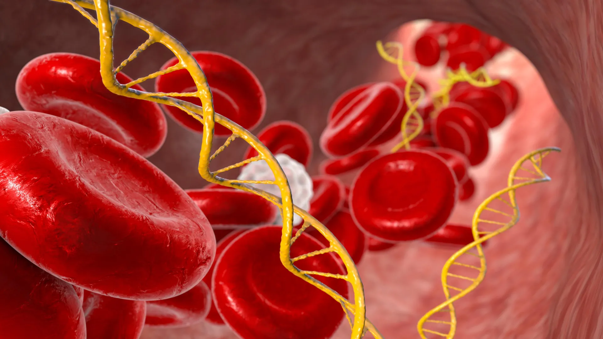
    Mitochondrial DNA disease

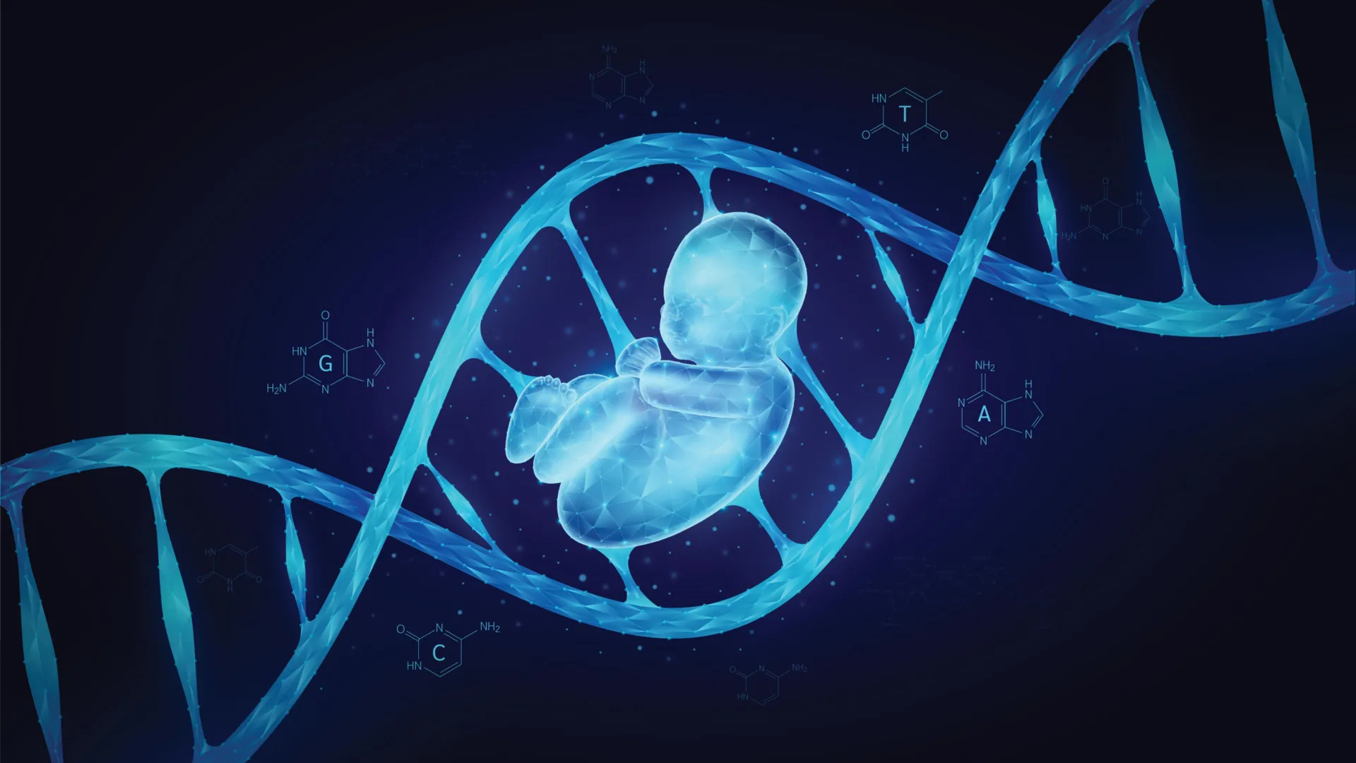
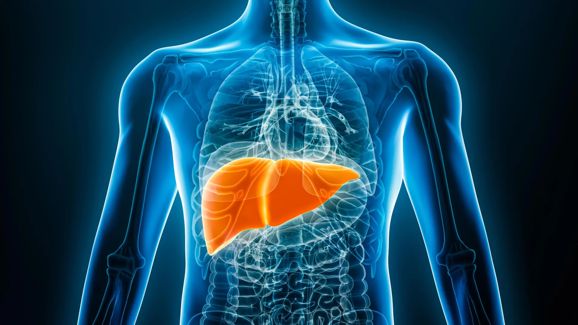
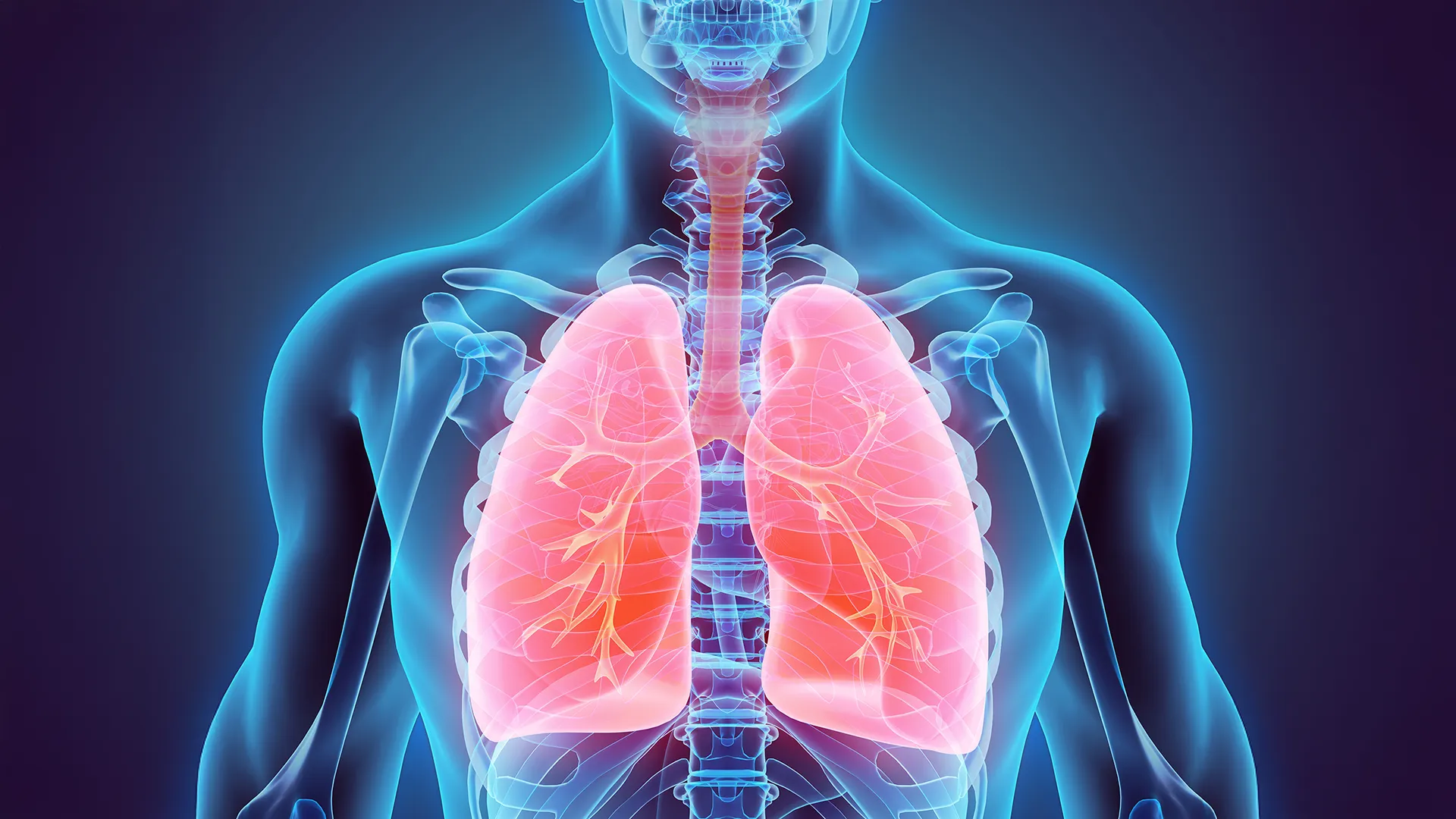
    Every year, around one in 5,000 children is born with mitochondrial DNA mutations that can cause devastating disease. Mitochondria produce the energy required for life and contain a small piece of DNA that only encodes some of the instructions required for energy production.  Harmful mutations in mitochondrial DNA can result in reduced availability of energy, particularly affecting tissues that have high energy demands – for example heart, muscle and brain. Mitochondrial DNA is maternally inherited, and these diseases are therefore passed from mother to child. Although males can be affected, they do not pass on the disease. Despite years of research there is still no cure for people with mitochondrial DNA disease.

    In the absence of a cure for mitochondrial DNA diseases, attention has focused on IVF-based technologies to reduce the risk of disease by limiting transmission of disease-causing mitochondrial DNA mutations from mother to child. The new IVF-based mitochondrial donation technology, pronuclear transfer, which was legalized in the UK in 2015, is designed to reduce the risk of mitochondrial DNA disease in children born to women who carry high levels of disease-causing mitochondrial DNA mutations. 

    The Newcastle team now include pronuclear transfer as part of a research study along with a range of reproductive options offered to women at risk of transmitting mitochondrial disease to their children.
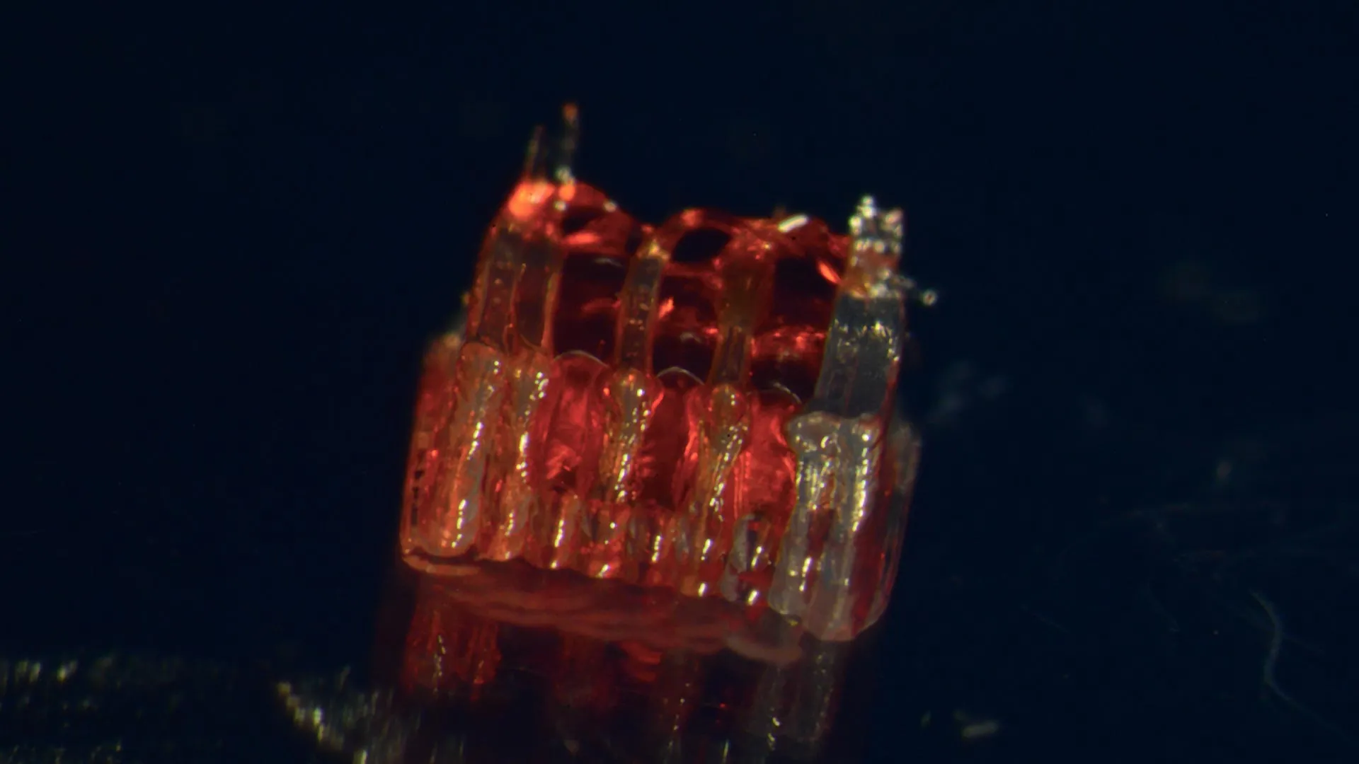
    Pronuclear transfer

    The technique, known as pronuclear transfer is performed after the egg is fertilized. It involves transplanting the nuclear genome (which contains all the genes essential for our individual characteristics, for example, hair color and height) from an egg carrying a mitochondrial DNA mutation to an egg donated by an unaffected woman that has had its nuclear genome removed. The resulting embryo inherits its parents’ nuclear DNA, but the mitochondrial DNA is inherited predominantly from the donated egg.

    The reproductive outcomes paper

    The UK-based Newcastle team who developed and optimized pronuclear transfer for use in fertilized human eggs now report on outcomes of pronuclear-transfer treatment to reduce the risk of mitochondrial DNA disease.

    Levels of disease-causing mitochondrial DNA detected in babies born after pronuclear transfer treatment ranged from undetectable to 16% in neonatal blood. The presence of mitochondrial DNA mutations in babies born after pronuclear transfer treatment results from carryover of maternal mitochondria surrounding the nuclear DNA at the time of transplantation. Carryover of maternal mitochondrial DNA is a known limitation of mitochondrial donation technologies.

    The team is seeking to better understand and address this issue as part of an underpinning research program.

    Professor Mary Herbert, lead author of the reproductive outcomes paper who carried out the research at Newcastle University said: “The findings give grounds for optimism. However, research to better understand the limitations of mitochondrial donation technologies, will be essential to further improve treatment outcomes.

    “Mitochondrial donation technologies are currently regarded as risk reduction treatments owing to carryover of maternal mitochondrial DNA during the mitochondrial donation procedure. Our ongoing research seeks to bridge the gap between risk reduction and prevention of mitochondrial DNA disease by addressing this problem.” 

    Pronuclear-transfer treatment is offered as part of an integrated program that includes preimplantation genetic testing (PGT) for reducing the risk of mitochondrial DNA disease. In accordance with HFEA regulations, pronuclear transfer is offered only to those women who are unlikely to benefit from PGT treatment.

    At the time of reporting the integrated program of PGT and pronuclear transfer, clinical pregnancies were confirmed in 8 of 22 (36%) patients who underwent pronuclear transfer and 16 of 39 (41%) of patients who underwent PGT. Pronuclear transfer has resulted in eight births and one further pregnancy. PGT has resulted in 18 births. In the children from pronuclear transfer, levels of disease-causing mitochondrial DNA mutations were either undetectable or well below the levels at which disease symptoms are observed. 

    The clinical outcomes paper

    The Newcastle team describe the pathway developed to provide the best possible care for women with pathogenic mitochondrial DNA mutations. It describes in detail how the mothers of the first children born with the technique were monitored and supported in pregnancy, and their babies closely followed from birth.

    Some of the mothers already had symptoms of mitochondrial disease including vision loss and heart problems. Others had family members with the disease and remain at risk of developing symptoms and passing it on.

    All eight babies, including a set of identical twins, were healthy at birth and are described as developing normally – five have had no medical problems since. In the paper, the team note that three babies overcame some early health issues that they believe they are not able to attribute directly to mitochondrial donation.

    The Newcastle team offers advice and treatment to women with harmful mitochondrial DNA mutations in the UK. They are carefully monitored during pregnancy and after mitochondrial donation, six of seven progressed without incident. One woman developed a rare complication of pregnancy with a high level of fats detected in her blood (hyperlipidaemia) which responded well to a reduced fat diet. 

    All eight babies, including the set of twins, were born by normal vaginal delivery or elective caesarean section. All babies had normal weight for gestational age. The level of disease-causing mitochondrial DNA mutation was measured in blood and urine cells and was undetectable in five babies. Three babies had low levels of disease-causing mitochondrial DNA mutations – 5 and 9%, 12 and 13%, 16 and 20% in blood and urine respectively.  These levels are well below the 80% level required for clinical disease for these mutations. The researchers note that at follow-up at 18 months, the level of the disease-causing mutation in the child with 5 and 9% was undetectable in blood and urine.

    All children are enrolled in an 18-month developmental study and at the date of reporting all the babies were meeting their relevant developmental milestones.

    One child developed some brief startles (involving neck flexion and eye blinking) at age 7 months, which resolved without treatment after 3 months. Another, a breast-fed baby, developed high blood fats (hyperlipidaemia) which had also affected the mother during pregnancy, and was successfully treated through a low-fat diet. This child was also diagnosed with an abnormal heart rhythm (cardiac arrhythmia) which is being successfully treated with a reducing amount of anti-arrhythmic medication. (Although the children born following PGT are not routinely followed-up, the team note that a cardiac anomaly was detected in one child.)  A third child had a urinary tract infection that responded quickly to antibiotic treatment.

    The authors say that the children’s health conditions are not thought to be related to the maternal mitochondrial DNA mutations as the low levels detected in these babies would not be expected to cause disease symptoms. Symptoms for these mutations are only seen with levels above 80%. Any effect of the pronuclear transfer procedure itself would be expected to have a more uniform clinical manifestation, that is, to affect children in the same way. However, follow-up studies will be of paramount importance in detecting any patterns in childhood conditions.

    The team emphasize that follow-up studies are essential for detecting any patterns in childhood conditions and say they will continue to offer assessments up to the age of 5 years.

    Professor Bobby McFarland, Director of the NHS Highly Specialised Service for Rare Mitochondrial Disorders (Newcastle Hospitals NHS Foundation Trust) and Professor of Paediatric Mitochondrial Medicine at Newcastle University is first author of one of the papers. He said: “While longer term follow-up of children born following mitochondrial donation is of paramount importance, these early results are very encouraging. Seeing the joy and relief these children have brought to their parents is such a privilege.

    “We believe the follow-up process we have put in place is thorough, since it allows us to detect and review even minor health conditions in children born after pronuclear transfer such as a urinary tract infection.” 

    The Lily Foundation, a charity dedicated to fighting mitochondrial disease has supported the Newcastle work. “We’re absolutely delighted with the results of these published papers,” said Liz Curtis, Lily founder and CEO. “We fought long and hard for this change so that families could have choices. After years of waiting, we now know that eight babies have been born using this technique, all showing no signs of mito. For many affected families, it’s the first real hope of breaking the cycle of this inherited condition.”

    FACT FILE

    Law – In a first worldwide and following extensive public debate and scientific and ethical review, UK legalization was press/articles/archive/2015/10/worldfirstledbynewcastleuniversity/”>approved in 2015 to enable the Human Fertilisation and Embryology Authority (HFEA) to allow mitochondrial donation treatments for women at high risk of transmitting serious mitochondrial DNA disease to their children. Following this, the law has now changed in Australia.

    Licence – Licences are regulated and granted by the HFEA. Newcastle Fertility Centre part of Newcastle Hospitals NHS Foundation Trust was granted the press/articles/archive/2017/03/mitochondrialicence/”>first license to perform clinical mitochondrial donation by pronuclear transfer in 2017.  A clinical pathway was established with mitochondrial clinicians as part of NHS England’s Highly Specialised Service.

    Mitochondrial disease refers to a group of genetic conditions that disrupt how our mitochondria – the energy producers in our cells – function.

    Pre-implantation genetic testing (PGT) is a procedure that helps couples avoid passing on genetic conditions to their children. This extra step tests embryos for genetic conditions.

    Pronuclear transfer (PNT) involves transferring the nuclear DNA of a fertilized egg into a fertilized donor egg to prevent the transmission of mitochondrial DNA (mtDNA) disease.

    Funding

    The team acknowledge that the Mitochondrial Reproductive Care Pathway is supported by the NHS at The Newcastle upon Tyne Hospitals NHS Foundation Trust (NUTH). Support was provided by Wellcome. Infrastructural support was provided by Newcastle University, a National Institute for Health and Care Research (NIHR) Biomedical Research Centre award to NUTH. The NHS Highly Specialised Services for Rare Mitochondrial Disorders is supported by NHS England and a career development award was made to Dr Hyslop from Health Education England and the NIHR.

    Cognitive collapse and the nuclear codes: When leaders lose control

    Many former leaders of the world’s nine nuclear-armed nations were impaired by health conditions while in office, raising concerns over their decision-making abilities while they had access to nuclear weapon launch codes, a study from the University of Otago, New Zealand, has found.

    The study analyzed the health information of 51 deceased leaders of nuclear-armed countries: China, France, India, Israel, North Korea, Pakistan, Russia, the United Kingdom and the United States. Eight of the leaders died from chronic disease while still in office, five from heart attacks or strokes. Many of the leaders had multiple serious health issues while in office, including dementia, personality disorders, depression and drug and alcohol abuse.

    The research was led by Professor Nick Wilson, from the Department of Public Health at the University of Otago, Wellington – Ōtākou Whakaihu Waka, Pōneke, with Associate Professor George Thomson and independent researcher Dr Matt Boyd. Professor Wilson says that of the leaders who left office while still alive, 15 had confirmed or possible health issues which likely hastened their departure.

    “Probably all of this group of 15 leaders had their performance in office impaired by their health conditions. In some cases, the degree of impairment was profound, such as in the case of two former Israeli Prime Ministers: Ariel Sharon, who became comatose after suffering a stroke in office, and Menachem Begin, whose depression was so severe he spent his last year as leader isolated in his home. Impairment during crises was also seen in the case of Richard Nixon’s bouts of heavy drinking – including during a nuclear crisis involving the Middle East.

    “There have also been occasions where health information about leaders has been kept secret at the time.”

    This was the case for multiple US presidents, including Dwight D Eisenhower, whose doctor described his 1955 heart attack as a digestive upset; John F Kennedy, whose aides lied about him having Addison’s disease, a serious, chronic condition; and Ronald Reagan, whose administration hid the extent of his injuries after he was shot in 1981, and the likely signs of his dementia near the end of his term.

    Professor Wilson says Kennedy was in poor health during his first two years in office in 1961 and 1962, with his performance likely impaired from Addison’s disease, back pain, and his use of anabolic steroids and amphetamines. It was in 1961 that he authorized the failed CIA-backed Bay of Pigs invasion of Cuba and that his poor performance at a Cold War summit with Soviet leader Nikita Khrushchev in Vienna was noted. In turn, Khrushchev’s poor mental health probably contributed to him triggering both the Berlin Crisis and the Cuban Missile Crisis.

    In France, long-serving President François Mitterrand clung onto power until the end of his term in 1995, despite keeping secret his advanced prostate cancer and after his doctor had concluded in late 1994 that he was no longer capable of carrying out his duties. This latest study follows previous research involving Professor Wilson on the health of former New Zealand Prime Ministers. It found the performance of at least four of the leaders was impaired, in three cases by poor health, and, in the case of Robert Muldoon, by his heavy drinking.

    Professor Wilson says with the rise in international instability following the Russian invasion of Ukraine in 2022 it has become even more important to ensure there is good leadership and governance in those countries with nuclear weapons.

    “This is particularly the case for the United States, where a leader can in principle authorize the release of nuclear weapons on their own, a situation referred to as a ‘nuclear monarchy’.”

    He says there are a range of measures which could reduce global security risks from leaders whose judgement is in question. They include removing nuclear weapons from ‘high alert’ status, adopting ‘no first use’ policies where nations refrain from using nuclear weapons except as a retaliatory second strike, ensuring any weapon launches need authorization by multiple people, and progressing nuclear disarmament treaties.

    Professor Wilson says democracies could consider introducing term limits for their leaders, as well as recall systems, so voters could petition for politicians to step down. Requirements for medical and psychological assessments could be introduced for leaders before they take office, and during their terms.

    “Maintaining a strong media with investigative journalists can also help expose impairment in leaders.”

    Professor Wilson says politicians in general are exposed to high levels of stress, which can affect their mental well-being. A study of UK Members of Parliament has found they were 34 per cent more likely to experience mental health problems than other high-income earners.

    “Finding ways to reduce stress on politicians and better address their mental health needs is another way global security risks can be reduced.”

    Fasting twice a week could be a game-changer for type 2 diabetes

    Intermittent energy restriction, time-restricted eating and continuous energy restriction can all improve blood sugar levels and body weight in people with obesity and type 2 diabetes, according to a study presented at ENDO 2025, the Endocrine Society’s annual meeting in San Francisco, California.

    “This study is the first to compare the effects of three different dietary interventions intermittent energy restriction (IER), time-restricted eating (TRE) and continuous energy restriction (CER) in managing type 2 diabetes with obesity,” said Haohao Zhang, Ph.D., chief physician at The First Affiliated Hospital of Zhengzhou University in Zhengzhou, China.

    Although researchers identified improved HbA1c levels, and adverse events were similar across the three groups, the IER group showed greater advantages in reducing fasting blood glucose, improving insulin sensitivity, lowering triglycerides, and strengthening adherence to the dietary interventions.

    “The research fills a gap in directly comparing 5:2 intermittent energy restriction with a 10-hour time-restricted eating in patients with obesity and type 2 diabetes. The findings provide scientific evidence for clinicians to choose appropriate dietary strategies when treating such patients,” Zhang said.

    Zhang and colleagues performed a single-center, randomized, parallel-controlled trial at the First Affiliated Hospital of Zhengzhou University from November 19, 2021 to November 7, 2024.

    Ninety patients were randomly assigned in a 1:1:1 ratio to the IER, TRE or CER group, with consistent weekly caloric intake across all groups. A team of nutritionists supervised the 16-week intervention.

    Of those enrolled, 63 completed the study. There were 18 females and 45 males, with an average age of 36.8 years, a mean diabetes duration of 1.5 years, a baseline BMI of 31.7 kg/m², and an HbA1c of 7.42%.

    At the end of the study, there were no significant differences in HbA1c reduction and weight loss between the IER, TRE and CER groups. However, the absolute decrease in HbA1c and body weight was greatest in the IER group.

    Compared to TRE and CER, IER significantly reduced fasting blood glucose and triglycerides and increased the Matsuda index, a measure of whole-body insulin sensitivity. Uric acid and liver enzyme levels exhibited no statistically significant changes from baseline in any study group.

    Two patients in the IER group and the TRE group, and three patients in the CER group, experienced mild hypoglycemia.

    The IER group had the highest adherence rate (85%), followed by the CER group at 84% and the TRE group at 78%. Both the IER and CER groups showed statistically significant differences compared with the TRE group.

    Zhang said these findings highlight the feasibility and effectiveness of dietary interventions for people who have obesity and type 2 diabetes.

    Sweet but risky: Common sweeteners may be accelerating puberty in kids

    Consuming certain sweeteners commonly found in foods and beverages may increase the risk of early puberty in children, particularly among those who are genetically predisposed, according to a study presented at ENDO 2025, the Endocrine Society’s annual meeting in San Francisco, Calif.

    The researchers found that consuming aspartame, sucralose, glycyrrhizin and added sugars was significantly associated with a higher risk of early puberty, especially in children with certain genetic traits. The more of these sweeteners the teens consumed, the higher their risk of central precocious puberty.

    “This study is one of the first to connect modern dietary habits — specifically sweetener intake — with both genetic factors and early puberty development in a large, real-world cohort,” said Yang-Ching Chen, M.D., Ph.D., of Taipei Municipal Wan Fang Hospital and Taipei Medical University in Taipei, Taiwan. “It also highlights gender differences in how sweeteners affect boys and girls, adding an important layer to our understanding of individualized health risks.”

    A type of early puberty known as central precocious puberty is increasingly common. It can lead to emotional distress, shorter adult height, and increased risk of future metabolic and reproductive disorders.

    Chen’s previous research found that certain sweeteners can directly influence hormones and gut bacteria linked to early puberty. For example, one artificial sweetener called acesulfame potassium or AceK was shown to trigger the release of puberty-related hormones by activating “sweet taste” pathways in brain cells and increasing stress-related molecules. Another sweetener, glycyrrhizin — found in licorice — was found to change the balance of gut bacteria and reduce the activity of genes involved in triggering puberty.

    “This suggests that what children eat and drink, especially products with sweeteners, may have a surprising and powerful impact on their development,” Chen said.

    The new findings come from the Taiwan Pubertal Longitudinal Study (TPLS), begun in 2018. The study included data from 1,407 teens. Central precocious puberty was diagnosed in 481 teens. The researchers assessed teens’ sweetener intake through validated questionnaires and testing of urine samples. Genetic predisposition was quantified using polygenic risk scores derived from 19 genes related to central precocious puberty. Early puberty was diagnosed based on medical exams, hormone levels and scans.

    Sucralose consumption was linked to a higher risk of central precocious puberty in boys and consumption of glycyrrhizin, sucralose and added sugars was associated with a higher risk of central precocious puberty in girls.

    “The findings are directly relevant to families, pediatricians and public health authorities,” Chen said. “They suggest that screening for genetic risk and moderating sweetener intake could help prevent early puberty and its long-term health consequences. This could lead to new dietary guidelines or risk assessment tools for children, supporting healthier development.”

    One shot, seven days: Long-acting levodopa gel tackles Parkinson’s tremors

    A new weekly injectable drug could transform the lives of more than eight million people living with Parkinson’s disease, potentially replacing the need for multiple daily tablets.

    Scientists from the University of South Australia (UniSA) have developed a long-acting injectable formulation that delivers a steady dose of levodopa and carbidopa – two key medications for Parkinson’s – over an entire week.

    Their findings have been reported in the journal Drug Delivery and Translational Research.

    The biodegradable formulation is injected under the skin or into muscle tissue, where it gradually releases the medication over seven days.

    Parkinson’s disease is the second most common neurological disorder, affecting more than 8.5 million people worldwide. Currently there is no cure and the symptoms – tremors, rigidity and slow movement – are managed with oral medications that must be taken several times a day.

    The frequent dosing is a burden, especially for elderly patients or those with swallowing difficulties, leading to inconsistent medication levels, more side effects, and reduced effectiveness.

    Lead researcher Professor Sanjay Garg, from UniSA’s Centre for Pharmaceutical Innovation, says the newly developed injectable could significantly improve treatment outcomes and patient adherence.

    “Our goal was to create a formulation that simplifies treatment, improves patient compliance, and maintains consistent therapeutic levels of medication. This weekly injection could be a game-changer for Parkinson’s care,” Prof Garg says.

    “Levodopa is the gold-standard therapy for Parkinson’s, but its short life span means it must be taken several times a day.”

    UniSA PhD student Deepa Nakmode says the in-situ implant is designed to release both levodopa and carbidopa steadily over one week, maintaining consistent plasma levels and reducing the risks associated with fluctuating drug concentrations.

    “After years of focused research, it’s incredibly rewarding to see our innovation in long-acting injectables for Parkinson’s disease reach this stage. Our invention has now been filed for an Australian patent,” Nakmode says.

    The injectable gel combines an FDA-approved biodegradable polymer PLGA with Eudragit L-100, a pH-sensitive polymer, to achieve a controlled and sustained drug release.

    Extensive lab tests confirmed the system’s effectiveness and safety:

    • More than 90% of the levodopa dose and more than 81% of the carbidopa dose was released over seven days.
    • The implant degraded by over 80% within a week and showed no significant toxicity in cell viability tests.
    • The formulation can be easily administered through a fine 22-gauge needle, minimising discomfort and eliminating the need for surgical implant.

    “The implications of this research are profound,” Prof Garg says. “By reducing the frequency of dosing from multiple times a day to a weekly injection is a major step forward in Parkinson’s therapy. We’re not just improving how the drug is delivered; we’re improving patients’ lives.”

    Prof Garg says the technology could also be adapted for other chronic conditions such as cancer, diabetes, neurodegenerative disorders, pain management, and chronic infections that require long-term drug delivery.

    The system can be tuned to release drugs over a period ranging from a few days to several weeks depending on therapeutic needs.

    UniSA scientists hope to start clinical trials in the near future and are exploring commercialisation opportunities.

    Scientists just found 200+ hidden proteins that may drive Alzheimer’s

    For decades, the story of Alzheimer’s research has been dominated by a battle between A-beta and tau amyloids, both of which can kill neurons and impact the brain’s ability to function. A new study suggests, however, that these sticky brain plaques may not be operating alone.

    Johns Hopkins University researchers have identified more than 200 types of misfolded proteins in rats that could be associated with age-related cognitive decline.

    The findings could lead the way to finding new therapeutic targets and treatments in humans that could provide relief for the millions of people over 65 who suffer from Alzheimer’s, dementia, or other diseases that rob them of their memories and independence as they age.

    “Amyloids are the buildup of misshapen proteins. They’re big and ugly and easy to see under the microscope, so it makes sense that they catch our attention. But we’re seeing hundreds of proteins misfolding in ways that don’t clump together in an amyloid and yet still seem to impact how the brain functions,” said Stephen Fried, an assistant professor of chemistry and protein scientist who studies how molecules in the brain change during aging. “Our research is showing that amyloids are just the tip of the iceberg.”

    The results were published on July 11 in Science Advances.

    To understand the molecular differences between older brains that are mentally sharp and those that are experiencing decline, Fried and his team studied 17 2-year-old rats that grew up in the same colony. Seven rats performed poorly on memory and problem-solving tests and were considered cognitively impaired, while 10 performed as well as 6-month-old rats.

    The researchers then measured more than 2,500 types of protein in the hippocampus, the part of the brain associated with spatial learning and memory. For the first time, scientists were able to determine for a large number of proteins whether individual proteins were misshapen or folded incorrectly, allowing the researchers to work out which proteins misfold for all the rats and are associated with aging in general versus which proteins specifically misfold in cognitively impaired rats.

    More than 200 proteins were misfolded in the cognitively impaired rats yet maintained their shapes in the cognitively healthy rats. The findings suggest that some of those proteins are contributing to cognitive decline, the researchers said.

    Misfolded proteins are unable to carry out tasks necessary for a cell to function properly, so cells have a natural surveillance system that identifies and destroys these misbehaving proteins. Previously, researchers thought misfolded proteins — specifically A-beta and tau proteins — were only disruptive when they clumped into amyloids.

    “We think there are a lot of proteins that can be misfolded, not form amyloids, and still be problematic,” Fried said. “And that suggests these misfolded proteins have ways of escaping this surveillance system in the cell.”

    But exactly how those misfolded proteins slip past a cell’s security system remains a mystery.

    Next, the team plans to look at misfolded proteins under high-resolution microscopes to get a more detailed picture of what their deformities look like at the molecular level.

    “A lot of us have experienced a loved one or a relative who has become less capable of doing those everyday tasks that require cognitive abilities,” Fried said. “Understanding what’s physically going on in the brain could lead to better treatments and preventive measures.”

    Florida cat sniffs out another new virus—and scientists are listening

    Pepper, the pet cat who made headlines last year for his role in the discovery of the first jeilongvirus found in the U.S., is at it again. This time, his hunting prowess contributed to the identification of a new strain of orthoreovirus.

    John Lednicky, Ph.D., Pepper’s owner and a University of Florida College of Public Health and Health Professions virologist, took Pepper’s catch — a dead Everglades short-tailed shrew — into the lab for testing as part of his ongoing work to understand transmission of the mule deerpox virus.

    Testing revealed the shrew had a previously unidentified strain of orthoreovirus. Viruses in this genus are known to infect humans, white-tailed deer, bats and other mammals. While orthoreoviruses’ effects on humans are not yet well understood, there have been rare reports of the virus being associated with cases of encephalitis, meningitis and gastroenteritis in children.

    “The bottom line is we need to pay attention to orthoreoviruses, and know how to rapidly detect them,” said Lednicky, a research professor in the PHHP Department of Environmental and Global Health and a member of UF’s Emerging Pathogens Institute.

    The UF team published the complete genomic coding sequences for the virus they named “Gainesville shrew mammalian orthoreovirus type 3 strain UF-1” in the journal Microbiology Resource Announcements.

    “There are many different mammalian orthoreoviruses and not enough is known about this recently identified virus to be concerned,” said the paper’s lead author Emily DeRuyter, a UF Ph.D. candidate in One Health. “Mammalian orthoreoviruses were originally considered to be ‘orphan’ viruses, present in mammals including humans, but not associated with diseases. More recently, they have been implicated in respiratory, central nervous system and gastrointestinal diseases.”

    The Lednicky lab’s jeilongvirus and orthoreovirus discoveries come on the heels of the team publishing their discovery of two other novel viruses found in farmed white-tailed deer. Given the propensity of viruses to constantly evolve, paired with the team’s sophisticated lab techniques, finding new viruses isn’t entirely surprising, Lednicky said.

    “I’m not the first one to say this, but essentially, if you look, you’ll find, and that’s why we keep finding all these new viruses,” Lednicky said.

    Like influenza virus, two different types of orthoreovirus can infect a host cell, causing the viruses’ genes to mix and match, in essence, creating a brand new virus, Lednicky said.

    In 2019, Lednicky and colleagues isolated the first orthoreovirus found in a deer. That strain’s genes were nearly identical to an orthoreovirus found in farmed mink in China and a deathly ill lion in Japan. How in the world, the scientific community wondered, could the same hybrid virus appear in a farmed deer in Florida and two species of carnivores across the globe? Some experts speculated that components of the animals’ feed could have come from the same manufacturer.

    With so many unanswered questions about orthoreoviruses and their modes of transmission, prevalence in human and animal hosts and just how sick they could make us, more research is needed, DeRuyter and Lednicky said.

    Next steps would include serology and immunology studies to understand the threat Gainesville shrew mammalian orthoreovirus type 3 strain UF-1 may hold for humans, wildlife and pets.

    For readers concerned about Pepper’s health, rest assured. He has shown no signs of illness from his outdoor adventures and will likely continue to contribute to scientific discovery through specimen collection.

    “This was an opportunistic study,” Lednicky said. “If you come across a dead animal, why not test it instead of just burying it? There is a lot of information that can be gained.”

    This tiny implant could save diabetics from silent, deadly crashes

    For people with Type 1 diabetes, developing hypoglycemia, or low blood sugar, is an ever-present threat. When glucose levels become extremely low, it creates a life-threatening situation for which the standard treatment of care is injecting a hormone called glucagon.

    As an emergency backup, for cases where patients may not realize that their blood sugar is dropping to dangerous levels, MIT engineers have designed an implantable reservoir that can remain under the skin and be triggered to release glucagon when blood sugar levels get too low.

    This approach could also help in cases where hypoglycemia occurs during sleep, or for diabetic children who are unable to administer injections on their own.

    “This is a small, emergency-event device that can be placed under the skin, where it is ready to act if the patient’s blood sugar drops too low,” says Daniel Anderson, a professor in MIT’s Department of Chemical Engineering, a member of MIT’s Koch Institute for Integrative Cancer Research and Institute for Medical Engineering and Science (IMES), and the senior author of the study. “Our goal was to build a device that is always ready to protect patients from low blood sugar. We think this can also help relieve the fear of hypoglycemia that many patients, and their parents, suffer from.”

    The researchers showed that this device could also be used to deliver emergency doses of epinephrine, a drug that is used to treat heart attacks and can also prevent severe allergic reactions, including anaphylactic shock.

    Siddharth Krishnan, a former MIT research scientist who is now an assistant professor of electrical engineering at Stanford University, is the lead author of the study, which was published on July 9 in Nature Biomedical Engineering.

    Emergency response

    Most patients with type 1 diabetes use daily insulin injections to help their body absorb sugar and prevent their blood sugar levels from getting too high. However, if their blood sugar levels get too low, they develop hypoglycemia, which can lead to confusion and seizures, and may be fatal if it goes untreated.

    To combat hypoglycemia, some patients carry preloaded syringes of glucagon, a hormone that stimulates the liver to release glucose into the bloodstream. However, it isn’t always easy for people, especially children, to know when they are becoming hypoglycemic.

    “Some patients can sense when they’re getting low blood sugar, and go eat something or give themselves glucagon,” Anderson says. “But some are unaware that they’re hypoglycemic, and they can just slip into confusion and coma. This is also a problem when patients sleep, as they are reliant on glucose sensor alarms to wake them when sugar drops dangerously low.”

    To make it easier to counteract hypoglycemia, the MIT team set out to design an emergency device that could be triggered either by the person using it, or automatically by a sensor.

    The device, which is about the size of a quarter, contains a small drug reservoir made of a 3D-printed polymer. The reservoir is sealed with a special material known as a shape-memory alloy, which can be programmed to change its shape when heated. In this case, the researcher used a nickel-titanium alloy that is programmed to curl from a flat slab into a U-shape when heated to 40 degrees Celsius.

    Like many other protein or peptide drugs, glucagon tends to break down quickly, so the liquid form can’t be stored long-term in the body. Instead, the MIT team created a powdered version of the drug, which remains stable for much longer and stays in the reservoir until released.

    Each device can carry either one or four doses of glucagon, and it also includes an antenna tuned to respond to a specific frequency in the radiofrequency range. That allows it to be remotely triggered to turn on a small electrical current, which is used to heat the shape-memory alloy. When the temperature reaches the 40-degree threshold, the slab bends into a U shape, releasing the contents of the reservoir.

    Because the device can receive wireless signals, it could also be designed so that drug release is triggered by a glucose monitor when the wearer’s blood sugar drops below a certain level.

    “One of the key features of this type of digital drug delivery system is that you can have it talk to sensors,” Krishnan says. “In this case, the continuous glucose-monitoring technology that a lot of patients use is something that would be easy for these types of devices to interface with.”

    Reversing hypoglycemia

    After implanting the device in diabetic mice, the researchers used it to trigger glucagon release as the animals’ blood sugar levels were dropping. Within less than 10 minutes of activating the drug release, blood sugar levels began to level off, allowing them to remain within the normal range and avert hypoglycemia.

    The researchers also tested the device with a powdered version of epinephrine. They found that within 10 minutes of drug release, epinephrine levels in the bloodstream became elevated and heart rate increased.

    In this study, the researchers kept the devices implanted for up to four weeks, but they now plan to see if they can extend that time up to at least a year.

    “The idea is you would have enough doses that can provide this therapeutic rescue event over a significant period of time. We don’t know exactly what that is — maybe a year, maybe a few years, and we’re currently working on establishing what the optimal lifetime is. But then after that, it would need to be replaced,” Krishnan says.

    Typically, when a medical device is implanted in the body, scar tissue develops around the device, which can interfere with its function. However, in this study, the researchers showed that even after fibrotic tissue formed around the implant, they were able to successfully trigger the drug release.

    The researchers are now planning for additional animal studies and hope to begin testing the device in clinical trials within the next three years.

    “It’s really exciting to see our team accomplish this, which I hope will someday help diabetic patients and could more broadly provide a new paradigm for delivering any emergency medicine,” says Robert Langer, the David H. Koch Institute Professor at MIT and an author of the paper.

    Other authors of the paper include Laura O’Keeffe, Arnab Rudra, Derin Gumustop, Nima Khatib, Claudia Liu, Jiawei Yang, Athena Wang, Matthew Bochenek, Yen-Chun Lu, Suman Bose, and Kaelan Reed.

    The research was funded by the Leona M. and Harry B. Helmsley Charitable Trust, the National Institutes of Health, a JDRF postdoctoral fellowship, and the National Institute of Biomedical Imaging and Bioengineering.

    How a hidden brain circuit fuels fibromyalgia, migraines, and PTSD

    Pain isn’t just a physical sensation — it also carries emotional weight. That distress, anguish, and anxiety can turn a fleeting injury into long-term suffering.

    Researchers at the Salk Institute have now identified a brain circuit that gives physical pain its emotional tone, revealing a new potential target for treating chronic and affective pain conditions such as fibromyalgia, migraine, and post-traumatic stress disorder (PTSD).

    Published on July 9, 2025, in Proceedings of the National Academy of Sciences, the study identifies a group of neurons in a central brain area called the thalamus that appears to mediate the emotional or affective side of pain in mice. This new pathway challenges the textbook understanding of how pain is processed in the brain and body.

    “For decades, the prevailing view was that the brain processes sensory and emotional aspects of pain through separate pathways,” says senior author Sung Han, associate professor and holder of the Pioneer Fund Developmental Chair at Salk. “But there’s been debate about whether the sensory pain pathway might also contribute to the emotional side of pain. Our study provides strong evidence that a branch of the sensory pain pathway directly mediates the affective experience of pain.”

    The physical sensation of pain is what allows you to immediately detect it, assess its intensity, and identify its source. The affective part of pain is what makes it so unpleasant. This emotional discomfort motivates you to take action and helps you learn to associate negative feelings with the situation so you can avoid it in the future.

    This is a critical distinction. Most people start to perceive pain at the same stimulus intensities, meaning we all process the sensory side of pain fairly similarly. In comparison, our ability to tolerate pain varies greatly. How much we suffer or feel threatened by pain is determined by our affective processing, and if that becomes too sensitive or lasts too long, it can result in a pain disorder. This makes it important to understand which parts of the brain control these different dimensions of pain.

    Sensory pain was thought to be mediated by the spinothalamic tract, a pathway that sends pain signals from the spinal cord to the thalamus, which then relays them to sensory processing areas across the brain.

    Affective pain was generally thought to be mediated by a second pathway called the spinoparabrachial tract, which sends pain information from the spinal cord into the brainstem.

    However, previous studies using older research methods have suggested the circuitry of pain may be more complex. This long-standing debate inspired Han and his team to revisit the question with modern research tools.

    Using advanced techniques to manipulate the activity of specific brain cells, the researchers discovered a new spinothalamic pathway in mice. In this circuit, pain signals are sent from the spinal cord into a different part of the thalamus, which has connections to the amygdala, the brain’s emotional processing center. This particular group of neurons in the thalamus can be identified by their expression of CGRP (calcitonin gene-related peptide), a neuropeptide originally discovered in Professor Ronald Evans’ lab at Salk.

    When the researchers “turned off” (genetically silenced) these CGRP neurons, the mice still reacted to mild pain stimuli, such as heat or pressure, indicating their sensory processing was intact. However, they didn’t seem to associate lasting negative feelings with these situations, failing to show any learned fear or avoidance behaviors in future trials. On the other hand, when these same neurons were “turned on” (optogenetically activated), the mice showed clear signs of distress and learned to avoid that area, even when no pain stimuli had been used.

    “Pain processing is not just about nerves detecting pain; it’s about the brain deciding how much that pain matters,” says first author Sukjae Kang, a senior research associate in Han’s lab. “Understanding the biology behind these two distinct processes will help us find treatments for the kinds of pain that don’t respond to traditional drugs.”

    Many chronic pain conditions — such as fibromyalgia and migraine — involve long, intense, unpleasant experiences of pain, often without a clear physical source or injury. Some patients also report extreme sensitivity to ordinary stimuli like light, sound, or touch, which others would not perceive as painful.

    Han says overactivation of the CGRP spinothalamic pathway may contribute to these conditions by making the brain misinterpret or overreact to sensory inputs. In fact, transcriptomic analysis of the CGRP neurons showed that they express many of the genes associated with migraine and other pain disorders.

    Notably, several CGRP blockers are already being used to treat migraines. This study may help explain why these medications work and could inspire new nonaddictive treatments for affective pain disorders.

    Han also sees potential relevance for psychiatric conditions that involve heightened threat perception, such as PTSD. Growing evidence from his lab suggests that the CGRP affective pain pathway acts as part of the brain’s broader alarm system, detecting and responding to not only pain but a wide range of unpleasant sensations. Quieting this pathway with CGRP blockers could offer a new approach to easing fear, avoidance, and hypervigilance in trauma-related disorders.

    Importantly, the relationship between the CGRP pathway and the psychological pain associated with social experiences like grief, loneliness, and heartbreak remains unclear and requires further study.

    “Our discovery of the CGRP affective pain pathway gives us a molecular and circuit-level explanation for the difference between detecting physical pain and suffering from it,” says Han. “We’re excited to continue exploring this pathway and enabling future therapies that can reduce this suffering.”

    Other authors include Shijia Liu, Jong-Hyun Kim, Dong-Il Kim, Tae Gyu Oh, Jiahang Peng, Mao Ye, Kuo-Fen Lee, Ronald M. Evans, and Martyn Goulding of Salk.

    The work was supported by the National Institutes of Mental Health (BRAINS grant 1R01MH116203) and the Simons Foundation (Bridge to Independence award SFARI #388708).

    In seconds, AI builds proteins to battle cancer and antibiotic resistance

    In the last year, there has been a surge in proteins developed by AI that will eventually be used in the treatment of everything from snakebites to cancer. What would normally take decades for a scientist to create — a custom-made protein for a particular disease — can now be done in seconds.

    For the first time, Australian scientists have used Artificial Intelligence (AI) to generate a ready-to-use biological protein, in this case, one that can kill antibiotic resistant bacteria like E. coli.

    This study, published in Nature Communications, provides a new way to combat the growing crisis caused by antibiotic resistant super bugs. By using AI in this way, Australian science has now joined countries like the US and China having developed AI platforms capable of rapidly generating thousands of ready-to-use proteins, paving the way for faster, more affordable drug development and diagnostics that could transform biomedical research and patient care.

    The Nature Communications paper is co-led by Dr. Rhys Grinter and Associate Professor Gavin Knott, a Snow Medical Fellow, who lead the new AI Protein Design Program with nodes at the University of Melbourne Bio21 Institute and Monash Biomedicine Discovery Institute.

    According to Dr. Grinter and A/Prof. Knott, the AI Protein Design Platform used in this work is the first in Australia that models the work done by David Baker (who won the Nobel Prize in Chemistry last year) developing an end-to-end approach that could create a wide range of proteins. “These proteins are now being developed as pharmaceuticals, vaccines, nanomaterials and tiny sensors, with many other applications yet to be tested” Associate Professor Knott said.

    For this study, the AI Protein Design Platform used AI-driven protein design tools that are freely available for scientists everywhere. “It’s important to democratize protein design so that the whole world has the ability to leverage these tools,” said Daniel Fox, the PhD student who performed most of the experimental work for the study. “Using these tools and those we are developing in-house, we can engineer proteins to bind a specific target site or ligand, as inhibitors, agonists or antagonists, or engineered enzymes with improved activity and stability.”

    According to Dr Grinter, currently proteins used in the treatment of diseases like cancer or infections are derived from nature and repurposed through rational design or in vitro evolution and selection. “These new methods in deep learning enable efficient de novo design of proteins with specific characteristics and functions, lowering the cost and accelerating the development of novel protein binders and engineered enzymes,” he said.

    Since the work of David Baker, new tools and software are being developed, such as Bindcraft and Chai which have been incorporated into an AI Protein Design Platform co-led by Dr. Grinter and A/Prof. Knott..

    Professor John Carroll, Director of the Monash Biomedicine Discovery Institute, said the new AI Protein Design Program ‘brings Australia “right up to speed in this exciting new modality for designing novel therapeutics and research tools. It is testament to the entrepreneurial spirit of two fabulous young scientists who have worked night and day to build this capability from scratch.”

    “The Program, based at Monash University and the University of Melbourne, is run by a team of talented structural biologists and computer scientists who understand the design process from end-to-end. This in-depth knowledge of protein structure and machine learning makes us a highly agile program capable of regularly onboarding cutting edge tools in AI-protein design,” Associate Professor Knott said.

    Lemurs age without inflammation—and it could change human health forever

    What can lemurs tell us about inflammation and aging, aka “inflammaging” in humans? That’s the question Elaine Guevara, a biological anthropologist who studies the evolution of life history and aging in primates, set out to understand.

    In newly published research on age-related inflammation in ring-tailed and sifaka lemurs, Guevara discovered that perhaps we should rethink the inevitability of inflammaging in humans.

    Although similar in many ways, ring-tailed and sifaka lemurs show differences in life pacing and lifespan, making useful comparisons. Because lemurs and humans are primates and share a common ancestor that lived millions of years ago, they offer valuable insights into human evolution.

    Her findings, she said, were “surprising.”

    “Contrary to our predictions, neither species showed age-related change in either marker of oxidative stress. Neither lemur species exhibited age-related change in inflammation; if anything, contrary to our prediction, ring-tailed lemurs showed marginal declines in inflammation with age,” Guevara said.

    This finding, consistent with a few recent studies of other non-human primates, suggests that lemurs avoid the phenomenon of “inflammaging” widely observed in humans.

    The study shows inflammaging is not a universal feature of primates, pointing to some differences that might suggest it turns out it’s not even a universal feature of humans, according to Christine Drea, a professor of evolutionary anthropology who was one of the researchers working with Guevara.

    What is Inflammaging?

    As we grow older, low-grade chronic inflammation sets in, which in turn can cause health problems such as heart disease, strokes, diabetes, cancer and osteoarthritis.

    Why inflammaging increases with age in humans, what causes it and how it can be prevented are answers to questions that can unlock critical information to help humans live longer and healthier lives.

    Collecting Data from Lemurs

    Drea said the team first had to find a way to measure oxidative stress, which can be found in blood, urine and saliva. They settled on urine.

    “Our role at the beginning was planning, designing, brainstorming, comparing and getting these samples,” said Drea, who has worked with the Duke Lemur Center since 1999. The Lemur Center does not allow research that will harm the animals.

    The next step says Guevara is to conduct similar research with lemurs in the wild.

    “There are a lot of good reasons to think that aging can be quite different in captivity and in the wild, and that in itself, is informative to evaluating the degree to which human inflammation is intrinsic versus environmental,” she said.

    In the meantime, Guevara says this study serves as the first step in unraveling the question of why humans are suffering from inflammatory-related and age-related conditions and finding ways to treat them.

    With a rapidly aging global population, “these insights are essential for mitigating disability and improving quality of life in later years,” she said.

    Bigger crops, fewer nutrients: The hidden cost of climate change

    New preliminary research suggests that a combination of higher atmospheric CO2 and hotter temperatures contribute to a reduction in nutritional quality in food crops, with serious implications for human health and wellbeing.

    Most research into the impact of climate change on food production has focused on crop yield, but the size of the harvest means little if the nutritional value is poor. “Our work looks beyond quantity to the quality of what we eat,” says Jiata Ugwah Ekele, a PhD student at Liverpool John Moores University, UK.

    The ongoing effects of climate change are posed to have devastating and irreversible consequences for plants across the globe. Ms Ekele’s research is primarily focused on exploring how the nutritional content of food crops may be affected by the interacting effects of rising CO2 levels and increasing temperatures associated with climate change. “These environmental changes can affect everything from photosynthesis and growth rates to the synthesis and storage of nutrients in crops,” says Ms Ekele.

    “It’s crucial to understand these impacts because we are what we eat, and plants form the foundation of our food network as the primary producers of the ecosystem,” says Ms Ekele. “By studying these interactions, we can better predict how climate change will shape the nutritional landscape of our food and work toward mitigating those effects.”

    Ms Ekele’s research is focused on popular leafy vegetables, including kale, rocket and spinach. For this project, these crops ae grown in environment-controlled growth chambers at Liverpool John Moores University, and the CO2 and temperature levels are changed to simulate the UK’s predicted future climate scenarios. “Photosynthetic markers such as chlorophyll fluorescence and quantum yield are assessed as the crops grow, while yield and biomass are recorded at harvest,” says Ms Ekele.

    After the plants have been grown under climate change conditions, their nutritional quality was analysed using high-performance liquid chromatography (HPLC) and X-Ray Fluorescence profiling to measure the concentrations of sugar, protein, phenolics, flavonoids, vitamins and antioxidants.

    Preliminary results from this project suggest that elevated levels of atmospheric CO2 can help crops grow faster and bigger, but certainly not healthier. “After some time, the crops showed a reduction in key minerals like calcium and certain antioxidant compounds,” says Ms Ekele.

    These changes were only exacerbated by increases in temperature. “The interaction between CO2 and heat stress had complex effects – the crops do not grow as big or fast and the decline in nutritional quality intensifies,” says Ms Ekele.

    A key early finding is that different crops have responded differently to these climate change stressors, with some species reacting more intensely than others. “This diversity in response highlights that we can’t generalise across crops. This complexity has been both fascinating and challenging and reminds us why it’s important to study multiple stressors together,” says Ms Ekele.

    This nutritional imbalance poses serious health implications for humanity. While higher CO2 levels can increase the concentration of sugars in crops, it can dilute essential proteins, minerals and antioxidants. “This altered balance could contribute to diets that are higher in calories but poorer in nutritional value,” says Ms Ekele. “Increased sugar content in crops, especially fruits and vegetables, could lead to greater risks of obesity and type 2 diabetes – particularly in populations already struggling with non-communicable diseases.”

    Crops with poor nutritional content can also lead to deficiencies in vital proteins and vitamins that compromise the human immune system and exacerbate existing health conditions – particularly in low or middle-income countries. “It’s not just about how much food we grow, but also what’s inside that food and how it supports long-term human wellbeing,” says Ms Ekele.

    Although this research simulates the UK’s projected climate changes, the implications are global. “Food systems in the Global North are already being challenged by shifting weather patterns, unpredictable growing seasons, and more frequent heatwaves,” says Ms Ekele. “In tropical and subtropical regions, these areas also contend with overlapping stressors such as drought, pests, and soil degradation – and are home to millions who depend directly on agriculture for food and income.”

    Ms Ekele and her team are open to collaborating further on this project with the wider research community, including those from agriculture, nutrition and climate policy. “It’s important to connect plant science with broader issues of human well-being. As the climate continues to change, we must think holistically about the kind of food system we’re building – one that not only produces enough food, but also promotes health, equity, and resilience,” says Ms Ekele. “Food is more than just calories; it’s a foundation for human development and climate adaptation.”

    This research is being presented at the Society for Experimental Biology Annual Conference in Antwerp, Belgium on July  8th, 2025.

    Matching your workouts to your personality could make exercising more enjoyable and give you better results

    Finding motivation to exercise can be the greatest challenge in working out. This might be part of the reason why less than a quarter of people achieve the activity goals recommended by the World Health Organization.

    But what if working out could be more enjoyable? One way of achieving this could be opting for types of exercise that fit our personalities. To this end, researchers in the UK now have examined how personality affects what types of exercise we prefer, and our commitment and engagement to them. The results were published in Frontiers in Psychology.

    “We found that our personality can influence how we engage with exercise, and particularly which forms of exercise we enjoy the most,” said first author Dr Flaminia Ronca from University College London’s (UCL) Institute of Sport, Exercise and Health.

    “Understanding personality factors in designing and recommending physical activity programs is likely to be very important in determining how successful a program is, and whether people will stick with it and become fitter,” added senior author Prof Paul Burgess from the UCL Institute of Cognitive Neuroscience.

    Different sports for different people

    The researchers recruited participants that attended lab testing for baseline fitness. They then split them into two groups; the first group was provided with an eight-week home-based fitness plan made up of cycling and strength training (intervention group), the other group continued their usual lifestyle (control group). During lab testing, the first intervention week, and after the intervention, all participants completed a questionnaire on how much they’d enjoyed each training session. The personality traits examined in the study included extraversion, conscientiousness, agreeableness, neuroticism, and openness.

    “Our brains are wired in different ways, which drives our behaviors and how we interact with our environment,” Ronca explained. “So it’s not surprising that personality would also influence how we respond to different intensities of exercise.”

    For example, people scoring high on extraversion enjoyed high intensity sessions with others around, including team sports. Contrary, people scoring high on neuroticism preferred private workouts. While they are fine with high intensity, they need short breaks in between. Others, scoring high on consciousness and openness were found to engage in exercise regardless of whether they particularly enjoyed it or were driven by curiosity, respectively.

    Stress less

    What was particularly interesting was the relationship between personality, change in fitness, and stress, the researchers said. Before the intervention, the stress levels of both groups were similar. After the intervention, however, especially people who scored high in neuroticism showed a strong reduction in stress. “It’s fantastic news, as it highlights that those who benefit the most from a reduction in stress respond very well to exercise,” Ronca said.

    The researchers pointed out that the most important part about exercising is finding something we enjoy and not to be discouraged if we don’t immediately find it. “It’s ok if we don’t enjoy a particular session,” Ronca said. “We can try something else.”

    “We hope that if people can find physical activities that they enjoy they will more readily choose to do them,” Burgess concluded. “After all, we don’t have to nag dogs to go for a walk: being so physically inactive that we start to feel miserable might be a peculiarly human thing to do. In effect, our body punishes us by making us miserable. But for some reason, many of us humans seem poor at picking up on these messages it is sending to our brain.”

    Pregnancy’s 100-million-year secret: Inside the placenta’s evolutionary power play

    An international research team led by scientists from the University of Vienna has uncovered new insights into how specialized cell types and communication networks at the interface between mother and fetus evolved over millions of years. These discoveries shed light on one of nature’s most remarkable innovations – the ability to sustain a successful pregnancy. The findings have just been published in Nature Ecology & Evolution.

    Pregnancy that lasts long enough to support full fetal development is a hallmark evolutionary breakthrough of placental mammals – a group that includes humans. At the center of this is the fetal-maternal interface: the site in the womb where a baby’s placenta meets the mother’s uterus, and where two genetically distinct organisms – mother and fetus – are in intimate contact and constant interaction. This interface has to strike a delicate balance: intimate enough to exchange nutrients and signals, but protected enough to prevent the maternal immune system from rejecting the genetically “foreign” fetus.

    To uncover the origins and mechanisms behind this intricate structure, the team analyzed single-cell transcriptomes – snapshots of active genes in individual cells – from six mammalian species representing key branches of the mammalian evolutionary tree. These included mice and guinea pigs (rodents), macaques and humans (primates), and two more unusual mammals: the tenrec (an early placental mammal) and the opossum (a marsupial that split off from placental mammals before they evolved complex placentas).

    A Cellular “Atlas of Mammal Pregnancy”

    By analyzing cells at the fetal-maternal interface, the researchers were able to trace the evolutionary origin and diversification of the key cell types involved. Their focus was on two main players: placenta cells, which originate from the fetus and invade maternal tissue, and uterine stromal cells, which are of maternal origin and respond to this invasion.

    Using molecular biology tools, the team identified distinct genetic signatures – patterns of gene activity unique to specific cell types and their specialized functions. Notably, they discovered a genetic signature associated with the invasive behavior of fetal placenta cells that has been conserved in mammals for over 100 million years. This finding challenges the traditional view that invasive placenta cells are unique to humans, and reveals instead that they are a deeply conserved feature of mammalian evolution. During this time, the maternal cells weren’t static, either. Placental mammals, but not marsupials, were found to have acquired new forms of hormone production, a pivotal step toward prolonged pregnancies and complex gestation, and a sign that the fetus and the mother could be driving each other’s evolution.

    Cellular Dialogue: Between Cooperation and Conflict

    To better understand how the fetal-maternal interface functions, the study tested two influential theories about the evolution of cellular communication between mother and fetus.

    The first, the “Disambiguation Hypothesis,” predicts that over evolutionary time, hormonal signals became clearly assigned to either the fetus or the mother – a possible safeguard to ensure clarity and prevent manipulation. The results confirmed this idea: certain signals, including WNT proteins, immune modulators, and steroid hormones, could be clearly traced back to one source tissue.

    The second, the “Escalation Hypothesis” (or “genomic Conflict”), suggests an evolutionary arms race between maternal and fetal genes – with, for example, the fetus boosting growth signals while the maternal side tries to dampen them. This pattern was observed in a small number of genes, notably IGF2, which regulates growth. On the whole, evidence pointed to fine-tuned cooperative signaling.

    “These findings suggest that evolution may have favored more coordination between mother and fetus than previously assumed,” says Daniel J. Stadtmauer, lead author of the study and now a researcher at the Department of Evolutionary Biology, University of Vienna. “The so-called mother-fetus power struggle appears to be limited to specific genetic regions. Rather than asking whether pregnancy as a whole is conflict or cooperation, a more useful question may be: where is the conflict?”

    Single-Cell Analysis: A Key to Evolutionary Discovery

    The team’s discoveries were made possible by combining two powerful tools: single-cell transcriptomics – which captures the activity of genes in individual cells – and evolutionary modeling techniques that help scientists reconstruct how traits might have looked in long-extinct ancestors. By applying these methods to cell types and their gene activity, the researchers could simulate how cells communicate in different species, and even glimpse how this dialogue has evolved over millions of years.

    “Our approach opens a new window into the evolution of complex biological systems – from individual cells to entire tissues,” says Silvia Basanta, co-first author and researcher at the University of Vienna. The study not only sheds light on how pregnancy evolved, but also offers a new framework for tracking evolutionary innovations at the cellular level – insights that could one day improve how we understand, diagnose, or treat pregnancy-related complications.

    The research was conducted in the labs of Mihaela Pavličev at the Department of Evolutionary Biology, University of Vienna, and Günter Wagner at Yale University. Wagner is Professor Emeritus at Yale and a Senior Research Fellow at the University of Vienna. The study was supported by the John Templeton Foundation and the Austrian Science Fund (FWF).

    New research shows Monday stress is etched into your biology

    A research study led by Professor Tarani Chandola from the Department of Sociology, Faculty of Social Sciences at the University of Hong Kong (HKU) has revealed that Mondays uniquely drive long-term biological stress, regardless of working status, with implications for heart health.

    The research has identified a striking biological phenomenon: older adults who feel anxious on Mondays exhibit significantly higher long-term stress hormone levels, up to two months later. This “Anxious Monday” effect, observed in both working and retirees, points to a deep-rooted link between the start of the week and dysregulation of the body’s stress response system, a known driver of cardiovascular disease (CVD). Published in the Journal of Affective Disorders, the study analyzed data from over 3,500 older adults participating in the English Longitudinal Study of Ageing (ELSA).

    Key Findings

    • 23% Higher Cortisol Levels: Older adults reporting Monday anxiety had 23% elevated cortisol levels in hair samples (reflecting cumulative exposure over two months) compared to peers anxious on other days.
    • Non-Workers Not Spared: The effect persisted among retirees, challenging assumptions that workplace stress alone explains Monday’s toll.
    • CVD Connection: Mondays are linked to a 19% spike in heart attacks — this study identifies HPA-axis dysregulation as a potential biological bridge.
    • Not just higher levels of Monday anxiety: Only 25% of the Monday effect was due to greater feelings of anxiety on Mondays. The rest was because of the greater effect of feeling anxious on Mondays compared to other days.

    The hypothalamic-pituitary-adrenal (HPA) axis regulates stress hormones like cortisol, which, when chronically elevated, contribute to hypertension, insulin resistance, and immune dysfunction. While prior research noted higher cortisol on weekdays versus weekends, this is the first study to pinpoint Mondays as uniquely disruptive. The findings suggest societal rhythms — not just job demands — embed themselves in human physiology, with lasting health risks.

    “Mondays act as a cultural ‘stress amplifier,'” said Professor Chandola. “For some older adults, the week’s transition triggers a biological cascade that lingers for months. This isn’t about work — it’s about how deeply ingrained Mondays are in our stress physiology, even after careers end.

    The study underscores how the “Monday blues” can become biologically embedded, with chronic stress hormone dysregulation posing long-term cardiovascular risks. Addressing Monday-specific stress could unlock new strategies to combat heart disease in aging populations.

    Feeling mental exhaustion? These two areas of the brain may control whether people give up or persevere
    • Two areas of the brain may work in combination to tell the brain when it’s “feeling” tired.
    • People with depression and post-traumatic stress disorder (PTSD) often experience cognitive fatigue.
    • Results of the study may provide a way for physicians to better evaluate and treat people who experience such fatigue.

    In experiments with healthy volunteers undergoing functional MRI imaging, scientists have found increased activity in two areas of the brain that work together to react to, and possibly regulate, the brain when it’s “feeling” tired and either quits or continues exerting mental effort.

    The experiments, designed to help detect various aspects of brain fatigue, may provide a way for physicians to better evaluate and treat people who experience overwhelming mental exhaustion, including those with depression and post-traumatic stress disorder (PTSD), the scientists say.

    A report on the NIH-funded study was published online June 11 in the Journal of Neuroscience, detailing results on 18 female and 10 male healthy adult volunteers given tasks to exercise their memory.

    “Our lab focuses on how [our minds] generate value for effort,” says Vikram Chib, Ph.D., associate professor of biomedical engineering at the Johns Hopkins University School of Medicine and a research scientist at Kennedy Krieger Institute. “We understand less about the biology of cognitive tasks, including memory and recall, than we do about physical tasks, even though both involve a lot of effort.” Anecdotally, Chib says, scientists know cognitive tasks are tiring, and relatively less about why and how such fatigue develops and plays out in the brain.

    The 28 study participants, who ranged in age from 21 to 29, were paid $50 to participate in the study, and were told they could receive additional payments based on their performance and choices. All participants received a baseline MRI scan before the experiments began.

    The tests of their working memory, which took place while undergoing subsequent MRI scans of their brains, included looking at a series of letters, in sequence, on a screen and recalling the position of certain letters. The farther back a letter was in the series of letters, the harder it was to recall its position, increasing the cognitive effort expended. The participants were given feedback on their performance after each test and opportunities to receive increasing payments ($1-$8) with more difficult recall exercises. The participants also were asked before and after each test to self-rate their level of cognitive fatigue.

    Overall, the test results found increased activity and connectivity in two brain areas when participants reported cognitive fatigue: the right insula, an area deep in the brain that has been associated with feelings of fatigue, and the dorsal lateral prefrontal cortex, areas on both sides of the brain that control working memory. For each participant, activity in both brain locations during cognitive fatigue increased by more than twice the level of baseline measurements taken before starting the tests.

    “Our study was designed to induce cognitive fatigue and see how people’s choices to exert effort change when they feel fatigue, as well as identify locations in the brain where these decisions are made,” says Chib.

    Notably, Chib and his research team members Grace Steward and Vivian Looi found that the financial incentives need to be high in order for participants to exert increased cognitive effort, suggesting that external incentives prompt such effort.

    “That outcome wasn’t entirely surprising, given our previous work finding the same need for incentives in spurring physical effort,” says Chib.

    “The two areas of the brain may be working together to decide to avoid more cognitive effort unless there are more incentives offered. However, there may be a discrepancy between perceptions in cognitive fatigue and what the human brain is actually capable of doing,” says Chib.

    Fatigue is linked with many neurological conditions, including PTSD and depression, says Chib. “Now that we’ve likely identified some of the neural circuits for cognitive effort in healthy people, we need to look at how fatigue manifests in the brains of people with these conditions,” he adds.

    Chib says it may be possible to use medication or cognitive behavior therapy to combat cognitive fatigue, and the current study using decision tasks and functional MRI could be a framework for objectively classifying cognitive fatigue.

    Functional MRI uses blood flow to measure broad areas of activity in the brain; however, it does not directly measure neuron activation, nor more subtle nuances in brain activity.

    “This study was performed in an MRI scanner and with very specific cognitive tasks. It will be important to see how these results generalize to other cognitive effort and real-world tasks,” says Chib.

    Funding for the research was provided by the National Institutes of Health (R01HD097619, R01MH119086).

    Study finds tummy-tuck patients still shedding pounds five years later

    Most patients undergoing “tummy tuck” surgery (abdominoplasty) to remove excess skin and tissue after weight loss continue to lose weight in the months and years after surgery, suggests a follow-up study in the July issue of Plastic and Reconstructive Surgery®, the official medical journal of the American Society of Plastic Surgeons (ASPS). The journal is published in the Lippincott portfolio by Wolters Kluwer.

    “We found that patients not only maintained their weight loss after abdominoplasty, but also continued to lose weight over time – up to ten pounds, on average,” comments senior author John Y.S. Kim of Northwestern University Feinberg School of Medicine, Chicago. “This postoperative weight loss appears greater, and increases at later follow-up times, in patients with initially higher body mass index [BMI].”

    Continued weight loss up to five years after tummy tuck

    Abdominoplasty is a cosmetic surgical procedure to improve the appearance of the abdomen. In 2023, ASPS Member Surgeons performed more than 170,000 abdominoplasties, according to ASPS statistics. Many of these procedures are performed in patients with massive weight loss that leaves them with excess, sagging skin.

    Plastic surgeons have observed that patients may continue to lose weight after abdominoplasty. However, there is little research evidence on this issue, including whether the abdominoplasty procedure itself contributes to long-term weight loss.

    Dr. Kim and colleagues performed a study to assess changes in body weight in 188 patients who underwent abdominoplasty between 2018 and 2022. Ninety-seven percent of patients were women. The average preoperative weight was about 168 pounds with a BMI of 27.7. Most patients underwent liposuction or a further procedure to remove excess fat (lipectomy) at the same time as abdominoplasty. Trends in body weight were assessed through up to five years after surgery.

    The results showed continued weight loss after abdominoplasty. At three to six months, average weight loss was between five and six pounds, with about a three percent decrease in BMI. From one to four years, weight loss was about five pounds, for a BMI reduction of about two percent. By five years (in a limited number of patients), average weight loss was nearly ten pounds, with more than a five percent decrease in BMI.

    ‘Near-constant negative change in body weight’ after abdominoplasty

    Overall, about 60% of patients lost weight during follow-up. Further analysis showed a “near constant negative change in body weight that did not significantly change over time,” the researchers write.

    After adjustment for other factors, continued weight loss was more likely for older patients, for those who underwent liposuction/lipectomy, and those who had never smoked. Weight loss was greater for patients who had higher body weight and BMI before surgery, and for a small number of patients who used the newer weight loss medication semaglutide.

    The study adds new evidence that “post-abdominoplasty weight reduction is a quantifiable phenomenon and that patients undergoing abdominoplasty continue to lose a significant amount of weight for up to five years after surgery,” the researchers write. They note some key limitations of their study, including varying follow-up times and potential confounding factors.

    The study cannot definitively explain why patients continue to lose weight after surgery. However, Dr. Kim and coauthors write, “We have found that patients who were able to achieve weight loss after their abdominoplasty succeeded in developing healthy habits that centered around nutrition and exercise.” They highlight the need for an “evidence-based platform” to assess weight changes after abdominoplasty and to identify factors associated with long-term weight loss.

    Multisensory VR forest reboots your brain and lifts mood—study confirms

    In Japan, Shinrin Yoku or forest bathing has already been used for therapeutic applications, for instance, to lower blood pressure and stress levels. For their study, the researchers wanted to find out whether forest bathing – consciously immersing oneself in nature – can also be effective when done virtually, and focused on whether the positive effect is stronger when several senses are addressed simultaneously.

    For the project, a high-quality 360° VR video was produced in Europe’s largest Douglas fir forest, the Sonnenberg nature reserve near Parchim – complete with original sounds and the scent of essential oils from the Douglas fir. The participants experienced the virtual forest scenery either as a full sensory experience (with images, sound and scent) or in a reduced form whereby forest stimuli appealing to just a single sense – visual, auditory or olfactory – were used. In variants where only hearing or scent was activated, participants were placed in a neutral virtual environment to minimize visual stimuli and the influence of VR technology.

    Significantly better effect with sensory combination

    More than 130 participants were first put into an acute stress situation using stress-inducing images. Then, equipped with VR glasses, they experienced one of the four forest stimulation/ bathing variants. The results show that the combination of all three sensory stimuli led to a significantly greater improvement in mood and a stronger feeling of connection with nature compared to when individual sensory stimuli were presented. In addition to positive effects on mood, there were also limited improvements in working memory – the cognitive function that enables us to store, process and retrieve information in the short term.

    However, the researchers point out that the effects are area-specific and cannot yet be considered universally valid. Further studies with larger samples are needed to confirm the results and provide a better understanding of the mechanisms behind the restorative effects of virtual nature experiences.

    “We can already say that digital nature experiences can absolutely produce an emotional effect – even if they don’t replace actual nature,” reports Leonie Ascone, lead author of the study and researcher in the Neuronal Plasticity working group at the University Medical Center Hamburg-Eppendorf (UKE).

    Potential for clinics, waiting rooms and urban spaces

    Simone Kühn, head of the study and Director of the Center for Environmental Neuroscience at the Max Planck Institute for Human Development, adds: “Especially in places with limited access to nature – such as clinics, waiting areas or urban interiors – multisensory VR applications or targeted nature staging could support mental well-being. The images, sounds and scents of nature offer previously underestimated potential for improving mood and mental performance in everyday situations.” Kühn conducts intensive research into the effects of the environment on the human brain and, together with colleagues from universities in Vienna, Exeter and Birmingham, was recently able to prove that just from watching nature videos, patients perceive physical pain as less intense (Steininger et al., 2025).

    In brief:

    • Forest bathing in Virtual Reality improves emotional well-being and increases connectedness to nature, particularly when several senses (sight, hearing, smell) are simultaneously engaged
    • The study used a 360° VR forest video complete with original sounds and the scent of Douglas fir essential oils
    • There is potential for application especially in clinical, urban and other environments with limited access to nature
    Cough medicine turned brain protector? Ambroxol may slow Parkinson’s dementia

    Dementia poses a major health challenge with no safe, affordable treatments to slow its progression.

    Researchers at Lawson Research Institute (Lawson), the research arm of St. Joseph’s Health Care London, are investigating whether Ambroxol — a cough medicine used safely for decades in Europe — can slow dementia in people with Parkinson’s disease.

    Published on June 30 in the prestigious JAMA Neurology, this 12-month clinical trial involving 55 participants with Parkinson’s disease dementia (PDD) monitored memory, psychiatric symptoms and GFAP, a blood marker linked to brain damage. Parkinson’s disease dementia causes memory loss, confusion, hallucinations and mood changes. About half of those diagnosed with Parkinson’s develop dementia within 10 years, profoundly affecting patients, families and the health care system.

    Led by Cognitive Neurologist Dr. Stephen Pasternak, the study gave one group daily Ambroxol while the other group received a placebo. “Our goal was to change the course of Parkinson’s dementia,” says Pasternak. “This early trial offers hope and provides a strong foundation for larger studies.”

    Key findings from the clinical trial include:

    • Ambroxol was safe, well-tolerated and reached therapeutic levels in the brain
    • Psychiatric symptoms worsened in the placebo group but remained stable in those taking Ambroxol.
    • Participants with high-risk GBA1 gene variants showed improved cognitive performance on Ambroxol
    • A marker of brain cell damage (GFAP) increased in the placebo group but stayed stable with Ambroxol, suggesting potential brain protection.

    Although Ambroxol is approved in Europe for treating respiratory conditions and has a long-standing safety record — including use at high doses and during pregnancy — it is not approved for any use in Canada or the U.S.

    “Current therapies for Parkinson’s disease and dementia address symptoms but do not stop the underlying disease,” explains Pasternak. “These findings suggest Ambroxol may protect brain function, especially in those genetically at risk. It offers a promising new treatment avenue where few currently exist.”

    Ambroxol supports a key enzyme called glucocerebrosidase (GCase), which is produced by the GBA1 gene. In people with Parkinson’s disease, GCase levels are often low. When this enzyme doesn’t work properly, waste builds up in brain cells, leading to damage. Pasternak learned about Ambroxol during a fellowship at The Hospital for Sick Children (SickKids) in Toronto, where it was identified as a treatment for Gaucher disease — a rare genetic disorder in children caused by a deficiency of GCase.

    He is now applying that research to explore whether boosting GCase with Ambroxol could help protect the brain in Parkinson’s-related diseases. “This research is vital because Parkinson’s dementia profoundly affects patients and families,” says Pasternak. “If a drug like Ambroxol can help, it could offer real hope and improve lives.”

    Funded by the Weston Foundation, this study is an important step toward developing new treatments for Parkinson’s disease and other cognitive disorders, including dementia with Lewy bodies. Pasternak and his team plan to start a follow-up clinical trial focused specifically on cognition later this year.

    Scientists discovered how a scent can change your mind

    Mice taught to link smells with tastes, and later fear, revealed how the amygdala teams up with cortical regions to let the brain draw powerful indirect connections. Disabling this circuit erased the links, hinting that similar pathways in humans could underlie disorders like PTSD and psychosis, and might be tuned with future brain-modulation therapies.

    Frozen light switches: How Arctic microbes could revolutionize neuroscience

    Imagine the magnificent glaciers of Greenland, the eternal snow of the Tibetan high mountains, and the permanently ice-cold groundwater in Finland. As cold and beautiful these are, for the structural biologist Kirill Kovalev, they are more importantly home to unusual molecules that could control brain cells’ activity.

    Kovalev, EIPOD Postdoctoral Fellow at EMBL Hamburg’s Schneider Group and EMBL-EBI’s Bateman Group, is a physicist passionate about solving biological problems. He is particularly hooked by rhodopsins, a group of colorful proteins that enable aquatic microorganisms to harness sunlight for energy.

    “In my work, I search for unusual rhodopsins and try to understand what they do,” said Kovalev. “Such molecules could have undiscovered functions that we could benefit from.”

    Some rhodopsins have already been modified to serve as light-operated switches for electrical activity in cells. This technique, called optogenetics, is used by neuroscientists to selectively control neuronal activity during experiments. Rhodopsins with other abilities, such as enzymatic activity, could be used to control chemical reactions with light, for example.

    Having studied rhodopsins for years, Kovalev thought he knew them inside out – until he discovered a new, obscure group of rhodopsins that were unlike anything he had seen before.

    As it often happens in science, it started serendipitously. While browsing online protein databases, Kovalev spotted an unusual feature common to microbial rhodopsins found exclusively in very cold environments, such as glaciers and high mountains. “That’s weird,” he thought. After all, rhodopsins are something you typically find in seas and lakes.

    These cold-climate rhodopsins were almost identical to each other, even though they evolved thousands of kilometres apart. This couldn’t be a coincidence. They must be essential for surviving in the cold, concluded Kovalev, and to acknowledge this, he named them ‘cryorhodopsins’.

    Rhodopsins out of the blue

    Kovalev wanted to know more: what these rhodopsins look like, how they work, and, in particular, what color they are.

    Color is the key feature of each rhodopsin. Most are pink-orange – they reflect pink and orange light, and absorb green and blue light, which activates them. Scientists strive to create a palette of different colored rhodopsins, so they could control neuronal activity with more precision. Blue rhodopsins have been especially sought-after because they are activated by red light, which penetrates tissues more deeply and non-invasively.

    To Kovalev’s amazement, the cryorhodopsins he examined in the lab revealed an unexpected diversity of colors, and, most importantly, some were blue.

    The color of each rhodopsin is determined by its molecular structure, which dictates the wavelengths of light it absorbs and reflects. Any changes in this structure can alter the color.

    “I can actually tell what’s going on with cryorhodopsin simply by looking at its color,” laughed Kovalev.

    Applying advanced structural biology techniques, he figured out that the secret to the blue color is the same rare structural feature that he originally spotted in the protein databases.

    “Now that we understand what makes them blue, we can design synthetic blue rhodopsins tailored to different applications,” said Kovalev.

    Next, Kovalev’s collaborators examined cryorhodopsins in cultured brain cells. When cells expressing cryorhodopsins were exposed to UV light, it induced electric currents inside them. Interestingly, if the researchers illuminated the cells right afterwards with green light, the cells became more excitable, whereas if they used UV/red light instead, it reduced the cells’ excitability.

    “New optogenetic tools to efficiently switch the cell’s electric activity both ‘on’ and ‘off’ would be incredibly useful in research, biotechnology and medicine,” said Tobias Moser, Group Leader at the University Medical Center Göttingen who participated in the study. “For example, in my group, we develop new optical cochlear implants for patients that can optogenetically restore hearing in patients. Developing the utility of such a multi-purpose rhodopsin for future applications is an important task for the next studies.”

    “Our cryorhodopsins aren’t ready to be used as tools yet, but they’re an excellent prototype. They have all the key features that, based on our findings, could be engineered to become more effective for optogenetics,” said Kovalev.

    Evolution’s UV light protector

    When exposed to sunlight even on a rainy winter day in Hamburg, cryorhodopsins can sense UV light, as shown using advanced spectroscopy by Kovalev’s collaborators from Goethe University Frankfurt led by Josef Wachtveitl. Wachtveitl’s team showed that cryorhodopsins are in fact the slowest among all rhodopsins in their response to light. This made the scientists suspect that those cryorhodopsins might act like photosensors letting the microbes ‘see’ UV light – a property unheard of among other cryorhodopsins.

    “Can they really do that?” Kovalev kept asking himself. A typical sensor protein teams up with a messenger molecule that passes information from the cell membrane to the cell’s inside.

    Kovalev grew more convinced, when together with his collaborators from Alicante, Spain, and his EIPOD co-supervisor, Alex Bateman from EMBL-EBI, they noticed that the cryorhodopsin gene is always accompanied by a gene encoding a tiny protein of unknown function – likely inherited together, and possibly functionally linked.

    Kovalev wondered if this might be the missing messenger. Using the AI tool AlphaFold, the team were able to show that five copies of the small protein would form a ring and interact with the cryorhodopsin. According to their predictions, the small protein sits poised against the cryorhodopsin inside the cell. They believe that when cryorhodopsin detects UV light, the small protein could depart to carry this information into the cell.

    “It was fascinating to uncover a new mechanism via which the light-sensitive signal from cryorhodopsins could be passed on to other parts of the cell. It is always a thrill to learn what the functions are for uncharacterised proteins. In fact, we find these proteins also in organisms that do not contain cryorhodopsin, perhaps hinting at a much wider range of jobs for these proteins.”

    Why cryorhodopsins evolved their astonishing dual function – and why only in cold environments – remains a mystery.

    “We suspect that cryorhodopsins evolved their unique features not because of the cold, but rather to let microbes sense UV light, which can be harmful to them,” said Kovalev. “In cold environments, such as the top of a mountain, bacteria face intense UV radiation. Cryorhodopsins might help them sense it, so they could protect themselves. This hypothesis aligns well with our findings.”

    “Discovering extraordinary molecules like these wouldn’t be possible without scientific expeditions to often remote locations, to study the adaptations of the organisms living there,” added Kovalev. “We can learn so much from that!”

    Unique approach to unique molecules

    To reveal the fascinating biology of cryorhodopsins, Kovalev and his collaborators had to overcome several technical challenges.

    One was that cryorhodopsins are nearly identical in structure, and even a slight change in the position of a single atom can result in different properties. Studying molecules at this level of detail requires going beyond standard experimental methods. Kovalev applied a 4D structural biology approach, combining X-ray crystallography at EMBL Hamburg beamline P14 and cryo-electron microscopy (cryo-EM) in the group of Albert Guskov in Groningen, Netherlands, with protein activation by light.

    “I actually chose to do my postdoc at EMBL Hamburg, because of the unique beamline setup that made my project possible,” said Kovalev. “The whole P14 beamline team worked together to tailor the setup to my experiments – I’m very grateful for their help.”

    Another challenge was that cryorhodopsins are extremely sensitive to light. For this reason, Kovalev’s collaborators had to learn to work with the samples in almost complete darkness.

    Sweet-smelling molecule halts therapy-resistant pancreatic cancer

    Cancer cells have the capacity to multiply rapidly. The aggressive cancer cells undergo conversion from their tightly connected epithelial state into a mesenchymal state, which lacks contact restrictions and spreads easily to other parts of the body. Such epithelial-to-mesenchymal plasticity also makes the cancer cells resistant to elimination by anticancer therapies.

    The search is ongoing for newer anticancer agents that can overcome this acquired resistance to therapy and destroy the ‘rogue’ cancer cells. A group of researchers led by Dr. Hideyuki Saya, Director of the Oncology Innovation Center, Fujita Health University, Japan, has uncovered the mechanism of the anticancer activity of benzaldehyde, a compound responsible for the aroma of almonds, apricots, and figs.

    Giving insights into their motivation for this study, Dr. Saya explains, “In the 1980s, researchers demonstrated the anticancer activity of benzaldehyde and its derivatives. The first author of our study, Dr. Jun Saito, is the daughter of one of the researchers involved in those early studies, and she was driven by a strong desire to uncover the mechanism behind benzaldehyde’s anticancer effects.” This study, published online in the British Journal of Cancer on May 02, 2025, shows the impact of benzaldehyde on key signaling protein interactions within the cancer cells and the resulting cytotoxicity.

    Early studies reported the ability of benzaldehyde to inhibit the progressive development of mouse embryonic cells, indicating its potential in preventing rapid cell proliferation. Here, the anticancer effects of benzaldehyde were studied by using a mouse model grafted to have a growing pancreatic cancer.

    In cell culture studies, benzaldehyde inhibited the growth of cancer cells resistant to radiation therapy and also those resistant to treatment with osimertinib, an agent blocking tyrosine kinases in growth factor signaling. Benzaldehyde synergized with radiation to eliminate previously radiation-resistant cancer cells.

    The study findings revealed that benzaldehyde exerted its anticancer effects by preventing interactions of the signaling protein 14-3-3ζ with the Ser28-phosphorylated form of histone H3 (H3S28ph). This interaction, key to cancer cell survival, was also responsible for treatment resistance and the expression of genes related to epithelial-mesenchymal plasticity.

    Here, benzaldehyde prevented 14-3-3ζ-dependent phosphorylation of the serine28 amino acid of histone H3. Consequently, benzaldehyde treatment reduced the expression of genes responsible for treatment resistance. Treatment of mice with a benzaldehyde derivative inhibited the growth of pancreatic tumors and suppressed the epithelial-to-mesenchymal plasticity, thus preventing the spread of cancer to distant organs like the lungs.

    By blocking an interaction key to cancer cell survival, benzaldehyde overcomes therapy resistance and prevents metastasis. Sharing the implications of their findings, Dr. Saya concludes, “The 14-3-3ζ protein has long been considered a target for cancer therapy, but its direct inhibition is not feasible due to its important functions in normal cells. Our results suggest that inhibition of the interaction between 14-3-3ζ and its client proteins by benzaldehyde has the potential to overcome the problem.”

    The present study shows benzaldehyde is effective against cancer cells that have acquired resistance to radiation and tyrosine kinase inhibitors commonly used in cancer treatment. In the long term, this study suggests its potential as a combinatorial anticancer agent, alongside molecular-targeted therapies.

    Dr. Jun Saito is a researcher in the laboratory of Dr. Hideyuki Saya, Oncology Innovation Center, Fujita Health University. She obtained her Ph.D. from the Nihon University Graduate School of Medicine. Continuing the legacy of one of her parents, who pioneered the breakthrough research on the anticancer activity of benzaldehyde in the 1980s, Dr. Saito has uncovered the underlying mechanism of benzaldehyde’s anticancer effects. Her expertise includes oncology, pathophysiology, immunology, applied physics, and chemistry.

    This work was supported by Grants-in-Aid for Scientific Research from the Ministry of Education, Culture, Sports, Science, and Technology of Japan (KAKENHI 19K22568).

    Even low levels of air pollution may quietly scar your heart, MRI study finds

    Researchers using cardiac MRI have found that long-term exposure to air pollution is associated with early signs of heart damage, according to a study that was published today in Radiology, a journal of the Radiological Society of North America (RSNA). The research indicates that fine particulate matter in the air may contribute to diffuse myocardial fibrosis, a form of scarring in the heart muscle that can precede heart failure.

    Cardiovascular disease is the leading cause of death worldwide. There is a large body of evidence linking poor air quality with cardiovascular disease. However, the underlying changes in the heart resulting from air pollution exposure are unclear.

    “We know that if you’re exposed to air pollution, you’re at higher risk of cardiac disease, including higher risk of having a heart attack,” said the study’s senior author Kate Hanneman, M.D., M.P.H., from the Department of Medical Imaging at the Temerty Faculty of Medicine, University of Toronto and University Health Network in Toronto. “We wanted to understand what drives this increased risk at the tissue level.”

    Dr. Hanneman and colleagues used cardiac MRI, a noninvasive imaging technique, to quantify myocardial fibrosis and assess its association with long-term exposure to particles known as PM2.5. At 2.5 micrometers in diameter or less, PM2.5 particles are small enough to enter the bloodstream through the lungs. Common sources include vehicle exhaust, industrial emissions and wildfire smoke.

    The researchers wanted to evaluate the effects of air pollution on both healthy people and those with heart disease, so the study group included 201 healthy controls and 493 patients with dilated cardiomyopathy, a disease that makes it more difficult for the heart to pump blood.

    Higher long-term exposure to fine particulate air pollution was linked with higher levels of myocardial fibrosis in both the patients with cardiomyopathy and the controls, suggesting that myocardial fibrosis may be an underlying mechanism by which air pollution leads to cardiovascular complications. The largest effects were seen in women, smokers and patients with hypertension.

    The study adds to growing evidence that air pollution is a cardiovascular risk factor, contributing to residual risk not accounted for by conventional clinical predictors such as smoking or hypertension.

    “Even modest increases in air pollution levels appear to have measurable effects on the heart,” Dr. Hanneman said. “Our study suggests that air quality may play a significant role in changes to heart structure, potentially setting the stage for future cardiovascular disease.”

    Knowing a patient’s long-term air pollution exposure history could help refine heart disease risk assessment and address the health inequities that air pollution contributes to both in level of exposure and effect. For instance, Dr. Hanneman said, if an individual works outside in an area with poor air quality, healthcare providers could incorporate that exposure history into heart disease risk assessment.

    The air pollution exposure levels of the patients in the study were below many of the global air quality guidelines, reinforcing that there are no safe exposure limits.

    “Public health measures are needed to further reduce long-term air pollution exposure,” Dr. Hanneman said. “There have been improvements in air quality over the past decade, both in Canada and the United States, but we still have a long way to go.”

    In addition to illuminating the links between air pollution and myocardial fibrosis, the study highlights the important role that radiologists will play in research and clinical developments going forward.

    “Medical imaging can be used as a tool to understand environmental effects on a patient’s health,” Dr. Hanneman said. “As radiologists, we have a tremendous opportunity to use imaging to identify and quantify some of the health effects of environmental exposures in various organ systems.”

    Parkinson’s reversal? One drug brings dying brain cells back to life

    Putting the brakes on an enzyme might rescue neurons that are dying due to a type of Parkinson’s disease that’s caused by a single genetic mutation, according to a new Stanford Medicine-led study conducted in mice.

    The genetic mutation causes an enzyme called leucine-rich repeat kinase 2, or LRRK2, to be overactive. Too much LRRK2 enzyme activity changes the structure of brain cells in a way that disrupts crucial communication between neurons that make the neurotransmitter dopamine and cells in the striatum, a region deep in the brain that is part of the dopamine system and is involved in movement, motivation and decision making.

    “Findings from this study suggest that inhibiting the LRRK2 enzyme could stabilize the progression of symptoms if patients can be identified early enough,” said Suzanne Pfeffer, PhD, the Emma Pfeiffer Merner Professor in Medical Sciences and a professor of biochemistry. Researchers can mitigate overactive LRRK2 using MLi-2 LRRK2 kinase inhibitor, a molecule that attaches to the enzyme and decreases its activity.

    Pfeffer added that because the genetic mutation is not the only way to end up with overactive LRRK2 enzyme, the inhibitor treatment might help with other types of Parkinson’s disease or even other neurodegenerative diseases.

    Pfeffer is the senior author of the study published in Science Signaling on July 1. Ebsy Jaimon, PhD, a postdoctoral scholar in biochemistry, is the lead author. The work is part of a longstanding collaboration with Dario Alessi, PhD, at the University of Dundee in Scotland.

    Cellular antennae

    About 25% of Parkinson’s disease cases are caused by genetic mutations, and the single genetic mutation that makes the LRRK2 enzyme too active is one of the most common. An overactive LRRK2 enzyme causes cells to lose their primary cilia, a cellular appendage that acts like an antenna, sending and receiving chemical messages. A cell that has lost its primary cilia is like your mobile phone when the network is down — no messages come through or are sent.

    In a healthy brain, many messages are sent back and forth between dopamine neurons in a region of the brain called the substantia nigra and the striatum. These cellular “conversations” are possible because dopamine neuron axons, which are tubular extensions coming off the cell body, reach all the way to the striatum to communicate with neurons and glia, cells that support neuronal function.

    An important communication that is disrupted by too much LRRK2 enzyme activity occurs when dopamine neurons are stressed and release a signal in the striatum called sonic hedgehog (named after the cartoon character). In a healthy brain, it causes certain neurons and astrocytes, a type of glial support cell, in the striatum to produce proteins called neuroprotective factors. As their name suggests, these proteins help shield other cells from dying. When there is too much LRRK2 enzyme activity, many of the striatal cells lose their primary cilia — and their ability to receive the signal from dopamine neurons. This disruption in sonic hedgehog signaling means that needed neuroprotective factors are not produced.

    “Many kinds of processes necessary for cells to survive are regulated through cilia sending and receiving signals. The cells in the striatum that secrete neuroprotective factors in response to hedgehog signals also need hedgehog to survive. We think that when cells have lost their cilia, they are also on the pathway to death because they need cilia to receive signals that keep them alive,” Pfeffer explained.

    Restored cilia were unexpected

    The goal of the study was to test if the MLi-2 LRRK2 kinase inhibitor reversed the effects of too much LRRK2 enzyme activity. Because the neurons and glia that were examined in this study were fully mature and no longer reproducing through cell division, the researchers were initially unsure whether cilia could regrow. Working with mice with the genetic mutation that causes overactive LRRK2 and symptoms consistent with early Parkinson’s disease, the scientists first tried feeding the mice the inhibitor for two weeks. There were no changes detected in brain structure, signaling or the viability of the dopamine neurons.

    Recent findings on neurons involved in regulating circadian rhythms, or sleep-wake cycles, inspired the researchers to try again. The primary cilia on those cells — which were also no longer dividing — grew and shrank every 12 hours.

    “The findings that other non-dividing cells grow cilia made us realize that it was theoretically possible for the inhibitor to work,” Pfeffer said.

    The team decided to see what happened after mice with overactive LRRK2 enzyme consumed the inhibitor for a longer period of time; Pfeffer described the results as “astounding.”

    After three months of eating the inhibitor, the percentage of striatal neurons and glia typically affected by the overactive LRRK2 enzyme that had primary cilia in mice with the genetic mutation was indistinguishable from that in mice without the genetic mutation. In the same way moving from an area with spotty cell service to one with good service restores our ability to send and receive text messages, the increase in primary cilia restored communication between dopamine neurons and the striatum.

    The striatal neurons and glia were again secreting neuroprotective factors in response to hedgehog signaling from dopamine neurons in the same amounts as the brains of mice without the genetic mutation. The hedgehog signaling from dopamine neurons decreased, suggesting they were under less stress. And, indicators of the density of dopamine nerve endings within the striatum doubled, suggesting an initial recovery for neurons that had been in the process of dying.

    “These findings suggest that it might be possible to improve, not just stabilize, the condition of patients with Parkinson’s disease,” Pfeffer said.

    The earliest symptoms of Parkinson’s disease begin about 15 years before someone notices a tremor. Typically, these symptoms are a loss of smell, constipation and a sleep disorder in which people act out their dreams while still sleeping, according to Pfeffer. She said the hope is that people who have the LRRK2 genetic mutation can start a treatment that inhibits the enzyme as early as possible.

    The next step for the research team is to test whether other forms of Parkinson’s disease that are not associated with the LRRK2 genetic mutation could benefit from this type of treatment.

    “We are so excited about these findings. They suggest this approach has great promise to help patients in terms of restoring neuronal activity in this brain circuit,” Pfeffer said. “There are multiple LRRK2 inhibitor clinical trials underway, and our hope is that these findings in mice will hold true for patients in the future.”

    The study was funded by The Michael J. Fox Foundation for Parkinson’s Research, the Aligning Science Across Parkinson’s initiative and the United Kingdom Medical Research Council.

    AI spots deadly heart risk most doctors can’t see

    A new AI model is much better than doctors at identifying patients likely to experience cardiac arrest.

    The linchpin is the system’s ability to analyze long-underused heart imaging, alongside a full spectrum of medical records, to reveal previously hidden information about a patient’s heart health.

    The federally-funded work, led by Johns Hopkins University researchers, could save many lives and also spare many people unnecessary medical interventions, including the implantation of unneeded defibrillators.

    “Currently we have patients dying in the prime of their life because they aren’t protected and others who are putting up with defibrillators for the rest of their lives with no benefit,” said senior author Natalia Trayanova, a researcher focused on using artificial intelligence in cardiology. “We have the ability to predict with very high accuracy whether a patient is at very high risk for sudden cardiac death or not.”

    The findings are published today in Nature Cardiovascular Research.

    Hypertrophic cardiomyopathy is one of the most common inherited heart diseases, affecting one in every 200 to 500 individuals worldwide, and is a leading cause of sudden cardiac death in young people and athletes.

    Many patients with hypertrophic cardiomyopathy will live normal lives, but a percentage are at significant increased risk for sudden cardiac death. It’s been nearly impossible for doctors to determine who those patients are.

    Current clinical guidelines used by doctors across the United States and Europe to identify the patients most at risk for fatal heart attacks have about a 50% chance of identifying the right patients, “not much better than throwing dice,” Trayanova says.

    The team’s model significantly outperformed clinical guidelines across all demographics.

    Multimodal AI for ventricular Arrhythmia Risk Stratification (MAARS), predicts individual patients’ risk for sudden cardiac death by analyzing a variety of medical data and records, and, for the first time, exploring all the information contained in the contrast-enhanced MRI images of the patient’s heart.

    People with hypertrophic cardiomyopathy develop fibrosis, or scarring, across their heart and it’s the scarring that elevates their risk of sudden cardiac death. While doctors haven’t been able to make sense of the raw MRI images, the AI model zeroed right in on the critical scarring patterns.

    “People have not used deep learning on those images,” Trayanova said. “We are able to extract this hidden information in the images that is not usually accounted for.”

    The team tested the model against real patients treated with the traditional clinical guidelines at Johns Hopkins Hospital and Sanger Heart & Vascular Institute in North Carolina.

    Compared to the clinical guidelines that were accurate about half the time, the AI model was 89% accurate across all patients and, critically, 93% accurate for people 40 to 60 years old, the population among hypertrophic cardiomyopathy patients most at-risk for sudden cardiac death.

    The AI model also can describe why patients are high risk so that doctors can tailor a medical plan to fit their specific needs.

    “Our study demonstrates that the AI model significantly enhances our ability to predict those at highest risk compared to our current algorithms and thus has the power to transform clinical care,” says co-author Jonathan Crispin, a Johns Hopkins cardiologist.

    In 2022, Trayanova’s team created a different multi-modal AI model that offered personalized survival assessment for patients with infarcts, predicting if and when someone would die of cardiac arrest.

    The team plans to further test the new model on more patients and expand the new algorithm to use with other types of heart diseases, including cardiac sarcoidosis and arrhythmogenic right ventricular cardiomyopathy.

    Authors include Changxin Lai, Minglang Yin, Eugene G. Kholmovski, Dan M. Popescu, Edem Binka, Stefan L. Zimmerman, Allison G. Hays, all of Johns Hopkins; Dai-Yin Luand M. Roselle Abrahamof the Hypertrophic Cardiomyopathy Center of Excellence at University of California San Francisco; and Erica Schererand Dermot M. Phelanof Atrium Health.

    The fatal mutation that lets cancer outsmart the human immune system

    New research from UC Davis Comprehensive Cancer Center has uncovered an evolutionary change that may explain why certain immune cells in humans are less effective at fighting solid tumors compared to non-human primates. This insight could lead to more powerful cancer treatments.

    The study was published in Nature Communications. It revealed a tiny genetic difference in an immune protein called Fas Ligand (FasL) between humans and non-human primates. This genetic mutation makes the FasL protein vulnerable to being disabled by plasmin, a tumor-associated enzyme. This vulnerability seems unique to humans and is not found in non-human primates, such as chimpanzees.

    “The evolutionary mutation in FasL may have contributed to the larger brain size in humans,” said Jogender Tushir-Singh, senior author for the study and an associate professor in the Department of Medical Microbiology and Immunology. “But in the context of cancer, it was an unfavorable tradeoff because the mutation gives certain tumors a way to disarm parts of our immune system.”

    Tumor environment neutralizes key immune protein

    FasL is an immune cell membrane protein that triggers a programmed cell death called apoptosis. Activated immune cells, including CAR-T cells made from a patient’s immune system, use apoptosis to kill cancer cells.

    The UC Davis team discovered that in human genes, a single evolutionary amino acid change — serine instead of proline at position 153 — makes FasL more susceptible to being cut and inactivated by plasmin.

    Plasmin is a protease enzyme that is often elevated in aggressive solid tumors like triple negative breast cancer, colon cancer and ovarian cancer.

    This means that even when human immune cells are activated and ready to attack the tumor cells, one of their key death weapons — FasL — can be neutralized by the tumor environment, reducing the effectiveness of immunotherapies.

    The findings may help explain why CAR-T and T-cell-based therapies can be effective in blood cancers but often fall short in solid tumors. Blood cancers often do not rely on plasmin to metastasize, whereas tumors like ovarian cancer rely heavily on plasmin to spread the cancer.

    Plasmin inhibitors may enhance immunotherapy

    Significantly, the study also showed that blocking plasmin or shielding FasL from cleavage can restore its cancer-killing power. That finding may open new doors for improving cancer immunotherapy.

    By combining current treatments with plasmin inhibitors or specially designed antibodies that protect FasL, scientists may be able to boost immune responses in patients with solid tumors.

    “Humans have a significantly higher rate of cancer than chimpanzees and other primates. There is a lot that we do not know and can still learn from primates and apply to improve human cancer immunotherapies,” said Tushir-Singh. “Regardless, this is a major step toward personalizing and enhancing immunotherapy for the plasmin-positive cancers that have been difficult to treat.”

    A midlife MRI that spots rapid aging and signals disease long before symptoms

    Any high school reunion is a sharp reminder that some people age more gracefully than others. Some enter their older years still physically spry and mentally sharp. Others start feeling frail or forgetful much earlier in life than expected.

    “The way we age as we get older is quite distinct from how many times we’ve traveled around the sun,” said Ahmad Hariri, professor of psychology and neuroscience at Duke University.

    Now, scientists at Duke, Harvard and the University of Otago in New Zealand have developed a freely available tool that can tell how fast someone is aging, and while they’re still reasonably healthy — by looking at a snapshot of their brain.

    From a single MRI brain scan, the tool can estimate your risk in midlife for chronic diseases that typically emerge decades later. That information could help motivate lifestyle and dietary changes that improve health.

    In older people, the tool can predict whether someone will develop dementia or other age-related diseases years before symptoms appear, when they might have a better shot at slowing the course of disease.

    “What’s really cool about this is that we’ve captured how fast people are aging using data collected in midlife,” Hariri said. “And it’s helping us predict diagnosis of dementia among people who are much older.”

    The results were published July 1 in the journal Nature Aging.

    Finding ways to slow age-related decline is key to helping people live healthier, longer lives. But first “we need to figure out how we can monitor aging in an accurate way,” Hariri said.

    Several algorithms have been developed to measure how well a person is aging. But most of these “aging clocks” rely on data collected from people of different ages at a single point in time, rather than following the same individuals as they grow older, Hariri said.

    “Things that look like faster aging may simply be because of differences in exposure” to things such as leaded gasoline or cigarette smoke that are specific to their generation, Hariri said.

    The challenge, he added, is to come up with a measure of how fast the process is unfolding that isn’t confounded by environmental or historical factors unrelated to aging.

    To do that, the researchers drew on data gathered from some 1,037 people who have been studied since birth as part of the Dunedin Study, named after the New Zealand city where they were born between 1972 and 1973.

    Every few years, Dunedin Study researchers looked for changes in the participants’ blood pressure, body mass index, glucose and cholesterol levels, lung and kidney function and other measures — even gum recession and tooth decay.

    They used the overall pattern of change across these health markers over nearly 20 years to generate a score for how fast each person was aging.

    The new tool, named DunedinPACNI, was trained to estimate this rate of aging score using only information from a single brain MRI scan that was collected from 860 Dunedin Study participants when they were 45 years old.

    Next the researchers used it to analyze brain scans in other datasets from people in the U.K., the U.S., Canada and Latin America.

    Faster aging and higher dementia risk

    Across data sets, they found that people who were aging faster by this measure performed worse on cognitive tests and showed faster shrinkage in the hippocampus, a brain region crucial for memory.

    More soberingly, they were also more likely to experience cognitive decline in later years.

    In one analysis, the researchers examined brain scans from 624 individuals ranging in age from 52 to 89 from a North American study of risk for Alzheimer’s disease.

    Those who the tool deemed to be aging the fastest when they joined the study were 60% more likely to develop dementia in the years that followed. They also started to have memory and thinking problems sooner than those who were aging slower.

    When the team first saw the results, “our jaws just dropped to the floor,” Hariri said.

    Links between body and brain

    The researchers also found that people whose DunedinPACNI scores indicated they were aging faster were more likely to suffer declining health overall, not just in their brain function.

    People with faster aging scores were more frail and more likely to experience age-related health problems such as heart attacks, lung disease or strokes.

    The fastest agers were 18% more likely to be diagnosed with a chronic disease within the next several years compared with people with average aging rates.

    Even more alarming, they were also 40% more likely to die within that timeframe than those who were aging more slowly, the researchers found.

    “The link between aging of the brain and body are pretty compelling,” Hariri said.

    The correlations between aging speed and dementia were just as strong in other demographic and socioeconomic groups than the ones the model was trained on, including a sample of people from Latin America, as well as United Kingdom participants who were low-income or non-White.

    “It seems to be capturing something that is reflected in all brains,” Hariri said.

    The work is important because people worldwide are living longer. In the coming decades, the number of people over age 65 is expected to double, reaching nearly one fourth of the world’s population by 2050.

    “But because we live longer lives, more people are unfortunately going to experience chronic age-related diseases, including dementia,” Hariri said.

    Dementia’s economic burden is already huge. Research suggests that the global cost of Alzheimer’s care, for example, will grow from $1.33 trillion in 2020 to $9.12 trillion in 2050 — comparable or greater than the costs of diseases like lung disease or diabetes that affect more people.

    Effective treatments for Alzheimer’s have proven elusive. Most approved drugs can help manage symptoms but fail to stop or reverse the disease.

    One possible explanation for why drugs haven’t worked so far is they were started too late, when the Alzheimer’s proteins that build up in and around nerve cells have already done too much damage.

    “Drugs can’t resurrect a dying brain,” Hariri said.

    But in the future, the new tool could make it possible to identify people who may be on the way to Alzheimer’s sooner, and evaluate interventions to stop it — before brain damage becomes extensive, and without waiting decades for follow-up.

    In addition to predicting our risk of dementia over time, the new clock will also help scientists better understand why people with certain risk factors, such as poor sleep or mental health conditions, age differently, said first author Ethan Whitman, who is working toward a Ph.D. in clinical psychology with Hariri and study co-authors Terrie Moffitt and Avshalom Caspi, also professors of psychology and neuroscience at Duke.

    More research is needed to advance DunedinPACNI from a research tool to something that has practical applications in healthcare, Whitman added.

    But in the meantime, the team hopes the tool will help researchers with access to brain MRI data measure aging rates in ways that aging clocks based on other biomarkers, such as blood tests, can’t.

    “We really think of it as hopefully being a key new tool in forecasting and predicting risk for diseases, especially Alzheimer’s and related dementias, and also perhaps gaining a better foothold on progression of disease,” Hariri said.

    The authors have filed a patent application for the work. This research was supported by the U.S. National Institute on Aging (R01AG049789, R01AG032282, R01AG073207), the UK Medical Research Council (MR/X021149/1), and the New Zealand Health Research Council (Program Grant 16-604).

    This virus infects millions—and we just discovered its secret weapon

    New research from the University of Pittsburgh School of Medicine and La Jolla Institute for Immunology, published today (June 30) in Nature Microbiology, reveals an opportunity for developing a therapy against cytomegalovirus (CMV), the leading infectious cause of birth defects in the United States.

    Researchers discovered a previously unappreciated mechanism by which CMV, a herpes virus that infects the majority of the world’s adult population, enters cells that line the blood vessels and contributes to vascular disease. In addition to using molecular machinery that is shared by all herpes viruses, CMV employs another molecular “key” that allows the virus to sneak through a side door and evade the body’s natural immune defenses.

    The finding might explain why efforts to develop prophylactic treatments against CMV have, so far, been unsuccessful. This research also highlights a new potential avenue for the development of future antiviral drugs and suggests that other viruses of the herpes family, such as Epstein-Barr and chickenpox, could use similar molecular structures to spread from one infected cell to the next while avoiding immune detection.

    “If we don’t know what weapons the enemy is using, it is hard to protect against it,” said senior author Jeremy Kamil, Ph.D., associate professor of microbiology and molecular genetics at Pitt. “We found a missing puzzle piece that represents one possible reason why immunization efforts against CMV have been unsuccessful.”

    In the United States, approximately one in every 200 babies is born with congenital CMV infection. Of the babies infected, one in five will have birth defects, such as hearing loss, or go on to have long-term health challenges. For most adults, CMV infections are asymptomatic. But a CMV infection during pregnancy presents significant health risks to the unborn child and could be deadly for people who are immunosuppressed, including organ transplant recipients.

    Because of the large size of its genome and its complicated molecular machinery, CMV long evaded attempts to develop prophylactic treatments. Similar to other herpes viruses, CMV relies on a protein called gH to enter cells of the vessel lining. But unlike other herpes viruses, which use a protein partner called gL to facilitate infection, the new study found that CMV replaces gL with another partner called UL116 and recruits a protein called UL141. The resulting complex of gH-UL116-UL141, called GATE by the authors, then becomes an alternative tool for breaking into cells lining the blood vessels and causing internal damage while simultaneously preventing the body’s own immune system from recognizing the signs of infection.

    The newly discovered GATE could become a potential vaccine target for CMV and other herpes viruses.

    “Previous attempts to generate a CMV vaccine have failed, but that was before we identified the GATE complex. We hope that new strategies targeting GATE will improve our chances to combat CMV infection, and also perhaps cleanse our bodies of this lifelong infection,” said Chris Benedict, Ph.D., associate professor at La Jolla Institute for Immunology and co-senior author of the study with Kamil and LJI professor, president & CEO Erica Ollmann Saphire, Ph.D., MBA. “If we can develop antiviral drugs or vaccines that inhibit CMV entry, this will allow us to combat the many diseases this virus causes in developing babies and immune-compromised people.”

    Other authors of this research are Michael Norris, Ph.D., of the University of Toronto; Lauren Henderson, Mohammed Siddiquey, Ph.D., both of Louisiana State University Health Shreveport; and Jieyun Yin, Ph.D., Kwangsun Yoo, Ph.D., Simon Brunel, Ph.D., Michael Mor, Ph.D., and Erica Ollmann Saphire, Ph.D., all of La Jolla Institute for Immunology.

    This research was supported by the National Institutes of Health (grants AI11685, AI139749, AI101423 and T32HL155022) and by ARPA-H APECx contract 1AY1AX000055.

    Scientists just mapped how the body rejects pig organs—and how to stop it

    A pioneering study has provided unprecedented insights into the immune response following pig-to-human kidney xenotransplantation.1

    The findings, presented today at the ESOT Congress 2025, mark a significant step forward in overcoming the biggest challenge in xenotransplantation: rejection by the human immune system.

    Using cutting-edge spatial molecular imaging, researchers mapped how human immune cells interact with pig kidney tissue in transplanted organs, revealing critical early markers of rejection and potential intervention strategies. The study, led by Dr. Valentin Goutaudier and a collaborative international research team (Paris Institute for Transplantation and Organ Regeneration & NYU Langone Transplant Institute), highlights key molecular mechanisms that could shape the future of xenotransplantation.

    One of the most striking discoveries was that human immune cells were found in every part of the pig kidney’s filtering system after the transplant. Researchers observed early molecular signs of antibody-mediated rejection as soon as Day 10 and peaking at Day 33, reinforcing previous findings that rejection begins rapidly but progresses over time.2 By tracking these immune responses for up to 61 days, the team identified a crucial window for targeted therapeutic intervention.

    “Our study provides the most detailed molecular map to date of how the human immune system engages with a transplanted pig kidney,” explained Dr. Goutaudier. “By pinpointing specific immune cell behaviours and gene expressions, we can refine anti-rejection treatments and improve transplant viability.”

    The study’s innovative approach used a bioinformatic pipeline to distinguish human immune cells from pig structural cells, allowing for precise mapping of immune infiltration patterns. Notably, macrophages and myeloid cells were the most prevalent immune cell types across all time points, further confirming their role as key mediators in xenograft rejection.

    When targeted therapeutic interventions were introduced, immune-mediated signs of rejection were successfully weakened. Combined with novel spatial insights into how immune cells interact with pig kidney tissue, this marks a major breakthrough — paving the way for more refined anti-rejection strategies. These advances come at a pivotal time as the first US-based clinical trials of pig kidney transplantation into living human recipients begin in 2025.

    With xenotransplantation poised to address the global organ shortage crisis, these findings bring researchers one step closer to making genetically modified pig kidneys a viable long-term solution. The next phase will focus on optimising anti-rejection treatments, refining genetic modifications in donor pigs, and developing early detection protocols to monitor and manage rejection responses.

    “Understanding the specific immune interactions at a molecular level allows us to develop targeted interventions that can prevent rejection before it escalates,” explained Dr. Goutaudier. “This research lays the groundwork for safer and more effective pig-to-human transplants in the near future.”

    As scientific progress accelerates, researchers remain cautiously optimistic that genetically modified pig kidneys could become a routine transplant option within the next decade. However, regulatory approvals will require consistent demonstration of safety and efficacy in diverse patient populations.

    References:

    1. Goutaudier V., Williams, C., Morgand, E., et al. Application of a Novel Spatial Transcriptomic 6000-Plex Panel in Pig-to-Human Xenotransplantation. Presented at ESOT Congress 2025; 30th June 2025; London, United Kingdom.
    2. Loupy, A., Goutaudier, V., Giarraputo, A. et al. (2023). Immune response after pig-to-human kidney xenotransplantation: A multimodal phenotyping study.The Lancet, 402(10408), 1158-1169. https://doi.org/10.1016/S0140-6736(23)01855-3
    3. Montgomery RA, Stern JM, Lonze BE, Tatapudi VS, Mangiola M, Wu M, Weldon E, Lawson N, Deterville C, Dieter RA, Sullivan B, Boulton G, Parent B, Piper G, Sommer P, Cawthon S, Duggan E, Ayares D, Dandro A, Fazio-Kroll A, Kokkinaki M, Burdorf L, Lorber M, Boeke JD, Pass H, Keating B, Griesemer A, Ali NM, Mehta SA, Stewart ZA. Results of Two Cases of Pig-to-Human Kidney Xenotransplantation. N Engl J Med. 2022 May 19;386(20):1889-1898. doi: 10.1056/NEJMoa2120238. PMID: 35584156.
    This AI tracks lung tumors as you breathe — and it might save lives

    In radiation therapy, precision can save lives. Oncologists must carefully map the size and location of a tumor before delivering high-dose radiation to destroy cancer cells while sparing healthy tissue. But this process, called tumor segmentation, is still done manually, takes time, varies between doctors — and can lead to critical tumor areas being overlooked.

    Now, a team of Northwestern Medicine scientists has developed an AI tool called iSeg that not only matches doctors in accurately outlining lung tumors on CT scans but can also identify areas that some doctors may miss, reports a large new study.

    Unlike earlier AI tools that focused on static images, iSeg is the first 3D deep learning tool shown to segment tumors as they move with each breath — a critical factor in planning radiation treatment, which half of all cancer patients in the U.S. receive during their illness.

    “We’re one step closer to cancer treatments that are even more precise than any of us imagined just a decade ago,” said senior author Dr. Mohamed Abazeed, chair and professor of radiation oncology at Northwestern University Feinberg School of Medicine.

    “The goal of this technology is to give our doctors better tools,” added Abazeed, who leads a research team developing data-driven tools to personalize and improve cancer treatment and is a member of the Robert H. Lurie Comprehensive Cancer Center of Northwestern University.

    The study was published today (June 30) in the journal npj Precision Oncology.

    How iSeg was built and tested

    The Northwestern scientists trained iSeg using CT scans and doctor-drawn tumor outlines from hundreds of lung cancer patients treated at nine clinics within the Northwestern Medicine and Cleveland Clinic health systems. That’s far beyond the small, single-hospital datasets used in many past studies.

    After training, the AI was tested on patient scans it hadn’t seen before. Its tumor outlines were then compared to those drawn by physicians. The study found that iSeg consistently matched expert outlines across hospitals and scan types. It also flagged additional areas that some doctors missed — and those missed areas were linked to worse outcomes if left untreated. This suggests iSeg may help catch high-risk regions that often go unnoticed.

    “Accurate tumor targeting is the foundation of safe and effective radiation therapy, where even small errors in targeting can impact tumor control or cause unnecessary toxicity,” Abazeed said.

    “By automating and standardizing tumor contouring, our AI tool can help reduce delays, ensure fairness across hospitals and potentially identify areas that doctors might miss — ultimately improving patient care and clinical outcomes,” added first author Sagnik Sarkar, a senior research technologist at Feinberg who holds a Master of Science in artificial intelligence from Northwestern.

    Clinical deployment possible ‘within a couple years’

    The research team is now testing iSeg in clinical settings, comparing its performance to physicians in real time. They are also integrating features like user feedback and working to expand the technology to other tumor types, such as liver, brain and prostate cancers. The team also plans to adapt iSeg to other imaging methods, including MRI and PET scans.

    “We envision this as a foundational tool that could standardize and enhance how tumors are targeted in radiation oncology, especially in settings where access to subspecialty expertise is limited,” said co- author Troy Teo, instructor of radiation oncology at Feinberg.

    “This technology can help support more consistent care across institutions, and we believe clinical deployment could be possible within a couple of years,” Teo added.

    This study is titled “Deep learning for automated, motion- resolved tumor segmentation in radiotherapy.”

    Scientists just found a sugar switch that protects your brain from Alzheimer’s

    A new study from scientists at the Buck Institute for Research on Aging has revealed a surprising player in the battle against Alzheimer’s disease and other forms of dementia: brain sugar metabolism. Published in Nature Metabolism, the research uncovers how breaking down glycogen — a stored form of glucose — in neurons may protect the brain from toxic protein buildup and degeneration.

    Glycogen is typically thought of as a reserve energy source stored in the liver and muscles. While small amounts also exist in the brain, particularly in support cells called astrocytes, its role in neurons has long been dismissed as negligible. “This new study challenges that view, and it does so with striking implications,” says Professor Pankaj Kapahi, PhD, senior scientist on the study. “Stored glycogen doesn’t just sit there in the brain; it is involved in pathology.”

    The research team, led by postdoc Sudipta Bar, PhD, discovered that in both fly and human models of tauopathy (a group of neurodegenerative diseases including Alzheimer’s), neurons accumulate excessive glycogen. More importantly, this buildup appears to contribute to disease progression. Bar says tau, the infamous protein that clumps into tangles in Alzheimer’s patients, appears to physically bind to glycogen, trapping it and preventing its breakdown.

    When glycogen can’t be broken down, the neurons lose an essential mechanism for managing oxidative stress, a key feature in aging and neurodegeneration. By restoring the activity of an enzyme called glycogen phosphorylase (GlyP) — which kicks off the process of glycogen breakdown — the researchers found they could reduce tau-related damage in fruit flies and human stem cell-derived neurons.

    Rather than using glycogen as a fuel for energy production, these enzyme-supported neurons rerouted the sugar molecules into the pentose phosphate pathway (PPP) — a critical route for generating NADPH (nicotinamide adenine dinucleotide phosphate) and Glutathione, molecules that protect against oxidative stress. “By increasing GlyP activity, the brain cells could better detoxify harmful reactive oxygen species, thereby reducing damage and even extending the lifespan of tauopathy model flies,” said Bar.

    Even more promising, the team demonstrated that dietary restriction (DR) — a well-known intervention to extend lifespan — naturally enhanced GlyP activity and improved tau-related outcomes in flies. They further mimicked these effects pharmacologically using a molecule called 8-Br-cAMP, showing that the benefits of DR might be reproduced through drug-based activation of this sugar-clearing system. “This work could explain why GLP-1 drugs, now widely used for weight loss, show promise against dementia, potentially by mimicking dietary restriction,” said Kapahi.

    Researchers also confirmed similar glycogen accumulation and protective effects of GlyP in human neurons derived from patients with frontotemporal dementia (FTD), strengthening the potential for translational therapies. Kapahi says the study emphasizes the power of the fly as a model system in uncovering how metabolic dysregulation impacts neurodegeneration. “Work in this simple animal allowed us to move into human neurons in a much more targeted way,” he said.

    Kapahi also acknowledges the Buck’s highly collaborative atmosphere as a major factor in the work. His lab, with expertise in fly aging and neurodegeneration, took advantage of proteomics expertise in the Schilling lab and the Seyfried lab (at Emory University) as well as the Ellerby lab which has expertise in human iPSCs and neurodegeneration.

    Kapahi says this study not only highlights glycogen metabolism as an unexpected hero in the brain but also opens up a new direction in the search for treatments against Alzheimer’s and related diseases. “By discovering how neurons manage sugar, we may have unearthed a novel therapeutic strategy: one that targets the cell’s inner chemistry to fight age-related decline,” he says. “As we continue to age as a society, findings like these offer hope that better understanding — and perhaps rebalancing — our brain’s hidden sugar code could unlock powerful tools for combating dementia.”

    Coauthors: Additional Buck collaborators include Kenneth A. Wilson, Tyler A.U. Hilsabeck, Sydney Alderfer, Jordan B Burton, Samah Shah, Anja Holtz, Enrique M. Carrera, Jennifer N. Beck, Jackson H Chen, Grant Kauwe, Tara E. Tracy, Birgit Schilling, and Lisa M. Ellerby. Other collaborators include Eric B. Dammer, Fatemeh Seifar and Nicholas T. Seyfried, Emory Center for Neurodegenerative Disease, Emory University School of Medicine, Atlanta, GA as well as Ananth Shantaraman, Department of Biochemistry, Emory University School of Medicine, Atlanta, GA

    Acknowledgments: The work was supported by NIH grants R01AG038688, R21AG054121, AG045835, R01AG071995, R01AG070193, T32AG000266-23, R01AG061879, P01AG066591 and 1S10 OD016281. Other support came from the Hevolution Foundation, American Federation of Aging Research, the Larry L. Hillblom Foundation and the CatalystX award from Alex and Bob Griswold

    Candy colors, THC inside: How cannabis edibles are tricking teen brains

    Bright colors, fruit imagery, and labels like “locally made” or “vegan” might seem harmless — but when used on cannabis edibles, they can send misleading messages to teens.

    That’s according to a new Washington State University-led study examining how adolescents perceive the packaging of cannabis-infused products such as gummies, chocolates and sodas. Despite regulations barring packaging that targets youth, many teens in the study found these products appealing — often likening them to everyday snacks or health foods.

    The research, conducted in collaboration with Public Health – Seattle & King County, is part of a broader effort to reduce accidental cannabis exposure among teens. The findings could help shape new rules aimed at limiting underage appeal.

    “What surprised us was how often these products were interpreted as healthy or natural,” said Jessica Willoughby, associate professor in WSU’s Murrow College of Communication and co- author of the study, published in the Journal of Health Communication. “When you combine that with vibrant packaging and familiar fruit flavors, it’s easy to see how these items start to look like snacks — not something potentially harmful or illegal for teens.”

    Researchers conducted virtual focus groups and interviews with 28 Washington teens, ages 13 to 17, using real product photos from stores to prompt discussion. With parental permission, participants shared which packaging elements caught their eye and why.

    The teens consistently pointed to bright, colorful designs and packaging that resembled healthy snacks as particularly appealing. Some said they’d display the packaging in their rooms or use it in social media posts. Others said terms like “locally made” and “vegan” made the products feel more aligned with their personal values — even if they knew the items contained cannabis.

    “Our findings suggest that teens are drawn not just to the look of these packages, but to what the design represents,” said Stacey Hust, a professor in WSU’s Murrow College and the study’s lead author. “They saw these products as trendy, natural and aspirational — qualities that resonate with their identities and beliefs.”

    The study also showed that teens with greater familiarity with cannabis — either through personal use or family exposure — were more likely to notice warning labels and dosage information. Those with less knowledge often overlooked health warnings or didn’t recognize cannabis symbols at all.

    The results raise concerns for health educators and policymakers as cannabis edibles become more prevalent. The researchers recommend incorporating teen perspectives into regulatory discussions and increasing cannabis literacy through targeted education efforts.

    “Teens are telling us what speaks to them — and sometimes it’s not what adults expect,” said Sarah Ross-Viles, youth cannabis prevention manager with King County and study co-author. “If we’re serious about making cannabis packaging less appealing to youth, we need to use their insights to guide smarter, more effective regulations.”

    The WSU team recently worked with Public Health – Seattle & King County health officials and the Washington State Liquor & Cannabis Board to conduct a follow-up quantitative study exploring how packaging elements correlate with perceived teen appeal and intent to use.

    While broad changes like plain packaging may ultimately be difficult to implement, the researchers say practical updates — such as clearer warnings and limiting branding that mimics health food — could help reduce youth attraction.

    “We’re not calling for a marketing ban,” Hust said. “We’re asking for thoughtful regulations that balance the rights of adult consumers with the need to protect kids.”

    Ross-Viles agreed: “This is about ensuring cannabis packaging serves its real purpose — informing adult consumers — without confusing or enticing teens. And now, for the first time, we are getting direct feedback from Washington youth to help make that possible.”

    Scientists turn beer yeast into mini factories for smart drugs

    Scientists at Ca’ Foscari University of Venice, in collaboration with researchers from Japan, China, Switzerland, and Italy, have developed an innovative method to produce and rapidly analyze a vast array of macrocyclic peptides, molecules increasingly used in modern medicine. The research, published in Nature Communications, harnesses the familiar brewer’s yeast, turning billions of these tiny organisms into miniature fluorescent factories, each capable of creating a unique peptide with potential therapeutic applications.

    Macrocyclic peptides are promising drugs because they combine precision targeting, stability, and safety, offering fewer side effects than traditional drugs. However, conventional methods for discovering and testing these peptides are often complex, difficult to control, slow, and environmentally unfriendly.

    To overcome these limitations, the researchers engineered common brewer’s yeast cells to individually produce different macrocyclic peptides. Each yeast cell acts like a tiny factory that lights up when prod-ucing the compound, allowing scientists to swiftly identify promising peptides. Using advanced fluorescence-based techniques, the team screened billions of these micro-factories in just a few hours, a process that is significantly faster and more ecofriendly than existing methods.

    Sara Linciano, lead author and postdoctoral researcher at Ca’ Foscari’s Department of Molecular Sciences and Nanosystems, explains: “We manipulated yeast cells so that each one functions as a ‘micro-factory’ that becomes fluorescent when producing a specific compound. This allowed us to analyze 100 million different peptides rapidly and effectively.”

    Ylenia Mazzocato, co-leader of the study, highlights the sustainability of their approach: “By exploiting the natural machinery of yeast, we produce peptide molecules that are biocompatible and biodegradable, making them safe for health and the environment, a truly ‘green pharma’ approach.”

    The team also clarified how these peptides precisely bind to their targets. Zhanna Romanyuk, who contributed to the structural analysis, says: “Using X-ray crystallography, we demonstrated the excellent binding properties of these peptides, confirming their precision and potency.”

    This new method offers significant advancements for drug discovery, especially for challenging targets that conventional drugs cannot easily address. Alessandro Angelini, associate professor and study coordinator, emphasizes: “We are pushing the boundaries of this technology to create macrocyclic peptides that can deliver advanced therapies directly to specific cells, potentially revolutionising treatments. This could greatly benefit patient health and have substantial scientific and economic impacts.”

    This work was part of the National Recovery and Resilience Plan (PNRR), supported by the European Union’s Next Generation EU initiative, involving multidisciplinary teams from Ca’ Foscari University of Venice, Kyoto Institute of Technology (KIT), Chinese Academy of Sciences, University of Padova, and École Polytechnique Fédérale de Lausanne (EPFL), including experts in chemistry, biophysics, biochemistry, and computational sciences.

    Part of this technology has already been patented by Ca’ Foscari and was recently acquired by the startup Arzanya S.r.l. “Seeing our technology gain international recognition makes me proud,” Angelini concludes. “I I hope Arzanya S.r.l. can provide our talented young researchers with the opportunity to pursue their passions here in Italy, without necessarily needing to move abroad.”

    A tiny implant just helped paralyzed rats walk again—is human recovery next?

    Spinal cord injuries are currently incurable with devastating effects on people’s lives, but now a trial at Waipapa Taumata Rau, University of Auckland offers hope for an effective treatment.

    Spinal cord injuries shatter the signal between the brain and body, often resulting in a loss of function.”Unlike a cut on the skin, which typically heals on its own, the spinal cord does not regenerate effectively, making these injuries devastating and currently incurable,” says lead researcher Dr Bruce Harland, a senior research fellow in the School of Pharmacy at Waipapa Taumata Rau, University of Auckland.

    Before birth, and to a lesser extent afterwards, naturally occurring electric fields play a vital role in early nervous system development, encouraging and guiding the growth of nerve tissue along the spinal cord. Scientists are now harnessing this same electrical guidance system in the lab.An implantable electronic device has restored movement following spinal cord injury in an animal study, raising hopes for an effective treatment for humans and even their pets.

    “We developed an ultra-thin implant designed to sit directly on the spinal cord, precisely positioned over the injury site in rats,” Dr Harland says.

    The device delivers a carefully controlled electrical current across the injury site. “The aim is to stimulate healing so people can recover functions lost through spinal-cord injury,” Professor Darren Svirskis, director of the CatWalk Cure Program at the University’s School of Pharmacy says.

    Unlike humans, rats have a greater capacity for spontaneous recovery after spinal cord injury, which allowed researchers to compare natural healing with healing supported by electrical stimulation.

    After four weeks, animals that received daily electric field treatment showed improved movement compared with those who did not.

    Throughout the 12-week study, they responded more quickly to gentle touch.

    “This indicates that the treatment supported recovery of both movement and sensation,” Harland says. “Just as importantly, our analysis confirmed that the treatment did not cause inflammation or other damage to the spinal cord, demonstrating that it was not only effective but also safe.”

    This new study, published in Nature Communications, has come out of a partnership between the University of Auckland and Chalmers University of Technology in Sweden.

    “Long term, the goal is to transform this technology into a medical device that could benefit people living with these life-changing spinal-cord injuries,” says Professor Maria Asplund of Chalmers University of Technology.

    “This study offers an exciting proof of concept showing that electric field treatment can support recovery after spinal cord injury,” says doctoral student Lukas Matter, also from Chalmers University.

    The next step is to explore how different doses, including the strength, frequency, and duration of the treatment, affect recovery, to discover the most effective recipe for spinal-cord repair.

    The brain’s sweet spot: How criticality could unlock learning, memory—and prevent Alzheimer’s

    In a new paper with implications for preventing Alzheimer’s disease and other neurological disorders, Keith Hengen, an associate professor of biology in Arts & Sciences at Washington University in St. Louis, suggests a new comprehensive approach to understanding how the brain works and the rules it must follow to reach optimal performance.

    “There’s a common perception that the human brain is the most complicated thing in the universe,” Hengen said. “The brain is immensely powerful, but that power may arise from a relatively simple set of mathematical principles.”

    Hengen starts with the premise that almost everything our brains do is learned or powerfully shaped by experience. In other words, we aren’t born with hard-wired circuits preprogrammed to help us read, drive cars or do anything else that we do every day. A healthy brain must be ready to learn anything and everything.

    But how is a collection of neurons capable of learning? Hengen suggests that brains become learning machines only when they reach a special state called “criticality.” A concept borrowed from physics, criticality describes a complex system that is at the tipping point between order and chaos. At this razor’s edge, brains are primed to gain new information, Hengen said. “Brains need to reach criticality to think, remember and learn.”

    Hengen proposed criticality as a unifying theory of brain function and disease in the prestigious journal Neuron. Woodrow Shew, a physicist at the University of Arkansas, is the co-author.

    A biologist and a physicist may seem like an odd pairing, but the new unifying theory blends both realms of science. Physicists often describe criticality using the classic example of a sand pile: As sand is added, the pile will grow steeper and steeper until it eventually avalanches. Right before that final grain triggered a moment of chaos, the pile was at a critical angle, one step away from instability.

    Shew explained that physicists first developed a deep understanding of criticality as a way to describe magnets and other materials. Around the turn of the 21st century, these ideas were expanded to explain a broader range of complex systems, including avalanches, earthquakes and, ultimately, living systems and the brain.

    A defining aspect of critical systems is that they look the same at any scale: A sand pile on the brink of an avalanche has the same slope whether the pile is tiny or mountainous. In the brain, criticality is constant whether it’s measured in a handful of neurons or an entire region. Likewise, brain patterns that unfold in time are startlingly similar when considered in milliseconds or hours. “This matches our intuitive understanding of how brains work,” Hengen said. “Our internal experiences span milliseconds to months. They don’t have a scale.”

    Hengen and Shew suggest that criticality isn’t just a theoretical concept; it’s a state that can be precisely measured and calculated through fMRI brain imaging technology. “Criticality is the optimal computational state of the brain,” Hengen said. “We’ve developed a mathematical way to measure how close the brain is to criticality, which should help us nail down the fundamental questions about how a human brain works.”

    A new understanding of disease

    The criticality framework offers a new perspective for understanding neurological disease. Rather than focusing on specific damaged brain regions or accumulated proteins, Hengen argues that diseases such as Alzheimer’s destroy something more basic: the brain’s ability to maintain criticality.

    “Alzheimer’s and other neurodegenerative diseases don’t just damage neurons, they break the brain’s general ability to compute by slowly dissolving criticality,” Hengen explained. “As a brain moves further and further from criticality, it loses the ability to adapt and process information effectively.”

    This framework explains a puzzling feature of brain diseases: Patients often appear completely normal until they’ve lost many neurons. “The brain has remarkable compensatory abilities that can mask functional problems even as criticality begins to erode,” Hengen said. “Traditional assessments miss the early stages because they focus on established endpoints that the brain tries to maintain through workarounds.”

    As criticality gradually deteriorates, the brain works harder to achieve the same cognitive outcomes, Hengen said. “It’s like an engine that still runs but requires more fuel and generates more heat. By the time we notice memory problems or other symptoms, criticality has likely been compromised for years.”

    Hengen’s collaboration with David M. Holtzman, MD, the Barbara Burton and Reuben M. Morriss III Distinguished Professor at WashU Medicine, has revealed that tau protein buildup in Alzheimer’s directly disrupts criticality, providing a clear link between the disease’s molecular hallmarks and cognitive collapse.

    This connection between criticality and Alzheimer’s opens exciting diagnostic possibilities. In theory, a simple fMRI could help detect breakdowns in criticality years before symptoms appear. “In combination with cutting-edge blood tests, we could identify people at risk and intervene before irreversible damage occurs,” Hengen said.

    In another collaboration, Hengen has teamed up with Deanna Barch, the Gregory B. Couch Professor of Psychiatry at WashU Medicine and a professor of psychological and brain sciences in Arts & Sciences, for an observational study to see how criticality at birth determines cognitive development and abilities in childhood. “From the beginning, some kids are closer to criticality than others, which, based on our theory, suggests they are going to be better learners,” Hengen said. “Many outside factors can affect their success in school, but criticality can explain an impressive amount of the variability between children.”

    The sleep-mind connection

    In early 2024, Hengen and co-author Ralf Wessel, a professor of physics in Arts & Sciences at WashU, used the concept of criticality to revisit an age-old question: Why do we need sleep? By tracking brain activity over multiple weeks, they found that sleep restores a state of criticality. “Being awake and active moves us away from criticality, and sleep is like a reset button,” Hengen explained.

    That insight could help researchers unlock the power of sleep as a therapy for Alzheimer’s and other neurological diseases that push the brain away from its optimal state. Previous studies by Holtzman and others have found that people who don’t get the sleep they need — perhaps due to shift work or chronic insomnia — are at a much higher risk for Alzheimer’s as they age. And there’s already some evidence that sleep interventions can help slow the progression of Alzheimer’s symptoms.

    Hengen believes that targeted, intensive sleep-based therapy could help restore criticality and improve learning and memory in people with brain disease. Studies of mice conducted by Holtzman and James McGregor, a postdoctoral researcher in Hengen’s lab, offer a glimpse of the possibilities: Mice specifically bred to have symptoms of Alzheimer’s become faster learners after a targeted sleep intervention reinforces criticality.

    Critical future

    There is much work to be done, but Hengen would eventually like to understand how criticality helps explain complex features of human neurobiology. “We may find that someone who is an amazing artist, for example, might be extremely close to criticality in parts of the brain involved in creative ideation,” he said. It’s also possible that a close look at criticality could point to undiscovered tendencies or talents that just need an outlet. “Maybe they never tried art, but we can see that the potential is there.”

    In the meantime, Hengen, Shew, and others are spreading the word about the importance of criticality. Hengen presented a TEDx talk on the subject in 2024 and shared his work at Arts & Sciences’ inaugural research pitch competition, where he took second place. He hopes the new Neuron paper will inspire conversations among neurologists, doctors, reporters and the general public.

    A unified theory of the mind could change the world, but first, it must unify the experts. “Woody (Shew) and I really think we’re on to something here,” Hengen said. “And, perhaps slowly, others are starting to agree.”

    WashU was the ideal place for a new concept of the brain to emerge, Hengen said. “We’re surrounded by brilliant people in diverse fields, including physics, biology, psychology, mathematics and neuroscience, and the community here is remarkably supportive,” he said. “Everyone is ready to help.”

    AI sees what doctors miss: Fatty liver disease hidden in chest x-rays

    Fatty liver disease, caused by the accumulation of fat in the liver, is estimated to affect one in four people worldwide. If left untreated, it can lead to serious complications, such as cirrhosis and liver cancer, making it crucial to detect early and initiate treatment.

    Currently, standard tests for diagnosing fatty liver disease include ultrasounds, CTs, and MRIs, which require costly specialized equipment and facilities. In contrast, chest X-rays are performed more frequently, are relatively inexpensive, and involve low radiation exposure. Although this test is primarily used to examine the condition of the lungs and heart, it also captures part of the liver, making it possible to detect signs of fatty liver disease. However, the relationship between chest X-rays and fatty liver disease has rarely been a subject of in-depth study.

    Therefore, a research group led by Associate Professor Sawako Uchida-Kobayashi and Associate Professor Daiju Ueda at Osaka Metropolitan University’s Graduate School of Medicine developed an AI model that can detect the presence of fatty liver disease from chest X-ray images.

    In this retrospective study, a total of 6,599 chest X-ray images containing data from 4,414 patients were used to develop an AI model utilizing controlled attenuation parameter (CAP) scores. The AI model was verified to be highly accurate, with the area under the receiver operating characteristic curve (AUC) ranging from 0.82 to 0.83.

    “The development of diagnostic methods using easily obtainable and inexpensive chest X-rays has the potential to improve fatty liver detection. We hope it can be put into practical use in the future,” stated Professor Uchida-Kobayashi.

    Parkinson’s may begin decades earlier — and your immune system might know first

    Your T cells work hard to fight disease. Unfortunately, “friendly fire” from T cells can sometimes harm the body’s healthy tissues.

    For people with autoimmune disease, T cell reactivity is a big problem. Haywire T cell responses lead to autoimmune diseases such as type 1 diabetes, rheumatoid arthritis, and inflammatory bowel disease.

    In recent years, scientists at La Jolla Institute for Immunology (LJI) have discovered that T cells may also contribute to the development of Parkinson’s disease. Researchers in the laboratory of LJI Professor Alessandro Sette, Dr.Biol.Sci., have found that many people with Parkinson’s disease have T cells that target key proteins, called alpha-synuclein and PINK1, on vulnerable brain cells.

    Earlier this year, Sette and his colleagues published a study in npj Parkinson’s Disease that sheds light on exactly which subtypes of T cells target alpha-synuclein. Their findings offered further clues that T cell reactivity plays a role in Parkinson’s disease. Still, the scientists didn’t have a timeline to show when T cells might contribute to disease development.

    “We can see these reactive T cells in people after they develop Parkinson’s, but what happens before that?” says LJI Visiting Scientist Emil Johansson, Ph.D., a researcher in the Sette Lab and co-author of the study.

    Now we have answers. In a new npj Parkinson’s Disease paper, Sette and his colleagues show that potentially harmful T cell reactivity is highest during the “prodromal” period in Parkinson’s — the years before patients receive a diagnosis.

    “This T cell immunity could be a marker for early Parkinson’s treatment, even before people show symptoms,” says Sette, who was senior author on the new paper. “And there’s reason to think that treating Parkinson’s in the very early stages can lead to a better outcome.”

    How the study worked

    The prodromal period in Parkinson’s disease can last for decades before a person develops noticeable symptoms such as tremors and cognitive impairments.

    Because prodromal Parkinson’s disease is very difficult to detect, the LJI team studied T cell reactivity in research volunteers at high risk of developing Parkinson’s disease. These volunteers had genetic risk factors for Parkinson’s and some had symptoms such as disrupted REM sleep cycles and loss of sense of smell, which can be early signs of Parkinson’s disease development.

    The researchers used a technique called Fluorospot to learn more about T cells found in blood samples from these study volunteers. This technique revealed which volunteers had high levels of T cells that reacted to alpha-synuclein or PINK1 — and when those T cell numbers were highest.

    Sette and his colleagues found that potentially harmful T cells show up early on, well before the onset of noticeable motor symptoms, such as tremors. “You can see that T cell reactivity before diagnosis,” says Sette.

    In fact, T cell reactivity to PINK1 was at an all-time high before diagnosis.

    Sette warns against jumping to conclusions. Parkinson’s is a complex disease, and the new research doesn’t prove that T cells are actually driving the inflammation associated with Parkinson’s disease.

    “Parkinson’s disease is associated with the destruction of nervous system cells. Does that destruction cause autoimmunity — or is the autoimmunity the cause of the disease? That’s the chicken-and-the-egg of inflammation in Parkinson’s disease,” says Sette.

    “Certainly, the fact that this T cell reactivity is highest when patients are closest to a diagnosis is intriguing,” Sette adds. “The finding suggests T cells could have something to do with it.”

    Next steps for helping patients

    The new research may guide the development of early diagnostic tools. In the meantime, LJI scientists are looking for ways to block inflammation and protect brain cells.

    As Johansson explains, some T cells actually help dial back inflammation to protect our tissues. “We want to see if there are specific T cells that are protective,” says Johansson. “Could they interfere in inflammation and maybe reduce the number of autoimmune T cells?”

    Sette and his colleagues are also working to understand the role of T cells in other neurodegenerative diseases.

    “We are very interested in diseases such as Alzheimer’s, for example, where a lot of progress has been made toward identifying people in very early stages of the disease progression,” says Sette.

    Additional authors of the study, “T cell responses towards PINK1 and α-synuclein are elevated in prodromal Parkinson’s disease,” included first author Antoine Freuchet, Gregory P. Williams, Tanner Michealis, April Frazier, Irene Litvan, Jennifer G. Goldman, Roy N. Alcalay, David G. Standaert, Amy W. Amara, Natividad Stover, Edward A. Fon, Ronald B. Postuma, John Sidney, David Sulzer, and Cecilia S. Lindestam Arlehamn.

    This study was supported by LJI & Kyowa Kirin, Inc. (KKNA- Kyowa Kirin North America), the Swedish Research Council (grant references 2024-00175), Aligning Science Across Parkinson’s (ASAP-000375), and the Michael J. Fox Foundation.

    Hot tubs outperform saunas in boosting blood flow and immune power

    Hot tubs and saunas can both soothe aching muscles and provide welcome warmth, but hot tubs might offer greater health benefits.

    That’s the takeaway from a new study done by researchers in the Bowerman Sports Science Center at the University of Oregon, which compared the physiological effects of soaking in a hot tub to sitting in a traditional dry heat sauna or a more modern far-infrared sauna.

    By raising core body temperatures, soaking in hot water can help lower blood pressure, stimulate the immune system and, over time, improve the body’s response to heat stress. Moreover, those effects can last beyond the minutes spent directly in heat treatment.

    “We compared the most commonly utilized modalities of passive heating as they’re used in everyday life and studied in scientific research,” said study lead author Jessica Atencio, a doctoral student in the lab of Christopher Minson. “No studies have compared the acute responses between the three.”

    The results were published in June in the American Journal of Physiology.

    Under the guidance of Minson, the Kenneth M. and Kenda H. Singer Endowed Professor of Human Physiology and director of the Bowerman Center, researchers monitored body temperature, blood pressure, heart rate, cardiac output (the amount of blood the heart pumps per minute) and immune cell populations and blood biomarkers of inflammation. Data were collected before, during and after subjects soaked in a hot tub and sat in traditional dry heat and far-infrared saunas.

    The study looked at 10 men and 10 women who exercised regularly and ranged in age from 20 to 28 years old. The goal was to isolate the physiological responses to each heating method in a young, healthy population.

    “We saw that hot water immersion was the most impactful in increasing core body temperature, which is the main stimulus for these subsequent responses,” Atencio said. “Increasing body temperature causes an increase in blood flow, and just the force of blood moving across your vessels is beneficial for your vascular health.”

    While the research team took blood samples from subjects after each kind of heat therapy, only hot-water immersion produced an inflammatory response as measured by the levels of inflammatory cytokines, a kind of immune signaling molecule, and immune cell populations.

    Atencio and her team were not surprised by those results.

    “Hot water immersion gives you the most robust changes in core temperature because you can’t effectively dissipate heat as you can if you have contact with the air and you’re sweating to cool the body,” she said. “When you’re submerged in water, the sweat mechanisms aren’t efficient.”

    Minson has studied heat therapies for more than two decades. He has focused on how heat interacts with factors such as age, exercise and illness in men and women.

    “There’s no doubt in my mind that if people are willing to do some heat therapy, it’s going to align with improved health, as long as it’s done in moderation,” Minson said. “If you repeat these stresses over time, our lab and many others have shown that they are consistent with improved health.”

    Regular exercise can provide benefits similar to and even better in some respects than those from heat therapy, he added, but individuals who are unable or unwilling to exercise may find that heat therapy provides an attractive option.

    “It can be a very peaceful, sometimes religious, sometimes cultural and sometimes social experience,” Minson said. “And I think those aspects contribute to the health benefits and are critically important.”

    “We want people to be smart and safe about it,” he added. “We need to make sure that they are cleared by their physicians or others for heat therapy or for exercise, whether it’s mild to moderate walking or jogging or strength training. Then they’ll be fine to do heat therapy.”

    As a runner herself, Atencio knows people who like to combine heat therapy with exercise.

    “We always say that exercise is the primary nonpharmacological treatment that people should be doing to promote health, but some people can’t or just won’t exercise,” she said. “Heat therapy is good supplementation.”

    Acid-busting diet triggers 13-pound weight loss in just 16 weeks

    Compared with a Mediterranean diet, dietary acid load decreased significantly on a low-fat vegan diet and was associated with weight loss, according to a randomized cross-over trial conducted by the Physicians Committee for Responsible Medicine and published in Frontiers in Nutrition.

    “Eating acid-producing foods like meat, eggs, and dairy can increase the dietary acid load, or the amount of acids consumed, causing inflammation linked to weight gain,” says Hana Kahleova, MD, PhD, director of clinical research at the Physicians Committee and lead author of the study. “But replacing animal products with plant-based foods like leafy greens, berries, and legumes can help promote weight loss and create a healthy gut microbiome.”

    This new research included 62 overweight adults who were randomized to a Mediterranean or a low-fat vegan diet for 16 weeks, separated by a four-week cleansing period, followed by an additional 16 weeks on the alternate diet.

    Participants’ dietary records were used to calculate dietary acid load, which is commonly estimated by two scores: Potential Renal Acid Load (PRAL) and Net Endogenous Acid Production (NEAP). A higher score indicates a higher dietary acid load.

    Animal products including meat, fish, eggs, and cheese cause the body to produce more acid, increasing dietary acid load, which is linked to chronic inflammation that disrupts metabolism and can lead to increased body weight. Plant-based diets, which are more alkaline, are associated with weight loss, improved insulin sensitivity, and lower blood pressure.

    In the new analysis, both PRAL and NEAP scores decreased significantly on the vegan diet, with no significant change on the Mediterranean diet. The reduction in dietary acid load was associated with weight loss, and this association remained significant even after adjustment for changes in energy intake. Body weight was reduced by 13.2 pounds on the vegan diet, compared with no change on the Mediterranean diet.

    The authors say that a vegan diet’s alkalizing effect, which increases the body’s pH level to make it less acidic, may also help promote weight loss. Top alkalizing foods include vegetables, particularly leafy greens, broccoli, beets, asparagus, garlic, carrots, and cabbage; fruits, such as berries, apples, cherries, apricots, or cantaloupe; legumes, for example lentils, chickpeas, peas, beans or soy; and grains, such as quinoa or millet.

    Founded in 1985, the Physicians Committee for Responsible Medicine is a nonprofit organization that promotes preventive medicine, conducts clinical research, and encourages higher standards for ethics and effectiveness in education and research.

    Scientists reveal your morning coffee flips an ancient longevity switch

    A new study from the Cellular Ageing and Senescence laboratory at Queen Mary University of London’s Cenfre for Molecular Cell Biology, reveals how caffeine — the world’s most popular neuroactive compound — might do more than just wake you up. The study in the journal Microbial Cell shows how caffeine could play a role in slowing down the ageing process at a cellular level.

    Caffeine has long been linked to potential health benefits, including reduced risk of age-related diseases. But how it works inside our cells, and what exactly are its connections with nutrient and stress responsive gene and protein networks has remained a mystery — until now.

    In new research published by scientists studying fission yeast — a single-celled organism surprisingly similar to human cells — researchers found that caffeine affects ageing by tapping into an ancient cellular energy system.

    A few years ago, the same research team found that caffeine helps cells live longer by acting on a growth regulator called TOR (Target of Rapamycin). TOR is a biological switch that tells cells when to grow, based on how much food and energy is available. This switch has been controlling energy and stress responses in living things for over 500 million years.

    But in their latest study, the scientists made a surprising discovery: caffeine doesn’t act on this growth switch directly. Instead, it works by activating another important system called AMPK, a cellular fuel gauge that is evolutionarily conserved in yeast and humans.

    “When your cells are low on energy, AMPK kicks in to help them cope,” explains Dr Charalampos (Babis) Rallis, Reader in Genetics, Genomics and Fundamental Cell Biology at Queen Mary University of London, the study’s senior author. “And our results show that caffeine helps flip that switch.”

    Interestingly, AMPK is also the target of metformin, a common diabetes drug that’s being studied for its potential to extend human lifespan together with rapamycin.

    Using their yeast model, the researchers showed that caffeine’s effect on AMPK influences how cells grow, repair their DNA, and respond to stress — all of which are tied to ageing and disease.

    “These findings help explain why caffeine might be beneficial for health and longevity,” said Dr John-Patrick Alao the postdoctoral research scientist leading this study. “And they open up exciting possibilities for future research into how we might trigger these effects more directly — with diet, lifestyle, or new medicines.”

    So, the next time you reach for your coffee, you might be doing more than just boosting your focus — you could also be giving your cells a helping hand.

    Scientists warn of bat virus just one mutation from infecting humans

    A group of bat viruses closely related to the deadly Middle East respiratory syndrome coronavirus (MERS-CoV) could be one small mutation away from being capable of spilling over into human populations and potentially causing the next pandemic.

    A recent study published in the journal Nature Communicationsexamined an understudied group of coronaviruses known as merbecoviruses — the same viral subgenus that includes MERS-CoV — to better understand how they infect host cells. The research team, which included scientists at Washington State University, the California Institute of Technology and the University of North Carolina, found that while most merbecoviruses appear unlikely to pose a direct threat to people, one subgroup known as HKU5 possesses concerning traits.

    “Merbecoviruses – and HKU5 viruses in particular – really hadn’t been looked at much, but our study shows how these viruses infect cells,” said Michael Letko, a virologist at WSU’s College of Veterinary Medicine who helped to spearhead the study. “What we also found is HKU5 viruses may be only a small step away from being able to spill over into humans.”

    During the past two decades, scientists have cataloged the genetic sequences of thousands of viruses in wild animals, but, in most cases, little is known about whether these viruses pose a threat to humans. Letko’s lab in WSU’s Paul G. Allen School for Global Health focuses on closing that gap and identifying potentially dangerous viruses.

    For their most recent study, Letko’s team targeted merbecoviruses, which have received limited attention apart from MERS-CoV, a zoonotic coronavirus first noted in 2012 that is transmitted from dromedary camels to humans. It causes severe respiratory disease and has a mortality rate of approximately 34%.

    Like other coronaviruses, merbecovirusesrely on a spike protein to bind to receptors and invade host cells. Letko’s team used virus-like particles containing only the portion of the spike responsible for binding to receptors and tested their ability to infect cells in the lab. While most merbecoviruses appear unlikely to be able to infect humans, HKU5 viruses – which have been found across Asia, Europe, Africa and the Middle East – were shown to use a host receptor known as ACE2, the same used by the more well-known SARS-CoV-2 virus that causes COVID-19. One small difference: HKU5 viruses, for now, can only use the ACE2 gene in bats, but do not use the human version nearly as well.

    Examining HKU5 viruses found in Asia where their natural host is the Japanese house bat (Pipistrellus abramus), the researchers demonstrated some mutations in the spike protein that may allow the viruses to bind to ACE2 receptors in other species, including humans. Researchers on another study that came out earlier this year analyzed one HKU5 virus in China that has already been documented to have jumped into minks, showing there is potential for these viruses to cross species-barriers.

    “These viruses are so closely related to MERS, so we have to be concerned if they ever infect humans,” Letko said. “While there’s no evidence they’ve crossed into people yet, the potential is there — and that makes them worth watching.”

    The team also used artificial intelligence to explore the viruses. WSU postdoctoral researcher Victoria Jefferson used a program called AlphaFold 3 to model how the HKU5 spike protein binds to ACE2 at the molecular level, which could help provide a better understanding of how antibodies might block the infection or how the virus could mutate.

    Up until this point, such structural analysis required months of lab work and specialized equipment. With AlphaFold, Jefferson generated accurate predictions in minutes. The results matched those recently documented by a research team that used traditional approaches.

    Letko noted the study and its methods could be used for future research projects and aid in the development of new vaccines and treatments.

    The research was funded through a research project grant from the National Institutes of Health. Jefferson’s work was supported by an NIH T32 training grant.

    The molecule that might save your sight—and your heart

    A new study from Washington University School of Medicine in St. Louis identifies a possible way to slow or block progression of age-related macular degeneration, a leading cause of blindness in people over age 50. The WashU Medicine researchers and their international collaborators implicated problems with cholesterol metabolism in this type of vision loss, perhaps helping to explain the links between macular degeneration and cardiovascular disease, which both worsen with age.

    The new findings — identified using human plasma samples and mouse models of macular degeneration — suggest that increasing the amount of a molecule called apolipoprotein M (ApoM) in the blood fixes problems in cholesterol processing that lead to cellular damage in the eyes and other organs. Various methods of dialing up ApoM could serve as new treatment strategies for age-related macular degeneration and perhaps some forms of heart failure triggered by similar dysfunctional cholesterol processing.

    The study appears June 24 in the journal Nature Communications.

    “Our study points to a possible way to address a major unmet clinical need,” said senior author Rajendra S. Apte, MD, PhD, the Paul A. Cibis Distinguished Professor of Ophthalmology and Visual Sciences at WashU Medicine. “Current therapies that reduce the chance of further vision loss are limited to only the most advanced stages of macular degeneration and do not reverse the disease. Our findings suggest that developing treatments that increase ApoM levels could treat or even prevent the disease and therefore preserve people’s vision as they age.”

    In macular degeneration, doctors can see cholesterol-rich deposits under the retina during an eye exam, according to Apte. In early stages, vision might still be normal, but the deposits increase inflammation and other damaging processes the lead to the gradual loss of central vision. In the most common type, “dry” macular degeneration, the cells in the central part of the retina can be damaged, causing a type of neurodegeneration called geographic atrophy, which is similar to what happens in the brain in conditions such as Alzheimer’s disease. Dry macular degeneration can turn into “wet” macular degeneration, in which abnormal blood vessel growth damages vision.

    Geographic atrophy and wet macular degeneration are advanced forms of the disease that are accompanied by vision loss. Although some approved therapies for advanced disease are available, the disease process itself is not reversible at that stage.

    A common culprit in eye disease and heart failure

    In recent years, evidence has emerged that ApoM can serve as a protective molecule with known anti-inflammatory effects and roles in maintaining healthy cholesterol metabolism. With that in mind, Apte and co-senior author Ali Javaheri, MD, PhD, an assistant professor of medicine, were interested assessing whether reduced ApoM levels, which fall with age, could be involved in the dysfunctional cholesterol metabolism that is at the root of multiple diseases of aging, including macular degeneration and heart disease. They showed that patients with macular degeneration have reduced levels of ApoM circulating in the blood compared with healthy controls. And past work by Javaheri, a WashU Medicine cardiologist, showed that patients with various forms of heart failure also had reduced levels of ApoM in the blood.

    This study revealed that ApoM is a key component in the “good cholesterol” pathways that mop up excess cholesterol — the bad kind that tends to drive inflammation — and clear it from the body through the liver.

    Apte and Javaheri’s research suggests that when ApoM is low, cells in the retina and heart muscle can’t correctly metabolize cholesterol deposits and have a hard time getting rid of these accumulating lipids. When these lipids build up, it leads to inflammation and cellular damage.

    To see if they could reverse the harmful effects of low ApoM, the researchers increased ApoM levels in mouse models of macular degeneration, using genetic modification or plasma transfer from other mice. The mice showed evidence of improved retinal health, improved function of light-sensing cells in the retina and reduced accumulation of cholesterol deposits. The researchers further found evidence that ApoM triggers a signaling pathway that breaks down the cholesterol in cellular compartments called lysosomes, which are known for playing important roles in disposing of cellular waste.

    The researchers also found that ApoM must be bound to a molecule called sphingosine-1-phosphate (S1P) to get the beneficial effects of ApoM treatment in the mice.

    Apte and Javaheri are working with Mobius Scientific, a WashU startup company that is working to harness this knowledge of the role of ApoM in macular degeneration to develop new approaches to treating or preventing the disease. Apte and Javaheri worked with WashU’s Office of Technology Management (OTM) to launch Mobius Scientific in 2022.

    The findings also could have implications for future interventions that raise ApoM in patients with heart failure.

    “One of the exciting things about this collaboration is realizing the links between retinal pigment epithelial cells and heart muscle cell, which are both vulnerable to low ApoM,” Javeheri said. “It is possible that the interaction between ApoM and S1P is regulating cholesterol metabolism in both cell types. We look forward to exploring strategies to increase ApoM in ways that could help the eye and the heart maintain healthy cholesterol metabolism over time and stave off two major diseases of aging.”

    This work was supported by National Institutes of Health (NIH), grant numbers R01 EY019287, P30 EY02687, 1T32GM1397740-1, K08HL138262, 1R01HL155344, P30DK020579 and P30DK056341; the Jeffrey T. Fort Innovation Fund; the Starr Foundation AMD Research Fund; the Siteman Retina Research Fund; a Research to Prevent Blindness/American Macular Degeneration Foundation Catalyst Award for Innovative Research Approaches for Age-Related Macular Degeneration; the Carl Marshall and Mildred Almen Reeves Foundation; the Retina Associates of St. Louis Research Fund; a pilot project grant from the Washington University Genome Technology Access Center; an unrestricted grant from Research to Prevent Blindness to the John F. Hardesty, MD Department of Ophthalmology and Visual Sciences at Washington University School of Medicine in St. Louis; a Vitreoretinal Surgery Foundation Fellowship, number VGR0023118; the Children’s Discovery Institute of Washington University and St. Louis Children’s Hospital, grant number MC-FR-2020- 919; the Longer Life Foundation; and by the Austrian Science Fund, grant number SFB 10.55776/F73.

    Apte and Javaheri have intellectual property applications licensed by Washington University to Mobius Scientific. Apte is currently the chief scientific officer at Mobius Scientific and both serve on the company’s advisory board.

    How brain cells meant to help may be making depression worse

    Major depressive disorder (MDD) is a mental health condition that negatively affects the mood of a person and causes a loss of interest in activities that were previously associated with happiness. In addition to cognitive impairments and forgetfulness, MDD can significantly affect social and occupational areas of functioning. Studies investigating the pathophysiology of MDD indicate that several immune factors and cells — such as brain glial cells — play a key role in driving neuroinflammation, ultimately contributing to the development of MDD.

    Microglial cells, the resident immune cells of the central nervous system (CNS), regulate inflammatory responses by releasing pro-inflammatory cytokines — chemical signaling molecules. While the neuroinflammatory functions of microglial cells are well-documented, the exact role of astrocytes (a specialized type of glial cell) in neural growth and development has remained unclear until recently. To shine light on the role of astrocytes in neuroinflammation and in the pathophysiology of MDD, a team of researchers, led by Dr. Gaurav Singhal from the Department of Surgery, University of Wisconsin, USA, conducted an in-depth review of literature. Their findings will be published in Neuroprotection.

    Explaining the motivation behind the present study, Dr. Singhal says, “MDD is one of the leading causes of disability worldwide and affects more than 280 million people across all age groups and regions. Moreover, the economic burden of MDD is substantial, with annual costs in the United States alone exceeding $326 billion. Gaining insights into the role of astrocytes in neuroinflammation can aid the development of therapeutic approaches to treat depression and other psychiatric disorders.”

    The research team began by conducting a comprehensive literature search using widely used online repositories such as PubMed and Google Scholar. They evaluated 226 research papers relevant to astrocytes, neuroinflammation, and depression. To ensure the high quality of their study, they followed the Preferred Reporting Items for Systematic Reviews and Meta-Analyses (PRISMA) guidelines.

    In their analysis, the researchers found that astrocytes were key to maintaining the structural integrity of synaptic junctions between neurons. The release of neurotrophic factors such as brain-derived neurotrophic factor and fibroblast growth factor-2 by astrocytes were critical for the promotion of neurite growth and synapse formation. Besides stabilizing the tripartite synapse comprising of neuron-astrocyte-neuron, astrocytes further facilitated the effective communication between neurons via regulation of the ionic environment. Notably, changes in astrocyte morphology and function were associated with poor synaptic connectivity, contributing to the development of depressive symptoms.

    Furthermore, they discovered a critical mechanism involving activated microglia and astrocytes that resulted in sustained neuroinflammation in MDD. The first step of the mechanism was the release of pro-inflammatory cytokines like tumor necrosis factor-α and interleukin-1 from activated microglia cells. These signals subsequently induced the secretion of additional inflammatory chemicals from astrocytes, thereby amplifying neuroinflammation.

    Elaborating on the molecular crosstalk between microglia and astrocytes during MDD, Dr. Singhal explains, “Increased intracellular calcium levels within astrocytes can induce the release of adenosine triphosphate (ATP), which, in turn, triggers a delayed calcium response in microglial cells. Following multiple cycles of astrocyte-released ATP-based activation,  microglial cells eventually undergo apoptosis or programed cell death.”

    Additionally, preclinical studies involving murine models showed that astrocytic lactate dehydrogenase A enzyme, responsible for lactate production, is important for maintaining neuronal excitability. A process known as histone lactylation — where lactate molecules are added to histone proteins in DNA — was found to alter gene expression, thereby contributing to astrocyte-driven neuroinflammation.

    Taken together, this study highlights the molecular mechanisms underlying astrocytic dysfunction, wherein astrocytes switch from a neuroprotective role to one that promotes neuroinflammation by increasing the expression and secretion of inflammatory cytokines.

    Affordances in the brain: The human superpower AI hasn’t mastered

    How do you intuitively know that you can walk on a footpath and swim in a lake? Researchers from the University of Amsterdam have discovered unique brain activations that reflect how we can move our bodies through an environment. The study not only sheds new light on how the human brain works, but also shows where artificial intelligence is lagging behind. According to the researchers, AI could become more sustainable and human-friendly if it incorporated this knowledge about the human brain.

    When we see a picture of an unfamiliar environment — a mountain path, a busy street, or a river — we immediately know how we could move around in it: walk, cycle, swim or not go any further. That sounds simple, but how does your brain actually determine these action opportunities?

    PhD student Clemens Bartnik and a team of co-authors show how we make estimates of possible actions thanks to unique brain patterns. The team, led by computational neuroscientist Iris Groen, also compared this human ability with a large number of AI models, including ChatGPT. “AI models turned out to be less good at this and still have a lot to learn from the efficient human brain,” Groen concludes.

    Viewing images in the MRI scanner

    Using an MRI scanner, the team investigated what happens in the brain when people look at various photos of indoor and outdoor environments. The participants used a button to indicate whether the image invited them to walk, cycle, drive, swim, boat or climb. At the same time, their brain activity was measured.

    “We wanted to know: when you look at a scene, do you mainly see what is there — such as objects or colors — or do you also automatically see what you can do with it,” says Groen. “Psychologists call the latter “affordances” — opportunities for action; imagine a staircase that you can climb, or an open field that you can run through.”

    Unique processes in the brain

    The team discovered that certain areas in the visual cortex become active in a way that cannot be explained by visible objects in the image. “What we saw was unique,” says Groen. “These brain areas not only represent what can be seen, but also what you can do with it.” The brain did this even when participants were not given an explicit action instruction. ‘These action possibilities are therefore processed automatically,” says Groen. “Even if you do not consciously think about what you can do in an environment, your brain still registers it.”

    The research thus demonstrates for the first time that affordances are not only a psychological concept, but also a measurable property of our brains.

    What AI doesn’t understand yet

    The team also compared how well AI algorithms — such as image recognition models or GPT-4 — can estimate what you can do in a given environment. They were worse at predicting possible actions. “When trained specifically for action recognition, they could somewhat approximate human judgments, but the human brain patterns didn’t match the models’ internal calculations,” Groen explains.

    “Even the best AI models don’t give exactly the same answers as humans, even though it’s such a simple task for us,” Groen says. “This shows that our way of seeing is deeply intertwined with how we interact with the world. We connect our perception to our experience in a physical world. AI models can’t do that because they only exist in a computer.”

    AI can still learn from the human brain

    The research thus touches on larger questions about the development of reliable and efficient AI. “As more sectors — from healthcare to robotics — use AI, it is becoming important that machines not only recognize what something is, but also understand what it can do,” Groen explains. “For example, a robot that has to find its way in a disaster area, or a self-driving car that can tell apart a bike path from a driveway.”

    Groen also points out the sustainable aspect of AI. “Current AI training methods use a huge amount of energy and are often only accessible to large tech companies. More knowledge about how our brain works, and how the human brain processes certain information very quickly and efficiently, can help make AI smarter, more economical and more human-friendly.”

    From cursed tomb fungus to cancer cure: Aspergillus flavus yields potent new drug

    Penn-led researchers have turned a deadly fungus into a potent cancer-fighting compound. After isolating a new class of molecules from Aspergillus flavus, a toxic crop fungus linked to deaths in the excavations of ancient tombs, the researchers modified the chemicals and tested them against leukemia cells. The result? A promising cancer-killing compound that rivals FDA-approved drugs and opens up new frontiers in the discovery of more fungal medicines.

    “Fungi gave us penicillin,” says Sherry Gao, Presidential Penn Compact Associate Professor in Chemical and Biomolecular Engineering (CBE) and in Bioengineering (BE) and senior author of a new paper in Nature Chemical Biologyon the findings. “These results show that many more medicines derived from natural products remain to be found.”

    From Curse to Cure

    Aspergillus flavus, named for its yellow spores, has long been a microbial villain. After archaeologists opened King Tutankhamun’s tomb in the 1920s, a series of untimely deaths among the excavation team fueled rumors of a pharaoh’s curse. Decades later, doctors theorized that fungal spores, dormant for millennia, could have played a role.

    In the 1970s, a dozen scientists entered the tomb of Casimir IV in Poland. Within weeks, 10 of them died. Later investigations revealed the tomb contained A. flavus, whose toxins can lead to lung infections, especially in people with compromised immune systems.

    Now, that same fungus is the unlikely source of a promising new cancer therapy.

    A Rare Fungal Find

    The therapy in question is a class of ribosomally synthesized and post-translationally modified peptides, or RiPPs, pronounced like the “rip” in a piece of fabric. The name refers to how the compound is produced — by the ribosome, a tiny cellular structure that makes proteins — and the fact that it is modified later, in this case, to enhance its cancer-killing properties.

    “Purifying these chemicals is difficult,” says Qiuyue Nie, a postdoctoral fellow in CBE and the paper’s first author. While thousands of RiPPs have been identified in bacteria, only a handful have been found in fungi. In part, this is because past researchers misidentified fungal RiPPs as non-ribosomal peptides and had little understanding of how fungi created the molecules. “The synthesis of these compounds is complicated,” adds Nie. “But that’s also what gives them this remarkable bioactivity.”

    Hunting for Chemicals

    To find more fungal RiPPs, the researchers first scanned a dozen strains of Aspergillus, which previous research suggested might contain more of the chemicals.

    By comparing chemicals produced by these strains with known RiPP building blocks, the researchers identified A. flavus as a promising candidate for further study.

    Genetic analysis pointed to a particular protein in A. flavus as a source of fungal RiPPs. When the researchers turned the genes that create that protein off, the chemical markers indicating the presence of RiPPs also disappeared.

    This novel approach — combining metabolic and genetic information — not only pinpointed the source of fungal RiPPs in A. flavus, but could be used to find more fungal RiPPs in the future.

    A Potent New Medicine

    After purifying four different RiPPs, the researchers found the molecules shared a unique structure of interlocking rings. The researchers named these molecules, which have never been previously described, after the fungus in which they were found: asperigimycins.

    Even with no modification, when mixed with human cancer cells, asperigimycins demonstrated medical potential: two of the four variants had potent effects against leukemia cells.

    Another variant, to which the researchers added a lipid, or fatty molecule, that is also found in the royal jelly that nourishes developing bees, performed as well as cytarabine and daunorubicin, two FDA-approved drugs that have been used for decades to treat leukemia.

    Cracking the Code of Cell Entry

    To understand why lipids enhanced asperigimycins’ potency, the researchers selectively turned genes on and off in the leukemia cells. One gene, SLC46A3, proved critical in allowing asperigimycins to enter leukemia cells in sufficient numbers.

    That gene helps materials exit lysosomes, the tiny sacs that collect foreign materials entering human cells. “This gene acts like a gateway,” says Nie. “It doesn’t just help asperigimycins get into cells, it may also enable other ‘cyclic peptides’ to do the same.”

    Like asperigimycins, those chemicals have medicinal properties — nearly two dozen cyclic peptides have received clinical approval since 2000 to treat diseases as varied as cancer and lupus — but many of them need modification to enter cells in sufficient quantities.

    “Knowing that lipids can affect how this gene transports chemicals into cells gives us another tool for drug development,” says Nie.

    Disrupting Cell Division

    Through further experimentation, the researchers found that asperigimycins likely disrupt the process of cell division. “Cancer cells divide uncontrollably,” says Gao. “These compounds block the formation of microtubules, which are essential for cell division.”

    Notably, the compounds had little to no effect on breast, liver or lung cancer cells — or a range of bacteria and fungi — suggesting that asperigimycins’ disruptive effects are specific to certain types of cells, a critical feature for any future medication.

    Future Directions

    In addition to demonstrating the medical potential of asperigimycins, the researchers identified similar clusters of genes in other fungi, suggesting that more fungal RiPPS remain to be discovered. “Even though only a few have been found, almost all of them have strong bioactivity,” says Nie. “This is an unexplored region with tremendous potential.”

    The next step is to test asperigimycins in animal models, with the hope of one day moving to human clinical trials. “Nature has given us this incredible pharmacy,” says Gao. “It’s up to us to uncover its secrets. As engineers, we’re excited to keep exploring, learning from nature and using that knowledge to design better solutions.”

    This study was conducted at the University of Pennsylvania School of Engineering and Applied Science; Rice University; the University of Pittsburgh; The University of Texas MD Anderson Cancer Center; Washington University School of Medicine, St. Louis; Baylor College of Medicine and the University of Porto.

    The study was supported by the U.S. National Institutes of Health (R35GM138207, R35CA274235, R35GM128779), the University of Pennsylvania, the Welch Foundation (C-2033-20200401), the Houston Area Molecular Biophysics Program (NIH Grant T32 GM008280), the Cancer Prevention and Research Institute of Texas (RR220087, RR210029) and the National Science Foundation (OAC-2117681, OAC-1928147, OAC-1928224).

    Additional co-authors include Fanglong Zhao, Xuerong Yu, Caleb Chang, Rory Sharkey, Bryce Kille, Hongzi Zheng, Kevin Yang, Alan Du, Todd Treangen, Yang Gao and Hans Renata of Rice University; Chunxiao Sun and Shuai Liu of Penn Engineering and Rice; Siting Li and Junjie Chen of MD Anderson; Mithun C. Madhusudhanan and Peng Liu of Pitt; Sandipan Roy Chowdhury, Dongyin Guan, Jin Wang, Xin Yu and Dishu Zhou of Baylor; Maria Zotova and Zichen Hu of Penn Engineering; Sandra A. Figueiredo and Pedro N. Leão of the University of Porto; and Andy Xu and Rui Tang of Wash U, St. Louis.

    Zapping aging cells: The fast, label-free test that could transform research

    Researchers from Tokyo Metropolitan University have created a new way of telling “aged” human cells apart from younger ones using electric fields. While key markers have been found for these “senescent” cells, current methods require biochemical “labels” which are difficult to apply and affect the cells themselves, making them difficult to study. The new method is label-free and less damaging. The team aims to diversify the method, extending it to other cell types.

    Aging starts at the cellular level. As we get older, aged or “senescent” cells accumulate in our body. Not only have these cells lost much of their original function, but they continue to emit compounds which trigger inflammation. There is a growing body of evidence for how they play a part in aging-related conditions like arterial hardening, Alzheimer’s disease, and type 2 diabetes.

    To understand and treat such ailments, scientists need to come to grips with how senescent cells affect our physiology. Naturally, this starts with identifying which of our cells are senescent, and which are not. Unfortunately, existing methods rely on selective “labeling,” e.g. the attachment of a fluorescent molecule to specific compounds known to be present in aged cells. Not only is this time-consuming and complex, but the process itself can change the properties of the very thing scientists want to study.

    To get around this issue, a team led by Assistant Professor Ippei Yagi from Tokyo Metropolitan University has come up with an entirely different approach to identifying senescent cells. Instead of chemical labels, they put cells under an alternating electric field. This causes a slight rearrangement of charge, where one end of the cell is more positively charged than the other. When the electric field is not uniform over space, the cell migrates; in the case of an alternating field, the cell wanders backwards and forwards between the electrodes. As the frequency of the field is changed, the motion of the cell changes significantly at a value known as the cutoff frequency. The method, known as frequency-modulated dielectrophoresis (FM-DEP), aims to characterize cell type by measuring this value.

    The team focused their efforts on human dermal fibroblasts, an important part of connective tissue in the skin. When they tested senescent cells against younger ones, they found that there was a marked difference in their cutoff frequencies. These changes come about from changes in the fatty (lipid) molecules which make up the membrane of the cells. Importantly, FM-DEP is rapid, easy to apply, and label-free.

    The new method is not only a convenient tool for research into aging, but may see application to regenerative medicine, and drug screening. The team hope to apply FM-DEP to other cell types as well, as a versatile new approach to cell identification.

    This work was supported by JSPS KAKENHI Grant Numbers JP23K28453 and JP23KK0260.

    HIV is surging in over-50s—But campaigns still target the young

    Prevention and treatment campaigns are not adequately targeting the particular needs of the 50+ years age group.

    Indeed, between 2000 and 2016, the number of adults aged 50 years and older living with HIV in sub-Saharan Africa doubled. At present, their HIV prevalence is exceeding that of younger adults.

    By 2040, one-quarter of people living with HIV in Africa will be aged 50 years and older; tailored awareness and treatment campaigns are pressing.

    Dr Luicer Olubayo, a researcher at the Sydney Brenner Institute for Molecular Bioscience (SBIMB) at Wits University and the first author of a study published in The Lancet Healthy Longevity journal, which investigated HIV in older people in Kenya and South Africa, noted that perceptions on who acquires HIV are limited. “We often think of HIV as a disease of younger people. It doesn’t help that intervention campaigns are mainly targeted at the youth.”

    Moreover, older adults are less likely to believe that they can get HIV. This misconception is pervasive and has consequences for reaching global targets to achieve UNAIDS’ 95-95-95 targets by 2030 (95% of people living with HIV know their status, 95% of people who know their status are on treatment, and 95% have a suppressed viral load).

    “While HIV prevalence among individuals over 50 years of age is similar to or even exceeds that of younger adults, HIV surveys focus on younger individuals, leaving considerable gaps in understanding HIV prevalence, incidence and treatment outcomes in older populations,” says Associate Professor F. Xavier Gómez-Olivé, at the MRC/Wits-Agincourt Research Unit.

    Stigma remains a barrier to treatment

    The uptake of HIV testing among older adults is poor, which delays diagnosis and limits access to care. This is, indeed, one of the signifiers of the pervasiveness of stigma surrounding the disease.

    “We know that there is significant social stigma related to HIV infection. This is why understanding HIV-related stigma in older adults remains crucial as a way to inform interventions to support older people’s mental health and overall well-being,” says Olubayo.

    Interventions could focus on repeated testing, the use of pre-exposure prophylaxis (PrEP), and campaigns to increase awareness and reduce infections among the elderly.

    “HIV can be managed alongside other chronic conditions, too, since HIV is managed as a long-term illness,” says Gómez-Olivé.

    Non-communicable diseases, such as hypertension, diabetes, and obesity, have dramatically increased in sub-Saharan Africa, particularly among older people. HIV treatment and intervention can be included in the healthcare ecosystem of long-term illnesses.

    Apart from stigma, a complex interplay of factors shapes HIV risk

    The study shows that age, education, gender, and where people live all affect their risk of HIV. Even though more people now have access to HIV treatment, older adults — especially in rural areas — still face significant challenges in preventing HIV, such as low education levels and gender inequality.

    Widowed women had the highest HIV rate (30.8%). This may be due to losing a partner to HIV, stigma, and a greater risk of unsafe behaviours like transactional sex and limited power to negotiate condom use. People without formal education and those with low income also had higher rates of HIV infection.

    The benefit of longitudinal data to make decisions

    An important added value of this study is the provision of longitudinal insights into the HIV epidemic among older adults in sub-Saharan Africa. “Our study is beneficial in that older populations are under-represented, and not much is known about them over time. What changes are occurring? We have to answer these kinds of questions. With longitudinal data, we can look at the effectiveness of antiretroviral therapy coverage in older people,” says Gómez-Olivé.

    The study used data collected in urban Kenya and in urban and rural sites across South Africa during two data collection waves: 2013-2016 and 2019-2022.

    Throughout a decade of research, the team has been gaining a deeper understanding of this ageing HIV epidemic. Numerous important insights about HIV in older populations have been achieved, and research gaps are being covered.

    Data for the study were drawn from the Africa Wits-INDEPTH Partnership for Genomic Research (AWI-Gen) from adults aged 40 years and older. AWI-Gen is a multicentre, longitudinal cohort study conducted at six research centres in four sub-Saharan African countries (South Africa, Kenya, Burkina Faso, and Ghana) to investigate various health determinants.

    Cold sore virus hijacks human genome in 3D–and scientists found its weak spot

    Viruses are entirely dependent on their hosts to reproduce. They ransack living cells for parts and energy and hijack the host’s cellular machinery to make new copies of themselves. Herpes simplex virus-1 (HSV-1), it turns out, also redecorates, according to a new study in Nature Communications.

    Researchers at the Center for Genomic Regulation (CRG) in Barcelona have discovered the cold sore virus reshapes the human genome’s architecture, rearranging its shape in three-dimensional space so that HSV-1 can access host genes most useful for its ability to reproduce.

    “HSV-1 is an opportunistic interior designer, reshaping the human genome with great precision and choosing which bits it comes into contact with. It’s a novel mechanism of manipulation we didn’t know the virus had to exploit host resources,” says Dr. Esther González Almela, first author of the study.

    While other herpes viruses have been seen compacting and reshaping host chromosomes, it was unclear whether it was a side effect of the virus invading and setting up its own viral replication factories. The study is the first proof that HSV-1 reshapes the human genome deliberately and within hours of infection.

    Crucially, the researchers found that blocking a single host enzyme, topoisomerase I, completely blocked HSV-1’s ability to rearrange the human genome during infection, bringing the hostile takeover to a halt. The discovery represents a new potential strategy to control a virus which infects nearly four billion people worldwide.

    “In cell culture, inhibiting this enzyme stopped the infection before the virus could make a single new particle,” says ICREA Research Professor Pia Cosma, corresponding author of the study at the Center for Genomic Regulation (CRG) in Barcelona. “That gives us a potential new therapeutic target to stop infection.”

    The researchers made the findings by combining super-resolution microscopy, an imaging technique which can see structures 20 nanometers wide, around 3,500 times thinner than a strand of hair, with Hi-C, a technique that reveals which bits of DNA are touching inside the nucleus. They used both techniques to gain new mechanistic insights into how HSV-1 hijacks human cells.

    They found the hostile takeover begins within the first hour, with the virus hijacking the human RNA-polymerase II enzyme to help synthesize its own proteins. Topoisomerase I, an enzyme that snips DNA to release torsional stress, and cohesin, a structural protein, followed human RNA-polymerase II into the newly forming viral replication compartments.

    Three hours after infection, most polymerase and a sizeable fraction of the other two factors had abandoned human genes. The wholesale theft causes transcription to collapse across the host genome, which in turn caused chromatin, the human genome’s natural state inside cells, to be crushed into a dense shell just 30% of its original volume.

    This was an unexpected finding, as the structure of chromatin is thought to dictate transcription. “We always thought dense chromatin shut genes down but here we see the opposite: stop enough transcription first and the DNA compacts afterwards. The relationship between activity and structure might be a two-way street,” says Dr. Álvaro Castells García, co-first author of the study.

    Two in every three people under age 50 live with HSV-1. Once infected, people have the virus for life, though most cases are asymptomatic or manifest as recurrent cold sores. Rarely, the virus can cause blindness or life-threatening disease in newborns and immunocompromised people.

    The findings of the study can help address the public health burden of HSV-1, which is considered a global health challenge because of its prevalence and ability to cause recurrent outbreaks. Though treatments are available to manage symptoms, drug-resistant strains are on the rise, and there is no cure

    Iron overload: The hidden culprit behind early Alzheimer’s in Down syndrome

    Scientists at the USC Leonard Davis School of Gerontology have discovered a key connection between high levels of iron in the brain and increased cell damage in people who have both Down syndrome and Alzheimer’s disease.

    In the study, researchers found that the brains of people diagnosed with Down syndrome and Alzheimer’s disease (DSAD) had twice as much iron and more signs of oxidative damage in cell membranes compared to the brains of individuals with Alzheimer’s disease alone or those with neither diagnosis. The results point to a specific cellular death process that is mediated by iron, and the findings may help explain why Alzheimer’s symptoms often appear earlier and more severely in individuals with Down syndrome.

    “This is a major clue that helps explain the unique and early changes we see in the brains of people with Down syndrome who develop Alzheimer’s,” said Max Thorwald, lead author of the study and a postdoctoral fellow in the laboratory of University Professor Emeritus Caleb Finch at the USC Leonard Davis School. “We’ve known for a long time that people with Down syndrome are more likely to develop Alzheimer’s disease, but now we’re beginning to understand how increased iron in the brain might be making things worse.”

    Down syndrome and Alzheimer’s

    Down syndrome is caused by having an extra third copy, or trisomy, of chromosome 21. This chromosome includes the gene for amyloid precursor protein, or APP, which is involved in the production of amyloid-beta (Aβ), the sticky protein that forms telltale plaques in the brains of people with Alzheimer’s disease.

    Because people with Down syndrome have three copies of the APP gene instead of two, they tend to produce more of this protein. By the age of 60, about half of all people with Down syndrome show signs of Alzheimer’s disease, which is approximately 20 years earlier than in the general population.

    “This makes understanding the biology of Down syndrome incredibly important for Alzheimer’s research,” said Finch, the study’s senior author.

    Key findings point to ferroptosis

    The research team studied donated brain tissue from individuals with Alzheimer’s, DSAD, and those without either diagnosis. They focused on the prefrontal cortex — an area of the brain involved in thinking, planning, and memory — and made several important discoveries:

    • Iron levels much higher in DSAD brains: Compared to the other groups, DSAD brains had twice the amount of iron in the prefrontal cortex. Scientists believe this buildup comes from tiny brain blood vessel leaks called microbleeds, which occur more frequently in DSAD than in Alzheimer’s and are correlated with higher amounts of APP.
    • More damage to lipid-rich cell membranes: Cell membranes are made of fatty compounds called lipids and can be easily damaged by chemical stress. In DSAD brains, the team found more byproducts of this type of damage, known as lipid peroxidation, compared to amounts in Alzheimer’s-only or control brains.
    • Weakened antioxidant defense systems: The team found that the activity of several key enzymes that protect the brain from oxidative damage and repair cell membranes was lower in DSAD brains, especially in areas of the cell membrane called lipid rafts.

    Together, these findings indicate increased ferroptosis, a type of cell death characterized by iron-dependent lipid peroxidation, Thorwald explained: “Essentially, iron builds up, drives the oxidation that damages cell membranes, and overwhelms the cell’s ability to protect itself.”

    Lipid rafts: a hotspot for brain changes

    The researchers paid close attention to lipid rafts — tiny parts of the brain cell membrane that play crucial roles in cell signaling and regulate how proteins like APP are processed. They found that in DSAD brains, lipid rafts had much more oxidative damage and fewer protective enzymes compared to Alzheimer’s or healthy brains.

    Notably, these lipid rafts also showed increased activity of the enzyme β-secretase, which interacts with APP to produce Aβ proteins. The combination of more damage and more Aβ production may promote the growth of amyloid plaques, thus speeding up Alzheimer’s progression in people with Down syndrome, Finch explained.

    Rare Down syndrome variants offer insight

    The researchers also studied rare cases of individuals with “mosaic” or “partial” Down syndrome, in which the third copy of chromosome 21 is only present in a smaller subset of the body’s cells. These individuals had lower levels of APP and iron in their brains and tended to live longer. In contrast, people with full trisomy 21 and DSAD had shorter lifespans and higher levels of brain damage.

    “These cases really support the idea that the amount of APP — and the iron that comes with it — matters a lot in how the disease progresses,” Finch said.

    Looking ahead

    The team says their findings could help guide future treatments, especially for people with Down syndrome who are at high risk of Alzheimer’s. Early research in mice suggests that iron-chelating treatments, in which medicine binds to the metal ions and allows them to leave the body, may reduce indicators of Alzheimer’s pathology, Thorwald noted.

    “Medications that remove iron from the brain or help strengthen antioxidant systems might offer new hope,” Thorwald said. “We’re now seeing how important it is to treat not just the amyloid plaques themselves but also the factors that may be hastening the development of those plaques.”

    The study was supported by the National Institute on Aging, National Institutes of Health (P30-AG066519, R01-AG051521, P50-AG05142, P01-AG055367, R01AG079806, P50-AG005142, P30-AG066530, P30-AG066509, U01-AG006781, T32AG052374, R01AG079806-02S1, and T32-AG000037); Cure Alzheimer’s Fund; Simons Collaboration on Plasticity in the Aging Brain (SF811217); Larry L. Hillblom Foundation (2022-A-010-SUP); Glenn Foundation for Medical Research; and the Navigage Foundation Award.

    Diabetes drug cuts migraines in half by targeting brain pressure

    A diabetes medication that lowers brain fluid pressure has cut monthly migraine days by more than half, according to a new study presented today at the European Academy of Neurology (EAN) Congress 2025.1

    Researchers at the Headache Center of the University of Naples “Federico II” gave the glucagon-like peptide-1 (GLP-1) receptor agonist liraglutide to 26 adults with obesity and chronic migraine (defined as ≥15 headache days per month). Patients reported an average of 11 fewer headache days per month, while disability scores on the Migraine Disability Assessment Test dropped by 35 points, indicating a clinically meaningful improvement in work, study, and social functioning.

    GLP-1 agonists have gained recent widespread attention, reshaping treatment approaches for several diseases, including diabetes and cardiovascular disease.2 In the treatment of type 2 diabetes, liraglutide helps lower blood sugar levels and reduce body weight by suppressing appetite and reducing energy intake.3,4,5

    Importantly, while participants’ body-mass index declined slightly (from 34.01 to 33.65), this change was not statistically significant. An analysis of covariance confirmed that BMI reduction had no effect on headache frequency, strengthening the hypothesis that pressure modulation, not weight loss, drives the benefit.

    “Most patients felt better within the first two weeks and reported quality of life improved significantly,” said lead researcher Dr Simone Braca. “The benefit lasted for the full three-month observation period, even though weight loss was modest and statistically non-significant.”

    Patients were screened to exclude papilledema (optic disc swelling resulting from increased intracranial pressure) and sixth nerve palsy, ruling out idiopathic intracranial hypertension (IIH) as a confounding factor. Growing evidence closely links subtle increases in intracranial pressure to migraine attacks.6 GLP-1-receptor agonists such as liraglutide reduce cerebrospinal fluid secretion and have already proved effective in treating IIH.7 Therefore, building on these observations, Dr Braca and colleagues hypothesized that exploiting the same mechanism of action might ultimately dampen cortical and trigeminal sensitization that underlie migraine.

    “We think that, by modulating cerebrospinal fluid pressure and reducing intracranial venous sinuses compression, these drugs produce a decrease in the release of calcitonin gene-related peptide (CGRP), a key migraine-promoting peptide,” Dr Braca explained. “That would pose intracranial pressure control as a brand-new, pharmacologically targetable pathway.”

    Mild gastrointestinal side effects (mainly nausea and constipation) occurred in 38% of participants but did not lead to treatment discontinuation.

    Following this exploratory 12-week pilot study, a randomized, double-blind trial with direct or indirect intracranial pressure measurement is now being planned by the same research team in Naples, led by Professor Roberto De Simone. “We also want to determine whether other GLP-1 drugs can deliver the same relief, possibly with even fewer gastrointestinal side effects,” Dr Braca noted.

    If confirmed, GLP-1-receptor agonists could offer a new treatment option for the estimated one in seven people worldwide who live with migraine,8 particularly those who do not respond to current preventives. Given liraglutide’s established use in type 2 diabetes and obesity, it may represent a promising case of drug repurposing in neurology.

    About the Expert:

    Dr Simone Braca is a neurology resident and clinical research fellow at the Headache Centre of the University of Naples “Federico II,” Italy. His work focuses on the interplay between applied pharmacodynamics, intracranial-pressure regulation and primary headache disorders. Dr Braca has authored or co-authored several peer-reviewed papers on migraine therapeutics and serves as an early-career representative in the European Academy of Neurology (EAN) Headache Scientific Panel. He combines hands-on patient care with translational research, aiming to bring novel, mechanism-based treatments from bench to bedside.

    References:

    1. Braca S., Russo C. et al.GLP-1R Agonists for the Treatment of Migraine: A Pilot Prospective Observational Study. Abstract A-25-13975. Presented at the 11th EAN Congress (Helsinki, Finland).
    2. Zheng, Z., Zong, Y., Ma, Y. et al. Glucagon-like peptide-1 receptor: mechanisms and advances in therapy. Sig Transduct Target Ther 9, 234 (2024).
    3. Lin, C. H. et al. An evaluation of liraglutide including its efficacy and safety for the treatment of obesity. Expert Opin. Pharmacother. 21, 275-285 (2020).
    4. Moon, S. et al. Efficacy and safety of the new appetite suppressant, liraglutide: A meta-analysis of randomized controlled trials. Endocrinol. Metab. (Seoul.) 36, 647-660 (2021).
    5. Jacobsen, L. V., Flint, A., Olsen, A. K. & Ingwersen, S. H. Liraglutide in type 2 diabetes mellitus: clinical pharmacokinetics and pharmacodynamics. Clin. Pharmacokinet. 55, 657-672 (2016).
    6. De Simone R, Sansone M, Russo C, Miele A, Stornaiuolo A, Braca S. The putative role of trigemino-vascular system in brain perfusion homeostasis and the significance of the migraine attack. Neurol Sci. 2022 Sep;43(9):5665-5672. doi: 10.1007/s10072-022-06200-x. Epub 2022 Jul 8. PMID: 35802218; PMCID: PMC9385793.
    7. Mitchell J.L., Lyons H.S., Walker J.K. et al. (2023). The effect of GLP-1RA exenatide on idiopathic intracranial hypertension: a randomised clinical trial. Brain. 146(5):1821-1830.
    8. Steiner T.J., Stovner L.J., Jensen, R. et al. (2020). Migraine remains second among the world’s causes of disability. The Journal of Headache and Pain. 21:137.
    Fitness trackers are failing millions — this fix could change everything

    For many, fitness trackers have become indispensable tools for monitoring how many calories they’ve burned in a day. But for those living with obesity, who are known to exhibit differences in walking gait, speed, energy burned and more, these devices often inaccurately measure activity — until now.

    h

    For many, fitness trackers have become indispensable tools for monitoring how many calories they’ve burned in a day. But for those living with obesity, who are known to exhibit differences in walking gait, speed, energy burned and more, these devices often inaccurately measure activity — until now.

    Scientists at Northwestern University have developed a new algorithm that enables smartwatches to more accurately monitor the calories burned by people with obesity during various physical activities.

    The technology bridges a critical gap in fitness technology, said Nabil Alshurafa, whose Northwestern lab, HABits Lab, created and tested the open-source, dominant-wrist algorithm specifically tuned for people with obesity. It is transparent, rigorously testable and ready for other researchers to build upon. Their next step is to deploy an activity-monitoring app later this year that will be available for both iOS and Android use.

    “People with obesity could gain major health insights from activity trackers, but most current devices miss the mark,” said Alshurafa, associate professor of behavioral medicine at Northwestern University Feinberg School of Medicine.

    Current activity-monitoring algorithms that fitness trackers use were built for people without obesity. Hip-worn trackers often misread energy burn because of gait changes and device tilt in people with higher body weight, Alshurafa said. And lastly, wrist-worn models promise better comfort, adherence and accuracy across body types, but no one has rigorously tested or calibrated them for this group, he said.

    “Without a validated algorithm for wrist devices, we’re still in the dark about exactly how much activity and energy people with obesity really get each day — slowing our ability to tailor interventions and improve health outcomes,” said Alshurafa, whose team tested his lab’s algorithm against 11 state-of-the-art algorithms designed by researchers using research-grade devices and used wearable cameras to catch every moment when wrist sensors missed the mark on calorie burn.

    The findings will be published on June 19 in Nature Scientific Reports.

    The exercise class that motivated the research

    Alshurafa was motivated to create the algorithm after attending an exercise class with his mother-in-law who has obesity.

    “She worked harder than anyone else, yet when we glanced at the leaderboard, her numbers barely registered,” Alshurafa said. “That moment hit me: fitness shouldn’t feel like a trap for the people who need it most.”

    Algorithm rivals gold-standard methods

    By using data from commercial fitness trackers, the new model rivals gold-standard methods of measuring energy burn and can estimate how much energy someone with obesity is using every minute, achieving over 95% accuracy in real-world situations. This advancement makes it easier for more people with obesity to track their daily activities and energy use, Alshurafa said.

    How the study measured energy burn

    In one group, 27 study participants wore a fitness tracker and metabolic cart — a mask that measures the volume of oxygen the wearer inhales and the volume of carbon dioxide the wearer exhales to calculate their energy burn (in kilocalories/kCals) and resting metabolic rate. The study participants went through a set of physical activities to measure their energy burn during each task. The scientists then looked at the fitness tracker results to see how they compared to the metabolic cart results.

    In another group, 25 study participants wore a fitness tracker and body camera while just living their lives. The body camera allowed the scientists to visually confirm when the algorithm over- or under-estimated kCals.

    At times, Alshurafa said he would challenge study participants to do as many pushups as they could in five minutes.

    “Many couldn’t drop to the floor, but each one crushed wall-pushups, their arms shaking with effort,” he said, “We celebrate ‘standard’ workouts as the ultimate test, but those standards leave out so many people. These experiences showed me we must rethink how gyms, trackers and exercise programs measure success — so no one’s hard work goes unseen.”

    The study is titled, “Developing and comparing a new BMI inclusive energy burn algorithm on wrist-worn wearables.”

    Other Northwestern authors include lead author Boyang Wei, and Christopher Romano and Bonnie Nolan. This work also was done in collaboration with Mahdi Pedram and Whitney A. Morelli, formerly of Northwestern.

    Funding for the study was provided by the National Institute of Diabetes and Digestive and Kidney Diseases (grants K25DK113242-01A1 and R01DK129843-01), the National Science Foundation (grant 1915847), the National Institute of Biomedical Imaging and Bioengineering (grant R21EB030305-01) and the National Institutes of Health’s National Center for Advancing Translational Sciences (grant UL1TR001422).

    How can we make fewer mistakes? US Navy invests $860k in placekeeping

    A team of cognitive psychologists from the Michigan State University Department of Psychology have received a $860,000 grant from the Office of Naval Research to develop assessments for identifying people who are good at performing complex procedural tasks, even under challenging conditions like sleep deprivation and frequent interruptions.

    “If we develop the right tools, we can identify people who are going to be better at performing a wide range of procedures. This is important because Navy personnel are increasingly called upon to do lots of different tasks as military systems become more complex,” said Erik Altmann, lead investigator of the study and professor in MSU’s psychology department. “The goal is to get the right person in the right job at the right time.”

    This multiyear study will look at individual differences in placekeeping, which is the cognitive ability to remember what step you are on in a procedural sequence. The researchers will also test whether incorporating task interruptions during training can help personnel develop cognitive strategies for placekeeping during deployment, when personnel may be sleep-deprived.

    “We know that under conditions of sleep deprivation, people make more procedural errors, especially when they’re interrupted in the middle of a task. Procedural errors can be catastrophic, so the Navy is interested in reducing them,” said Altmann.

    The research team, which also includes cognitive psychologists Kimberly Fenn and Zach Hambrick, has been funded since 2016 by the Office of Naval Research, with past studies looking at multitasking and the effect of sleep on cognitive performance.

    The results of this study could improve personnel selection and classification in the Navy and in other fields where procedural accuracy is critical; results also could suggest approaches to training that make people more resilient to effects of stressors like sleep deprivation and task interruption.

    Why this matters

    • This research will help to identify people who can successfully perform a wide range of procedures, namely military personnel, as they often are sought to undertake complex tasks.
    • Results from the study could offer new training approaches to help make people more resilient to effects of stressors, such as sleep deprivation or task interruption.
    Microscopic heist: How lung bacteria forge weapons to steal iron and survive

    Bacteria of the genus Pandoraea have not been studied much to date. Their name is reminiscent of Pandora’s box from Greek mythology, which is a symbol of uncontrollable dangers. “We have been working with an antibiotic-resistant bacterium,” says Elena Herzog. She is the first author of the publication and works as a doctoral researcher in the team of Christian Hertweck, the head of the study at the Leibniz Institute for Natural Product Research and Infection Biology (Leibniz-HKI). However, like so many things in nature, these pathogenic bacteria do not only have negative properties. “Pandoraea bacteria not only harbor risks. They also produce natural products with an antibacterial effect.”

    Despite the high health risk posed by Pandoraea, their molecular properties were hardly known until now. “We only knew that these bacteria occur in nature and that they can be pathogenic because they have been found in the lung microbiome of patients with cystic fibrosis or sepsis,” explains Herzog.

    The race for iron

    As for most living organisms, iron is also essential for bacteria. “Iron plays a central role in enzymes and the respiratory chain of living organisms, for example,” explains Herzog. Particularly in iron-poor environments such as the human body, the conditions for sufficient absorption of the element are anything but ideal. Many microorganisms therefore produce so-called siderophores: small molecules that bind iron from the environment and transport it into the cell.

    “However, there were no known virulence or niche factors in the Pandoraea bacteria that could help them survive,” says Herzog. The research team therefore wanted to find out how Pandoraea strains can survive in such a competitive environment.

    Using bioinformatic analyses, the team identified a previously unknown gene cluster called pan. It codes for a non-ribosomal peptide synthetase – a typical enzyme for the production of siderophores. “We started with a gene cluster analysis and specifically searched for genes that could be responsible for the production of siderophores,” reports Herzog.

    Through targeted inactivation of genes as well as culture-based methods and state-of-the-art analytical techniques – including mass spectrometry, NMR spectroscopy, chemical degradation and derivatization – the researchers from Jena succeeded in isolating two new natural products and elucidating their chemical structure: Pandorabactin A and B. Both are able to complex iron and could play an important role in how Pandoraea strains survive in difficult environments. “The molecules help the bacteria to take up iron when it is scarce in their environment,” says Herzog.

    Less iron, fewer competitors

    Bioassays have also shown that pandorabactins inhibit the growth of other bacteria such as Pseudomonas, Mycobacterium and Stenotrophomonas by removing iron from these competitors.

    Analyses of sputum samples from the lungs of cystic fibrosis patients further revealed that the detection of the pan gene cluster correlates with changes in the lung microbiome. Pandorabactins could therefore have a direct influence on microbial communities in diseased lungs.

    “However, it is still too early to derive medical applications from these findings,” emphasizes Herzog. Nevertheless, the discovery provides important information on the survival strategies of bacteria of the genus Pandoraea and on the complex competition for vital resources in the human body.

    The study was carried out in close cooperation between the Leibniz-HKI and the universities of Jena, Heidelberg and Hong Kong. It was conducted as part of the “Balance of the Microverse” Cluster of Excellence and the ChemBioSys Collaborative Research Center and was funded by the German Research Foundation. The imaging mass spectrometer used for the analyses was funded by the Free State of Thuringia and co-financed by the European Union.

    Single psilocybin trip delivers two years of depression relief for cancer patients

    New results from a clinical trial reveal that a single dose of psilocybin — a naturally occurring psychedelic compound found in mushrooms — can provide sustained reductions in depression and anxiety in individuals with cancer suffering from major depressive disorder. The findings are published by Wiley online in CANCER, a peer-reviewed journal of the American Cancer Society.

    People with cancer often struggle with depression. In this phase 2 trial, 28 patients with cancer and major depressive disorder received psychological support from a therapist prior to, during, and following a single 25-mg dose of psilocybin.

    During clinical interviews conducted 2 years later, 15 (53.6%) patients demonstrated a significant reduction in depression, and 14 (50%) had sustained depression reduction as well as remission. Similarly, psilocybin reduced anxiety for 12 (42.9%) patients at 2 years.

    An ongoing randomized, double-blind trial is currently evaluating up to two doses of 25 mg of psilocybin versus placebo as treatment for depression and anxiety in patients with cancer. This study is building on the single-dose study in an effort to bring a larger majority of the patients into remission of depression and anxiety.

    “One dose of psilocybin with psychological support to treat depression has a long-term positive impact on relieving depression for as much as 2 years for a substantial portion of patients with cancer, and we’re exploring whether repeating the treatment resolves depression for more than half of the patients,” said lead author Manish Agrawal, MD, of Sunstone Therapies. “If randomized testing shows similar results, this could lead to greater use of psilocybin to treat depression in patients with cancer.”

    Scientists discover llama antibodies that shut down COVID — and its future variants

    Scientists have discovered a unique class of small antibodies that are strongly protective against a wide range of SARS coronaviruses, including SARS-CoV-1 and numerous early and recent SARS-CoV-2 variants. The unique antibodies target an essential highly conserved site at the base of the virus’s spike protein, effectively clamping it shut and preventing the virus from infecting cells. The findings, published in Nature Communications, offer a promising route to developing broad-spectrum antiviral treatments that could remain effective against future viral variants.

    SARS-CoV-2, the virus behind COVID-19, continues to be a potential threat as it evolves into newer variants that are resistant to currently approved antibody therapies. Resistance largely emerges because antibodies typically target virus regions, such as the receptor binding domain of the spike protein, that also frequently mutate, enabling escape from antibody recognition.

    To address this, a research team led by Prof. Xavier Saelens and Dr. Bert Schepens at the VIB-UGent Center for Medical Biotechnology explored a different strategy by focusing on one of the more stable subunits of the spike protein. The so-called S2 subunit is critical for the virus’s ability to fuse with host cells, a process essential for infection, and it is more conserved across different coronaviruses.

    A molecular clamp on the virus

    The team turned to llamas (more specifically a llama called Winter). Llamas generate so-called single-domain antibodies, also known as VHHs or nanobodies, that are much smaller than the antibodies generated by most animals, including humans. The researchers identified several llama antibodies that strongly neutralize a broad panel of SARS coronaviruses.

    What makes these antibodies particularly promising is their unique mode of action: they act like a molecular clamp. They latch onto the poorly exposed, highly conserved region (a coiled coil of 3 alpha helices) at the base of the virus’s spike protein. In doing so, they lock the spike protein in its original shape, physically preventing it from unfolding into the form the virus needs to infect cells.

    The antibodies showed strong protection against infection in lab animals, even at low doses. And when researchers attempted to force the virus to evolve resistance, the virus struggled, producing only rare escape variants that were much less infectious. This points to a powerful, hard-to-evade treatment option.

    “This region is so crucial to the virus that it can’t easily mutate without weakening the virus itself,” explains Schepens, senior author of the study. “That gives us a rare advantage: a target that’s both essential and stable across variants.”

    Better treatments

    This discovery marks a significant advancement in the quest for durable and broadly effective antiviral therapies, offering hope for treatments that can keep pace with viral evolution.

    “The combination of high potency, broad activity against numerous viral variants, and a high barrier to resistance is incredibly promising,” adds Saelens. “This work provides a strong foundation for developing next-generation antibodies that could be vital in combating not only current but also future coronavirus threats.”

    This research was made possible with financial support from, among others, the Research Foundation – Flanders (FWO), EOS, EU Horizon 2021, and Exevir.

    Your brain has a hidden beat — and smarter minds sync to it

    When the brain is under pressure, certain neural signals begin to move in sync – much like a well-rehearsed orchestra. A new study from Johannes Gutenberg University Mainz (JGU) is the first to show how flexibly this neural synchrony adjusts to different situations and that this dynamic coordination is closely linked to cognitive abilities. “Specific signals in the midfrontal brain region are better synchronized in people with higher cognitive ability – especially during demanding phases of reasoning,” explained Professor Anna-Lena Schubert from JGU’s Institute of Psychology, lead author of the study recently published in the Journal of Experimental Psychology: General.

    The researchers focused on the midfrontal area of the brain and the measurable coordination of the so-called theta waves. These brainwaves oscillate between four and eight hertz and belong to the group of slower neural frequencies. “They tend to appear when the brain is particularly challenged such as during focused thinking or when we need to consciously control our behavior,” said Schubert, who heads the Analysis and Modeling of Complex Data Lab at JGU.

    Being able to focus even next to a buzzing phone

    The 148 participants in the study, aged between 18 and 60, first completed tests assessing memory and intelligence before their brain activity was recorded using electroencephalography (EEG). This method measures tiny electrical signals in the brain using electrodes placed on the scalp and is a well-established technique for gaining precise insights into cognitive processes. During EEG recording, participants completed three mentally demanding tasks designed to assess cognitive control.

    The researchers were interested in the participants’ ability to flexibly shift between changing rules, which is an essential aspect of intelligent information processing. For example, participants had to press a button to decide whether a number was even or odd, and moments later whether it was greater or less than five. Each switch of rules required rapid adjustment of mental strategies – a process that allowed researchers to closely observe how the brain’s networks coordinate in real time.

    As a result, individuals with higher cognitive abilities showed especially strong synchronization of theta waves during crucial moments, particularly when making decisions. Their brains were better at sustaining purposeful thought when it mattered most. “People with stronger midfrontal theta connectivity are often better at maintaining focus and tuning out distractions, be it that your phone buzzes while you’re working or that you intend to read a book in a busy train station,” explained Schubert.

    A flexible rhythm in the brain

    Professor Anna-Lena Schubert was particularly surprised by how closely this brain rhythm coordination was tied to cognitive abilities. “We did not expect the relationship to be this clear,” she said. What mattered most was not continuous synchronization, but the brain’s ability to adapt its timing flexibly and contextually – like an orchestra that follows a skilled conductor. The midfrontal region often sets the tone in this coordination but works in concert with other areas across the brain. This midfrontal theta connectivity appears to be particularly relevant during the execution of decisions, however not during the preparatory mental adjustment to new task rules.

    Previous EEG studies on cognitive ability mostly examined activity in isolated brain regions. In contrast, this study took a network-level approach, examining how different areas interact across multiple tasks to identify stable, overarching patterns. The findings show that individual differences in cognitive ability are linked to the brain’s dynamic network behavior.

    “Potential applications such as brain-based training tools or diagnostics are still a long way off,” emphasized Schubert. “But our study offers important groundwork for understanding how intelligence functions at a neural level.” A follow-up study, now seeking participants aged 40 and older from the Rhine-Main region, will explore which biological and cognitive factors further support this kind of efficient brain coordination and the role of additional cognitive abilities, such as processing speed and working memory.

    Black coffee, longer life: The science behind your morning perk

    While you’re probably not pouring your morning cup for the long-term health benefits, coffee consumption has been linked to lower risk of mortality. In a new observational study, researchers from the Gerald J. and Dorothy R. Friedman School of Nutrition Science and Policy at Tufts University found the association between coffee consumption and mortality risk changes with the amount of sweeteners and saturated fat added to the beverage.

    The study, published online in The Journal of Nutrition, found that consumption of 1-2 cups of caffeinated coffee per day was linked to a lower risk of death from all causes and death from cardiovascular disease. Black coffee and coffee with low levels of added sugar and saturated fat were associated with a 14% lower risk of all-cause mortality as compared to no coffee consumption. The same link was not observed for coffee with high amounts of added sugar and saturated fat.

    “Coffee is among the most-consumed beverages in the world, and with nearly half of American adults reporting drinking at least one cup per day, it’s important for us to know what it might mean for health,” said Fang Fang Zhang, senior author of the study and the Neely Family Professor at the Friedman School. “The health benefits of coffee might be attributable to its bioactive compounds, but our results suggest that the addition of sugar and saturated fat may reduce the mortality benefits.”

    The study analyzed data from nine consecutive cycles of the National Health and Nutrition Examination Survey (NHANES) from 1999 to 2018, linked to National Death Index Mortality Data. The study included a nationally representative sample of 46,000 adults aged 20 years and older who completed valid first-day 24-hour dietary recalls. Coffee consumption was categorized by type (caffeinated or decaffeinated), sugar, and saturated fat content. Mortality outcomes included all-cause, cancer, and cardiovascular disease. Low added sugar (from granulated sugar, honey, and syrup) was defined as under 5% of the Daily Value, which is 2.5 grams per 8-ounce cup or approximately half a teaspoon of sugar. Low saturated fat (from milk, cream, and half-and-half) was defined as 5% of the Daily Value, or 1 gram per 8-ounce cup or the equivalent of 5 tablespoons of 2% milk, 1 tablespoon of light cream, or 1 tablespoon of half-and-half.

    In the study, consumption of at least one cup per day was associated with a 16% lower risk of all-cause mortality. At 2-3 cups per day, the link rose to 17%. Consumption beyond three cups per day was not associated with additional reductions, and the link between coffee and a lower risk of death by cardiovascular disease weakened when coffee consumption was more than three cups per day. No significant associations were seen between coffee consumption and cancer mortality.

    “Few studies have examined how coffee additives could impact the link between coffee consumption and mortality risk, and our study is among the first to quantify how much sweetener and saturated fat are being added,” said first author Bingjie Zhou, a recent Ph.D. graduate from the nutrition epidemiology and data science program at the Friedman School. “Our results align with the Dietary Guidelines for Americans which recommend limiting added sugar and saturated fat.”

    Limitations of the study include the fact that self-reported recall data is subject to measurement error due to day-to-day variations in food intake. The lack of significant associations between decaffeinated coffee and all-cause mortality could be due to the low consumption among the population studied.

    Additional authors are Yongyi Pan and Lu Wang, both of the Friedman School, and Mengyuan Ruan, a graduate of the Friedman School.

    The study was supported by the National Institutes of Health’s National Institute on Minority Health and Health Disparities under award number R01MD011501. Complete information on methodology is available in the published paper. The content is the sole responsibility of the authors and does not necessarily represent the official views the National Institutes of Health.

    Only 13 % know: The one-minute self-exam that could save young men’s lives

    A recent survey commissioned by The Ohio State University Comprehensive Cancer Center — Arthur G. James Cancer Hospital and Richard J. Solove Research Institute (OSUCCC — James) focused on Americans’ perceptions of testicular cancer. The results suggest more can be done to educate the public about the disease, which affects nearly 10,000 adults in the United States each year according to the American Cancer Society.

    The OSUCCC — James survey found that only 13% of U.S. adults — just over 1 in 10 — correctly identified testicular cancer as most commonly affecting men under 40. This is significant, as the disease is most prevalent among men between the ages of 20 and 40.

    Additionally, two-thirds (65%) of respondents believe an evaluation should be part of an annual exam after age 40. However, cancer experts note that self-exams are most relevant between the ages of 20 and 40.

    “In my experience, a lot of men are surprised that testicular cancer is most common among young men,” said Shawn Dason, MD, urologic oncologist at the OSUCCC — James. “It’s something you’re just not expecting in your twenties or thirties. A lot of young men’s focus might be on developing their career, their day-to-day life. That’s a very different track of mind than perhaps your health.”

    Survey results In the survey of 1,008 respondents aged 18 and older, 6 in 10 (63%) correctly identified that testicular cancer is often curable if caught early, and just over half (54%) correctly said that monthly self-checks should be conducted.

    “We are really fortunate in testicular cancer that the vast majority of patients are diagnosed at an early stage,” said Dason, also an associate clinical professor of urology at Ohio State College of Medicine. “That means the vast majority of patients are actually diagnosed before the cancer has had an opportunity to spread to other parts of the body.”

    Younger Americans, age 18-29, and adults, age 30-49, were more likely than their older counterparts to say that testicular cancer affects fertility (68% and 61%, respectively). However, younger adults were also more likely than all other age groups to incorrectly agree with the statement that testicular cancer symptoms are always painful (18%).

    “Testicular cancer does not typically come with painful symptoms,” said Dason. “That’s why routine self-exams are so important to detect any lumps or changes to the testicle. If you feel something out of the ordinary, like a lump or bump, or if the testicle changes in size, call your doctor.”

    Survey methodology

    This survey was conducted by SSRS on its Opinion Panel Omnibus platform. The SSRS Opinion Panel Omnibus is a national, twice-per-month, probability-based survey. Data collection was conducted from May 2 — May 5, 2025, among a sample of 1,008 respondents. The survey was conducted via web (n=978) and telephone (n=30) and administered in English. The margin of error for total respondents is +/-3.6 percentage points at the 95% confidence level. All SSRS Opinion Panel Omnibus data are weighted to represent the target population of U.S. adults ages 18 or older.

    To learn more about cancer treatment and clinical trials at the OSUCCC — James, visit cancer.osu.edu or call 1-800-293-5066.

    The invisible killer: PM 1 pollution uncovered across America

    Air pollution causes health problems and is attributable to some 50,000 annual deaths in the United States, but not all air pollutants pack the same punch.

    Scientists have tracked the scope of “PM 2.5” pollution over decades. PM 2.5 is a size of “particulate matter” that is less than 2.5 microns in diameter. But less information was available about its even tinier cousin, described as “submicron” or “PM 1” particulate matter, which is less than 1 micron in diameter. Why does that matter? Because the “little guys” might be the source of worse health effects.

    With a study now published in The Lancet Planetary Health, researchers at Washington University in St. Louis have quantified the amount of PM 1 over the United States from the past 25 years.

    “This measurement serves as a starting point to understand which pollutants regulators could target to make the most effective health impact,” said Randall Martin, the Raymond R. Tucker Distinguished Professor of energy, environmental and chemical engineering in the McKelvey School of Engineering. “This effort builds upon WashU’s strengths in satellite remote sensing and modeling atmospheric aerosols that were leveraged in this study,” he added.

    Chi Li, research assistant professor in Martin’s atmospheric composition analysis group, is the first author of the work. Li said these estimates will enable further investigation into both the health and environmental effects of submicron particles.

    Li said the very small particles quantified in this study generally come from direct air emissions, such as the black carbon particles released by diesel engines or the smoke from wildfires. Sometimes PM 1 can also form through secondary processes when sulfur dioxide or nitrogen oxides are spit out through fuel combustion and burning coal.

    It makes intuitive sense that smaller particles of air pollution could do more damage to the human body because they are able to slip past the body’s innate defenses. These submicron particles are at least 6 times smaller than blood cells.

    Air particles are not always one single thing, but mixtures of other materials stacked together.

    The larger sizes of particles are critically more dominated by components that are not easily modifiable like mineral dust, noted Li.

    The researchers were able to calculate their submicron estimates based on the known ratios of what makes up PM 2.5 particles, which include seven main components such as sulfate, nitrate and mineral dust.

    “Putting the seven species together, we can calculate the total PM 1 concentration over the country,” Li said.

    This research sets the stage for further analysis of where, how and why certain types of particles congregate, and how they can affect the environment and human body.

    “When EPA first promulgated a fine PM air quality standard in 1997, there was considerable discussion about regulating PM 1 or PM2.5,” said Jay Turner, the James McKelvey Professor of Engineering Education and co-author on the study. “For numerous reasons, including but not limited to the lack of health impacts studies for PM 1 compared to studies for PM 2.5, the latter was chosen. This study provides a comprehensive, nationwide dataset to examine PM1 impacts on health.”

    A next step will involve working with epidemiologists to assess the association of PM 1 with health outcomes.

    The new dataset revealed another notable fact: pollution regulation does help. Across the contiguous U.S., average PM 1 levels in the air people breathe dropped sharply from 1998 to 2022, thanks to decades of environmental regulations like the Clean Air Act. However, this progress has slowed since 2010, mainly because of rising wildfire activity. Future pollution controls will need to address emerging, non-fossil fuel sources, study authors said.

    Other countries like China have a head start tracking nationwide PM 1, but now the U.S. can quickly catch up.

    “This dataset offers unprecedented information for the United States about an important pollutant for which few other measurements exist,” Martin said.

    Funding from National Institute of Environmental Health Sciences, National Institutes of Health.

    Brain food fight: Rutgers maps the hidden switch that turns cravings on and off

    Scientists know the stomach talks to the brain, but two new studies from Rutgers Health researchers suggest the conversation is really a tug-of-war, with one side urging another bite, the other signaling “enough.”

    Together, the papers in Nature Metabolism and Nature Communications trace the first complementary wiring diagram of hunger and satiety in ways that could refine today’s blockbuster weight-loss drugs and blunt their side effects.

    One study, led by Zhiping Pang of Robert Wood Johnson Medical School’s Center for NeuroMetabolism, pinpointed a slender bundle of neurons that runs from the hypothalamus to the brainstem.

    The cells bristle with GLP-1 receptors, the proteins mimicked by weight-loss drugs such as Ozempic. When Pang’s team hit the pathway with pulses of light, well-fed mice quit eating; when they silenced the circuit or deleted the receptor, the animals packed on weight. Fasting weakened the connection until a burst of natural or synthetic GLP-1 restored it.

    “The synapse is a volume knob that only turns up when energy stores are low,” Pang said, warning that drugs that keep the signal high around the clock could disrupt the brain’s normal rhythm and create some of the side effects of GLP-1 drugs such as nausea, vomiting, constipation or diarrhea and muscle wasting.

    For the other paper, Mark Rossi, who co-leads the Center for NeuroMetabolism with Pang, charted the circuit that triggers hunger. His group traced inhibitory neurons in the stria terminalis to similar cells in the lateral hypothalamus.

    When researchers triggered the connection, a suddenly hungry mouse would sprint for sugar water; when they blocked it, the animals lounged even after a long fast.

    Hormones modulated the effect. An injection of ghrelin, the gut’s hunger messenger, revved food seeking, while leptin, the satiety signal, slammed it shut. Overfed mice gradually lost the response, but it returned after diets made them thin again.

    “Pang’s pathway shuts things down,” Rossi said. “Ours steps on the accelerator.”

    Although the circuits sit in different corners of the brain, members of both teams saw the same principle: Energy state rewires synapses quickly. During a fast, the hunger circuit gains sensitivity while the satiety circuit loosens; after a meal, the relationship flips.

    It is the first time researchers have watched the push-pull mechanism operate in parallel pathways, a yin-yang arrangement that may explain why diets and drugs that treat only one side of the equation often lose power over time and may help in making drugs that work even better than today’s generation of GLP-1 medications.

    GLP-1 mimics such as Wegovy and Zepbound can trigger double-digit weight loss but also nausea, diarrhea and, in some cases, muscle wasting. Pang’s data suggest a therapy targeting only the brainstem circuit and sparing peripheral organs might curb eating without the side effects. Conversely, Rossi’s work hints that restoring the body’s response to the hunger-regulating hormone ghrelin could help dieters who plateau after months of calorie cutting.

    Both projects relied on the modern toolkit of neural biology – optogenetics to fire axons with laser light, chemogenetics to silence them, fiber-optic photometry to watch calcium pulses and old-fashioned patch-clamp recordings to monitor single synapses. Those techniques allowed the researchers to tune individual pathways with a precision that has only recently become possible.

    Follow-up work from both teams will explore more questions that could improve drug design. Pang wants to measure GLP-1 release in real time to see whether short bursts, rather than constant exposure, are enough to calm appetite. Rossi is cataloging the molecular identity of his hunger-trigger cells in hopes of finding drug targets that steer craving without crushing the joy of eating.

    “You want to keep the system’s flexibility,” Rossi said. “It’s the difference between dimming the lights and flicking them off.”

    Allowing the brain to correctly rebalance the desire to eat or stop eating throughout the day, rather than using drugs to keep desire constantly low, may be an important ingredient in tomorrow’s weight-loss prescriptions.

    Space-laser AI maps forest carbon in minutes—a game-changer for climate science

    Satellite data used by archaeologists to find traces of ancient ruins hidden under dense forest canopies can also be used to improve the speed and accuracy to measure how much carbon is retained and released in forests.

    Understanding this carbon cycle is key to climate change research, according to Hamdi Zurqani, an assistant professor of geospatial science for the Arkansas Forest Resources Center and the College of Forestry, Agriculture and Natural Resources at the University of Arkansas at Monticello. The center is headquartered at UAM and conducts research and extension activities through the Arkansas Agricultural Experiment Station and the Cooperative Extension Service, the University of Arkansas System Division of Agriculture’s research and outreach arms.

    “Forests are often called the lungs of our planet, and for good reason,” Zurqani said. “They store roughly 80 percent of the world’s terrestrial carbon and play a critical role in regulating Earth’s climate.”

    To measure a forest’s carbon cycle, a calculation of forest aboveground biomass is needed. Though effective, traditional ground-based methods for estimating forest aboveground biomass are labor-intensive, time-consuming and limited in spatial coverage abilities, Zurqani said.

    In a study recently published in Ecological Informatics, Zurqani shows how information from open-access satellites can be integrated on Google Earth Engine with artificial intelligence algorithms to quickly and accurately map large-scale forest aboveground biomass, even in remote areas where accessibility is often an issue.

    Zurqani’s novel approach uses data from NASA’s Global Ecosystem Dynamics Investigation LiDAR, also known as GEDI LiDAR, which includes three lasers installed on the International Space Station. The system can precisely measure three-dimensional forest canopy height, canopy vertical structure and surface elevation. LiDAR stands for “light detection and ranging” and uses light pulses to measure distance and create 3D models.

    Zurqani also used imagery data from the European Space Agency’s collection of Earth observation Copernicus Sentinel satellites — Sentinel-1 and Sentinel-2. Combining the 3D imagery from GEDI and the optical imagery from the Sentinels, Zurqani improved the accuracy of biomass estimations.

    The study tested four machine learning algorithms to analyze the data: Gradient tree boosting, random forest, classification and regression trees, or CART, and support vector machine. Gradient tree boosting achieved the highest accuracy score and the lowest error rates. Random forest came in second, proving reliable but slightly less precise. CART provided reasonable estimates but tended to focus on a smaller subset. The support vector machine algorithm struggled, Zurqani said, highlighting that not all AI models are equally suited for estimating aboveground forest biomass in this study.

    The most accurate predictions, Zurqani said, came from combining Sentinel-2 optical data, vegetation indices, topographic features, and canopy height with the GEDI LiDAR dataset serving as the reference input for both training and testing the machine learning models, showing that multi-source data integration is critical for reliable biomass mapping.

    Why it matters

    Zurqani said that accurate forest biomass mapping has real-world implications for better accounting of carbon and improved forest management on a global scale. With more accurate assessments, governments and organizations can more precisely track carbon sequestration and emissions from deforestation to inform policy decisions.

    The road ahead

    While the study marks a leap forward in measuring aboveground forest biomass, Zurqani said the challenges remaining include the impact weather can have on satellite data. Some regions still lack high-resolution LiDAR coverage. He added that future research may explore deeper AI models, such as neural networks, to refine predictions further.

    “One thing is clear,” Zurqani said. “As climate change intensifies, technology like this will be indispensable in safeguarding our forests and the planet.”

    Sugar shield restored: The breakthrough reversing brain aging and memory loss

    In a compelling Genomic Press Interview published today in Brain Medicine, Sophia Shi, PhD, unveils her pioneering research that fundamentally changes our understanding of brain aging and opens revolutionary therapeutic pathways for Alzheimer’s disease and related neurodegenerative conditions.

    Uncovering the Brain’s Hidden Shield

    Dr. Shi’s groundbreaking work focuses on the glycocalyx, a complex “forest” of sugar molecules coating blood-brain barrier endothelial cells. Her research, recently published in Nature, demonstrates that this protective layer deteriorates dramatically with age, leading to blood-brain barrier dysfunction and neuroinflammation, key drivers of cognitive decline and neurodegenerative diseases.

    “The glycocalyx acts like a protective shield for the brain’s blood vessels,” Dr. Shi explains. “When we restored these critical sugar molecules in aged mice, we saw remarkable improvements in both barrier integrity and cognitive function.” This discovery represents the first time scientists have successfully reversed age-related blood-brain barrier dysfunction through glycocalyx restoration.

    From Puzzles to Proteins: A Scientific Journey

    Dr. Shi’s path to this breakthrough began with childhood fascinations with puzzles and pattern recognition, skills that would later prove invaluable in decoding the complex language of glycosylation. Working under the mentorship of Nobel laureate Carolyn Bertozzi and renowned neurobiologist Tony Wyss-Coray at Stanford, she bridged two distinct fields, glycobiology and neuroscience, to tackle questions others had overlooked.

    Her interdisciplinary approach faced significant challenges. How do you study molecules so structurally complex that they’ve resisted traditional analysis methods? What techniques can capture the dynamic nature of glycosylation in living brain tissue? Dr. Shi’s innovative solutions to these problems exemplify the power of cross-disciplinary thinking in modern biomedical research.

    Recognition and Research Excellence

    The impact of Dr. Shi’s work extends far beyond the laboratory. Her research garnered the prestigious David S. Miller Young Scientist Award at the Cerebral Vascular Biology Conference, recognizing her as one of the field’s most promising young investigators. Perhaps more remarkably, she is launching her independent laboratory at Harvard’s prestigious Rowland Institute directly from doctoral training — a rare achievement that speaks to the transformative potential of her discoveries.

    “Post-translational modifications like glycosylation have been understudied for too long,” Dr. Shi notes. “These modifications can completely transform protein function, yet we’re only beginning to understand their role in brain health and disease.” Her work positions glycoscience at the forefront of neurodegeneration research, challenging long-held assumptions about therapeutic targets.

    Therapeutic Implications and Future Directions

    The therapeutic implications of Dr. Shi’s findings are profound. By identifying specific mucin-type O-glycans as critical for blood-brain barrier integrity, her research provides concrete molecular targets for drug development. This precision approach could lead to treatments that address the root causes of neurodegeneration rather than merely managing symptoms.

    Intriguing questions emerge from this work: Can glycocalyx restoration prevent or slow Alzheimer’s disease progression in humans? How early in the aging process do these protective molecules begin to deteriorate? What environmental or genetic factors influence glycocalyx health throughout the lifespan? These questions will drive the next phase of Dr. Shi’s research program at Harvard.

    Building an Inclusive Scientific Future

    Beyond her scientific contributions, Dr. Shi is committed to fostering diversity in science. “It’s easy to feel isolated or like you don’t belong in science, especially without early exposure or role models,” she reflects. Her dedication to mentoring and creating inclusive research environments promises to amplify her impact by inspiring the next generation of interdisciplinary scientists.

    The interview reveals how personal experiences shape scientific pursuits. Dr. Shi’s appreciation for hiking and trail running mirrors her approach to research: seeking new perspectives from challenging vantage points. This blend of rigorous science with human experience characterizes the new generation of biomedical researchers.Implications for Brain Medicine

    Dr. Shi’s discoveries raise fundamental questions about how we approach brain aging and disease. If glycocalyx deterioration is a common pathway in multiple neurodegenerative conditions, could targeting these molecules provide a unified therapeutic strategy? How might lifestyle factors influence glycocalyx health? These considerations could reshape preventive medicine approaches for brain health.

    The transition from viewing the blood-brain barrier as a simple wall to understanding it as a dynamic, sugar-coated interface represents a paradigm shift in neuroscience. This new perspective demands innovative research approaches and may explain why previous therapeutic strategies targeting the barrier have shown limited success.

    Dr. Sophia Shi’s Genomic Press interview is part of a larger series called Innovators & Ideas that highlights the people behind today’s most influential scientific breakthroughs. Each interview in the series offers a blend of cutting-edge research and personal reflections, providing readers with a comprehensive view of the scientists shaping the future. By combining a focus on professional achievements with personal insights, this interview style invites a richer narrative that both engages and educates readers. This format provides an ideal starting point for profiles that explore the scientist’s impact on the field, while also touching on broader human themes.

    CRISPR-edited stem cells reveal hidden causes of autism

    To allow studying the genetic causes of autism spectrum disorder, a Kobe University research team created a bank of 63 mouse embryonic stem cell lines containing the mutations most strongly associated with the disorder. The achievement was made possible by developing a new and more efficient method for changing the genome of embryonic stem cells.

    Although it is well understood that genetics influence the development of autism spectrum disorder, no one could yet pinpoint the precise cause and mechanism. To study the biological background of diseases, researchers use models: Cell models allow us to study how changes in the genes affect the shape and function of the cell, while animal models show how the change in its cellular components affects health and behavior. Despite significant differences between mice and humans, many disease-causing genes are very similar and cause similar conditions across these species. “One of the problems, however, is the lack of a standardized biological model to study the effects of the different mutations associated with autism spectrum disorder. This makes it difficult to find out, for example, whether they have common effects or what is specific to certain cell types,” explains Kobe University neuroscientist TAKUMI Toru.

    Thus, twelve years ago, Takumi and his team embarked on a journey to change that. Being experts in studying mouse models of the disorder, they combined a conventional manipulation technique for mouse embryonic stem cells — cells that can be made to develop into almost any kind of cell in the body — with the then-newly discovered, highly specific and easy-to-handle CRISPR gene editing system. This new method proved highly efficient in making genetic variants of these cells and allowed the Kobe University team to produce a bank of 63 mouse embryonic stem cell lines of the genetic variants most strongly associated with autism spectrum disorder.

    In the journal Cell Genomics, Takumi and his team now published that they were able to develop their cells into a broad range of cell types and tissues, and even generate adult mice with their genetic variations. The analysis of these alone proved that their cell lines were adequate models for studying autism spectrum disorder. However, the cell lines also allowed them to conduct large-scale data analyses to clearly identify genes that are abnormally active, and in which cell types this is the case. 

    One of the things the data analysis brought to light is that autism-causing mutations often result in neurons being unable to eliminate misshapen proteins. “This is particularly interesting since the local production of proteins is a unique feature in neurons, and a lack of quality control of these proteins may be a causal factor of neuronal defects,” explains Takumi.

    The Kobe University neuroscientist expects that his team’s achievement, which has been made available to other researchers and can be flexibly integrated with other lab techniques and adjusted to other targets, will be an invaluable resource for the scientific community studying autism and trying to find drug targets. He adds: “Interestingly, the genetic variants we studied are also implicated in other neuropsychiatric disorders such as schizophrenia and bipolar disorder. So, this library may be useful for studying other conditions as well.”

    This research was funded by the Japan Society for the Promotion of Science (grants 16H06316, 16F16110, 21H00202, 21H04813, 23KK0132, 23H04233, 24H00620, 24H01241, 24K22036, 17K07119 and 21K07820), the Japan Agency for Medical Research and Development (grant JP21wm0425011), the Japan Science and Technology Agency (grants JPMJPF2018, JPMJMS2299 and JPMJMS229B), the National Center of Neurology and Psychiatry (grant 6-9), the Takeda Science Foundation, the Smoking Research Foundation, the Tokyo Biochemical Research Foundation, the Kawano Masanori Memorial Public Interest Incorporated Foundation for Promotion of Pediatrics, the Taiju Life Social Welfare Foundation, the Tokumori Yasumoto Memorial Trust for Researches on Tuberous Sclerosis Complex and Related Rare Neurological Diseases, and Takeda Pharmaceutical Company Ltd. It was conducted in collaboration with researchers from the RIKEN Center for Brain Science, Radboud University, the RIKEN Center for Integrative Medical Sciences, the Agency for Science, Technology and Research, the RIKEN Center for Biosystems Dynamics Research, and Hiroshima University.

    Kobe University is a national university with roots dating back to the Kobe Higher Commercial School founded in 1902. It is now one of Japan’s leading comprehensive research universities with nearly 16,000 students and nearly 1,700 faculty in 11 faculties and schools and 15 graduate schools. Combining the social and natural sciences to cultivate leaders with an interdisciplinary perspective, Kobe University creates knowledge and fosters innovation to address society’s challenges.

    Johns Hopkins blood test detects tumor dna three years early

    Genetic material shed by tumors can be detected in the bloodstream three years prior to cancer diagnosis, according to a study led by investigators at the Ludwig Center at Johns Hopkins, Johns Hopkins Kimmel Cancer Center, the Johns Hopkins University School of Medicine and the Johns Hopkins Bloomberg School of Public Health.

    The study, partly funded by the National Institutes of Health, was published May 22 in Cancer Discovery.

    Investigators were surprised they could detect cancer-derived mutations in the blood so much earlier, says lead study author Yuxuan Wang, M.D., Ph.D., an assistant professor of oncology at the Johns Hopkins University School of Medicine. “Three years earlier provides time for intervention. The tumors are likely to be much less advanced and more likely to be curable.”

    To determine how early cancers could be detected prior to clinical signs or symptoms, Wang and colleagues assessed plasma samples that were collected for the Atherosclerosis Risk in Communities (ARIC) study, a large National Institutes of Health-funded study to investigate risk factors for heart attack, stroke, heart failure and other cardiovascular diseases. They used highly accurate and sensitive sequencing techniques to analyze blood samples from 26 participants in the ARIC study who were diagnosed with cancer within six months after sample collection, and 26 from similar participants who were not diagnosed with cancer.

    At the time of blood sample collection, eight of these 52 participants scored positively on a multicancer early detection (MCED) laboratory test. All eight were diagnosed within four months following blood collection. For six of the eight individuals, investigators also were able to assess additional blood samples collected 3.1-3.5 years prior to diagnosis, and in four of these cases, tumor-derived mutations could also be identified in samples taken at the earlier timepoint.

    “This study shows the promise of MCED tests in detecting cancers very early, and sets the benchmark sensitivities required for their success,” says Bert Vogelstein, M.D., Clayton Professor of Oncology, co-director of the Ludwig Center at Johns Hopkins and a senior author on the study.

    “Detecting cancers years before their clinical diagnosis could help provide management with a more favorable outcome,” adds Nickolas Papadopoulos, Ph.D., professor of oncology, Ludwig Center investigator and senior author of the study. “Of course, we need to determine the appropriate clinical follow-up after a positive test for such cancers.”

    The study was supported in part by National Institutes of Health grant #s R21NS113016, RA37CA230400, U01CA230691, P30 CA 06973, DRP 80057309, and U01 CA164975. Additional funding was provided by the Virginia and D.K. Ludwig Fund for Cancer Research, the Commonwealth Fund, the Thomas M Hohman Memorial Cancer Research Fund, The Sol Goldman Sequencing Facility at Johns Hopkins, The Conrad R. Hilton Foundation, the Benjamin Baker Endowment, Swim Across America, Burroughs Wellcome Career Award for Medical Scientists, Conquer Cancer — Fred J. Ansfield, MD, Endowed Young Investigator Award, and The V Foundation for Cancer Research. The Atherosclerosis Risk in Communities study has been funded in whole or in part with federal funds from the National Heart, Lung, and Blood Institute, National Institutes of Health, Department of Health and Human Services, under contract numbers 75N92022D00001, 75N92022D00002, 75N92022D00003, 75N92022D00004, and 75N92022D00005.

    Sleep-in science: How 2 extra weekend hours can calm teen anxiety

    A new study to be presented at the SLEEP 2025 annual meeting found that teens who get moderate — but not excessive — catch-up sleep on weekends have fewer symptoms of anxiety.

    Results show that teens who got up to two more hours of sleep on weekends than on weekdays exhibited fewer anxiety symptoms compared with those who did not sleep longer on weekends. However, longer durations of catch-up sleep on weekends were associated with slightly more internalizing symptoms.

    “The results show that both sleeping less on weekends than weekdays and sleeping substantially more on weekends were associated with higher anxiety symptoms,” said lead author Sojeong Kim, a doctoral candidate in the department of clinical psychology and psychology graduate advisor at the University of Oregon in Eugene. “In contrast, moderate catch-up sleep — defined as less than two hours — was associated with lower anxiety symptoms, suggesting that some weekend recovery sleep may be beneficial.”

    The American Academy of Sleep Medicine recommends that teenagers 13 to 18 years of age should sleep 8 to 10 hours on a regular basis to promote optimal health. However, CDC data show that only 23% of high school students get sufficient sleep on an average school night.

    “Many teens try to make up for lost sleep by sleeping in on weekends,” Kim said.

    Consistently getting sufficient sleep is associated with better health outcomes including improved attention, behavior, learning, memory, emotional regulation, quality of life, and mental and physical health. In contrast, insufficient sleep in teenagers is associated with increased risks of problems such as depression and suicidal thoughts.

    The study involved 1,877 adolescents with a mean age of 13.5 years. Sleep duration was estimated using Fitbit devices, while internalizing symptoms were assessed using the Child Behavior Checklist survey. Weekend catch-up sleep was calculated as the difference between weekend and weekday sleep duration.

    Kim noted that it is important to identify the right amount of catch-up sleep that is beneficial to teens who restrict their sleep during the week.

    “Too little or too much sleep variability from weekday to weekend may contribute to the symptoms someone is trying to combat, like physical or mental fatigue and feelings of anxiety,” she said.

    The research abstract was published recently in an online supplement of the journal Sleep and will be presented Wednesday, June 11, during SLEEP 2025 in Seattle. SLEEP is the annual meeting of the Associated Professional Sleep Societies, a joint venture of the American Academy of Sleep Medicine and the Sleep Research Society.

    Abstract Title: The Sweet Spot of Weekend Catch-Up Sleep: A Protective Factor Against Depressive Symptoms?

    Abstract ID: 0263

    Smart nanoparticles launch genetic attack on lung cancer and cystic fibrosis

    Scientists have made a key breakthrough for treating respiratory diseases by developing a new drug delivery system that transports genetic therapies directly to the lungs, opening promising possibilities for patients with conditions like lung cancer and cystic fibrosis.

    The research, led by Gaurav Sahay of Oregon State University’s College of Pharmacy, was conducted in collaboration with Oregon Health & Science University and the University of Helsinki. Findings were published in a pair of papers, in Nature Communications and the Journal of the American Chemical Society.

    Scientists created and tested more than 150 different materials and discovered a new type of nanoparticle that can safely and effectively carry messenger RNA and gene-editing tools to lung cells. In studies with mice, the treatment slowed the growth of lung cancer and helped improve lung function that had been limited by cystic fibrosis, a condition caused by one faulty gene.

    Researchers also developed a chemical strategy to build a broad library of lung-targeting lipids used in the nanocarriers. These materials form the foundation for the new drug delivery system and could be customized to reach different organs in the body, Sahay said.

    “The streamlined synthesis method makes it easier to design future therapies for a wide range of diseases,” he said. “These results demonstrate the power of targeted delivery for genetic medicines. We were able to both activate the immune system to fight cancer and restore function in a genetic lung disease, without harmful side effects.”

    Oregon State’s K. Yu Vlasova, D.K. Sahel, Namratha Turuvekere Vittala Murthy, Milan Gautam and Antony Jozic were co-authors of the Nature Communications paper, which also included scientists from OHSU and the University of Helsinki. OSU’s Murthy, Jonas Renner, Milan Gautam, Emily Bodi and Antony Jozic teamed with Sahay on the other study.

    “Our long-term goal is to create safer, more effective treatments by delivering the right genetic tools to the right place,” said Sahay. “This is a major step in that direction.”

    These studies were funded by the Cystic Fibrosis Foundation, the National Cancer Institute and the National Heart, Lung and Blood Institute.

    Atom-thin tech replaces silicon in the world’s first 2D computer

    UNIVERSITY PARK, Pa. — Silicon is king in the semiconductor technology that underpins smartphones, computers, electric vehicles and more, but its crown may be slipping according to a team led by researchers at Penn State. In a world first, they used two-dimensional (2D) materials, which are only an atom thick and retain their properties at that scale, unlike silicon, to develop a computer capable of simple operations.

    The development, published today (June 11) in Nature, represents a major leap toward the realization of thinner, faster and more energy-efficient electronics, the researchers said. They created a complementary metal-oxide semiconductor (CMOS) computer — technology at the heart of nearly every modern electronic device — without relying on silicon. Instead, they used two different 2D materials to develop both types of transistors needed to control the electric current flow in CMOS computers: molybdenum disulfide for n-type transistors and tungsten diselenide for p-type transistors.

    “Silicon has driven remarkable advances in electronics for decades by enabling continuous miniaturization of field-effect transistors (FETs),” said Saptarshi Das, the Ackley Professor of Engineering and professor of engineering science and mechanics at Penn State, who led the research. FETs control current flow using an electric field, which is produced when a voltage is applied. “However, as silicon devices shrink, their performance begins to degrade. Two-dimensional materials, by contrast, maintain their exceptional electronic properties at atomic thickness, offering a promising path forward.”

    Das explained that CMOS technology requires both n-type and p-type semiconductors working together to achieve high performance at low power consumption — a key challenge that has stymied efforts to move beyond silicon. Although previous studies demonstrated small circuits based on 2D materials, scaling to complex, functional computers had remained elusive, Das said.

    “That’s the key advancement of our work,” Das said. “We have demonstrated, for the first time, a CMOS computer built entirely from 2D materials, combining large area grown molybdenum disulfide and tungsten diselenide transistors.”

    The team used metal-organic chemical vapor deposition (MOCVD) — a fabrication process that involves vaporizing ingredients, forcing a chemical reaction and depositing the products onto a substrate — to grow large sheets of molybdenum disulfide and tungsten diselenide and fabricate over 1,000 of each type of transistor. By carefully tuning the device fabrication and post-processing steps, they were able to adjust the threshold voltages of both n- and p-type transistors, enabling the construction of fully functional CMOS logic circuits.

    “Our 2D CMOS computer operates at low-supply voltages with minimal power consumption and can perform simple logic operations at frequencies up to 25 kilohertz,” said first author Subir Ghosh, a doctoral student pursuing a degree in engineering science and mechanics under Das’s mentorship.

    Ghosh noted that the operating frequency is low compared to conventional silicon CMOS circuits, but their computer — known as a one instruction set computer — can still perform simple logic operations.

    “We also developed a computational model, calibrated using experimental data and incorporating variations between devices, to project the performance of our 2D CMOS computer and benchmark it against state-of-the-art silicon technology,” Ghosh said. “Although there remains scope for further optimization, this work marks a significant milestone in harnessing 2D materials to advance the field of electronics.”

    Das agreed, explaining that more work is needed to further develop the 2D CMOS computer approach for broad use, but also emphasizing that the field is moving quickly when compared to the development of silicon technology.

    “Silicon technology has been under development for about 80 years, but research into 2D materials is relatively recent, only really arising around 2010,” Das said. “We expect that the development of 2D material computers is going to be a gradual process, too, but this is a leap forward compared to the trajectory of silicon.”

    Ghosh and Das credited the 2D Crystal Consortium Materials Innovation Platform (2DCC-MIP) at Penn State with providing the facilities and tools needed to demonstrate their approach. Das is also affiliated with the Materials Research Institute, the 2DCC-MIP and the Departments of Electrical Engineering and of Materials Science and Engineering, all at Penn State. Other contributors from the Penn State Department of Engineering Science and Mechanics include graduate students Yikai Zheng, Najam U. Sakib, Harikrishnan Ravichandran, Yongwen Sun, Andrew L. Pannone, Muhtasim Ul Karim Sadaf and Samriddha Ray; and Yang Yang, assistant professor. Yang is also affiliated with the Materials Research Institute and the Ken and Mary Alice Lindquist Department of Nuclear Engineering at Penn State. Joan Redwing, director of the 2DCC-MIP and distinguished professor of materials science and engineering and of electrical engineering, and Chen Chen, assistant research professor, also co-authored the paper. Other contributors include Musaib Rafiq and Subham Sahay, Indian Institute of Technology; and Mrinmoy Goswami, Jadavpur University.

    The U.S. National Science Foundation, the Army Research Office and the Office of Naval Research supported this work in part.

    This “Healthy” Fat May Secretly Be Fueling Obesity

    Eating a high-fat diet containing a large amount of oleic acid – a type of fatty acid commonly found in olive oil – could drive obesity more than other types of dietary fats, according to a study published in the journal Cell Reports.

    The study found that oleic acid, a monounsaturated fat associated with obesity, causes the body to make more fat cells. By boosting a signaling protein called AKT2 and reducing the activity of a regulating protein called LXR, high levels of oleic acid resulted in faster growth of the precursor cells that form new fat cells.

    “We know that the types of fat that people eat have changed during the obesity epidemic. We wanted to know whether simply overeating a diet rich in fat causes obesity, or whether the composition of these fatty acids that make up the oils in the diet is important. Do specific fat molecules trigger responses in the cells?” said Michael Rudolph, Ph.D., assistant professor of biochemistry and physiology at the University of Oklahoma College of Medicine and member of OU Health Harold Hamm Diabetes Center.

    Rudolph and his team, including Matthew Rodeheffer, Ph.D., of Yale University School of Medicine and other collaborators at Yale and New York University School of Medicine, fed mice a variety of specialized diets enriched in specific individual fatty acids, including those found in coconut oil, peanut oil, milk, lard and soybean oil. Oleic acid was the only one that caused the precursor cells that give rise to fat cells to proliferate more than other fatty acids.

    “You can think of the fat cells as an army,” Rudolph said. “When you give oleic acid, it initially increases the number of ‘fat cell soldiers’ in the army, which creates a larger capacity to store excess dietary nutrients. Over time, if the excess nutrients overtake the number of fat cells, obesity can occur, which can then lead to cardiovascular disease or diabetes if not controlled.”

    Unfortunately, it’s not quite so easy to isolate different fatty acids in a human diet. People generally consume a complex mixture if they have cream in their coffee, a salad for lunch and meat and pasta for dinner. However, Rudolph said, there are increasing levels of oleic acid in the food supply, particularly when access to food variety is limited and fast food is an affordable option.

    “I think the take-home message is moderation and to consume fats from a variety of different sources,” he said. “Relatively balanced levels of oleic acid seem to be beneficial, but higher and prolonged levels may be detrimental. If someone is at risk for heart disease, high levels of oleic acid may not be a good idea.”

    Scientists found the brain glitch that makes you think you’re still hungry

    Researchers identify “meal memory” neurons in laboratory rats that could explain why forgetting lunch leads to overeating.

    Scientists have discovered a specific group of brain cells that create memories of meals, encoding not just what food was eaten but when it was eaten. The findings, published today in Nature Communications, could explain why people with memory problems often overeat and why forgetting about a recent meal can trigger excessive hunger and lead to disordered eating.

    During eating, neurons in the ventral hippocampus region of the brain become active and form what the team of researchers call “meal engrams” — specialized memory traces that store information about the experience of food consumption. While scientists have long studied engrams for their role in storing memories and other experiences in the brain, the new study identified engrams dedicated to meal experiences.

    “An engram is the physical trace that a memory leaves behind in the brain,” said Scott Kanoski, professor of biological sciences at the USC Dornsife College of Letters, Arts and Sciences and corresponding author of the study. “Meal engrams function like sophisticated biological databases that store multiple types of information such as where you were eating, as well as the time that you ate.”

    Distracted eating implications

    The discovery has immediate relevance for understanding human eating disorders. Patients with memory impairments, such as those with dementia or brain injuries that affect memory formation, may often consume multiple meals in quick succession because they cannot remember eating.

    Furthermore, distracted eating — such as mindlessly snacking while watching television or scrolling on a phone — may impair meal memories and contribute to overconsumption.

    Based on the experiment’s findings, meal engrams are formed during brief pauses between bites when the brain of laboratory rats naturally survey the eating environment. These moments of awareness allow specialized hippocampal neurons to integrate multiple streams of information.

    Kanoski said it can be assumed a human’s brain would undergo a similar phenomenon. When someone’s attention is focused elsewhere — on phone or television screens — these critical encoding moments are compromised. “The brain fails to properly catalog the meal experience,” said Lea Decarie-Spain, postdoctoral scholar at USC Dornsife and the study’s first author, “leading to weak or incomplete meal engrams.”

    Mechanism of ‘meal memories’

    The research team used advanced neuroscience techniques to observe the brain activity of laboratory rats as they ate, providing the first real-time view of how meal memories form.

    The meal memory neurons are distinct from brain cells involved in other types of memory formation. When researchers selectively destroyed these neurons, lab rats showed impaired memory for food locations but retained normal spatial memory for non-food-related tasks, indicating a specialized system dedicated to meal-related information processing. The study revealed that meal memory neurons communicate with the lateral hypothalamus, a brain region long known to control hunger and eating behavior. When this hippocampus-hypothalamus connection was blocked, the lab rats overate and could not remember where meals were consumed.

    Eating management implications

    Kanoski said the findings could eventually inform new clinical approaches for treating obesity and weight management. Current weight management strategies often focus on restricting food intake or increasing exercise, but the new research suggests that enhancing meal memory formation could be equally important.

    “We’re finally beginning to understand that remembering what and when you ate is just as crucial for healthy eating as the food choices themselves,” Kanoski said.

    In addition to Kanoski, other study authors include Lea Decarie-Spain, Cindy Gu, Logan Tierno Lauer, Alicia E. Kao, Iris Deng, Molly E. Klug, Alice I. Waldow, Ashyah Hewage Galbokke, Olivia Moody, Kristen N. Donohue, Keshav S. Subramanian, Serena X. Gao, Alexander G. Bashaw and Jessica J. Rea of USC; and Samar N. Chehimi, Richard C. Crist, Benjamin C. Reiner and Matthew R. Hayes from the University of Pennsylvania’s Perelman School of Medicine; and Mingxin Yang and Guillaume de Lartigue from the Monell Chemical Senses Center; and Kevin P. Myers from the Department of Psychology at Bucknell University.

    The study was supported by a Quebec Research Funds Postdoctoral Fellowship (315201), an Alzheimer’s Association Research Fellowship (AARFD-22-972811), a National Science Foundation Graduate Research Fellowship (DK105155), and a National Institute of Diabetes and Digestive and Kidney Diseases grant (K104897).

    This overlooked supplement could help you think sharper and age better

    Creatine, the supplement popular with athletes for its ability to help build strength and power, is increasingly being recognized for its broad health benefits.

    The compound’s usefulness extends well beyond the gym, according to Dr. Richard Kreider, professor and director of the Exercise & Sport Nutrition Lab at Texas A&M University. Kreider has spent more than 30 years investigating the effects of creatine, a naturally occurring compound stored in the muscle that combines with phosphate to form creatine phosphate, which is needed for cellular energy.

    “When the body is stressed, like in exercise or under metabolic conditions like some diseases, creatine phosphate is needed to maintain energy in the cell, and therefore has a lot of protective and health benefits, in addition to the exercise performance effects that have been seen,” Kreider said.

    How Much Creatine Do We Need?

    Our bodies create about a gram per day, but it’s recommended to get two to four grams of creatine per day, depending on muscle mass and activity levels. According to Kreider, most people fall short of getting enough creatine from diet alone. The best sources of creatine in the diet are meat and fish.

    “You only get about a gram of creatine per pound of red meat or fish, like salmon, so it’s expensive and takes a lot of calories to get a gram,” Kreider said. This is why supplementation matters, especially for vegetarians or vegans who do not consume enough creatine in their diet.

    For athletes with performance-related goals, Kreider said it’s recommended to supplement 5 grams, four times a day for a week. Supplementation “helps load the muscle up with more energy,” which makes for improved high-intensity exercise, recovery and even cognitive function. After that, consuming 5 to 10 grams per day will maintain creatine stores and provide enough creatine for the brain.

    Beyond boosting athletic performance, creatine is important for everyone as they age throughout their lives, Kreider said. It can help older adults who lose muscle mass and cognitive function as they age, he said, and in adolescents, low dietary creatine intake is associated with slower growth, less muscle mass and higher body fat.

    Is Creatine Safe?

    In a comprehensive review published in February in the Journal of the International Society of Sports Nutrition, Kreider and colleagues analyzed 685 clinical trials on creatine supplementation to assess its safety and the frequency of reported side effects. The analysis showed there were no significant differences in the rate of side effects for participants taking a placebo and those taking creatine.

    As for anecdotal concerns like bloating or cramping, Kreider says those claims don’t hold up under scrutiny, and studies have shown creatine can actually prevent cramping because it helps the body retain more fluid.

    Despite the strong evidence base, Kreider said creatine has long been the subject of misconceptions and misinformation. He’s among the members of the International Society of Sports Nutrition who recently issued a letter affirming the safety and efficacy of creatine, urging lobbyists and policymakers not to restrict access to it.

    “There’s absolutely no data supporting any negative side effect anecdotally reported about creatine on the internet and in the media,” he said. “Creatine is safe, and it’s important for everybody, not just bodybuilders and athletes.”

    New discovery: Tylenol stops pain at the nerves, before it hits the brain

    A new study from Hebrew University reveals that acetaminophen doesn’t just work in the brain — it also blocks pain at its source by acting on nerve endings in the body. The researchers found that its active metabolite, AM404, shuts down specific sodium channels in pain-sensing neurons, stopping pain signals before they reach the brain. This discovery not only reshapes our understanding of how one of the world’s most common painkillers works, but also opens the door to developing safer, more targeted pain treatments.

    A breakthrough study from the Hebrew University of Jerusalem, published this week in the prestigious journal PNAS (Proceedings of the National Academy of Sciences USA), reveals a previously unknown peripheral mechanism by which acetaminophen (also known as paracetamol, Tylenol®, or Panadol®) relieves pain.

    The study was led by Prof. Alexander Binshtok from the Hebrew University’s Faculty of Medicine and Center for Brain Sciences (ELSC) and Prof. Avi Priel from its School of Pharmacy. Together, they uncovered a surprising new way that acetaminophen — one of the world’s most common painkillers — actually works.

    For decades, scientists believed that acetaminophen relieved pain by working only in the brain and spinal cord. But this new research, published in PNAS, shows that the drug also works outside the brain, in the nerves that first detect pain.

    Their discovery centers on a substance called AM404, which the body makes after taking acetaminophen. The team found that AM404 is produced right in the pain-sensing nerve endings — and that it works by shutting off specific channels (called sodium channels) that help transmit pain signals. By blocking these channels, AM404 stops the pain message before it even starts.

    “This is the first time we’ve shown that AM404 works directly on the nerves outside the brain,” said Prof. Binshtok. “It changes our entire understanding of how acetaminophen fights pain.”

    This breakthrough could also lead to new types of painkillers. Because AM404 targets only the nerves that carry pain, it may avoid the numbness, muscle weakness, and side effects that come with traditional local anesthetics.

    “If we can develop new drugs based on AM404, we might finally have pain treatments that are highly effective but also safer and more precise,” added Prof. Priel.

    Shocking brain cancer breakthrough: Electric fields supercharge immune assault

    A new study led by Keck Medicine of USC researchers may have uncovered an effective combination therapy for glioblastoma, a brain tumor diagnosis with few available effective treatments. According to the National Brain Tumor Society, the average survival for patients diagnosed with glioblastoma is eight months.

    The study finds that using Tumor Treating Fields therapy (TTFields), which delivers targeted waves of electric fields directly into tumors to stop their growth and signal the body’s immune system to attack cancerous tumor cells, may extend survival among patients with glioblastoma, when combined with immunotherapy (pembrolizumab) and chemotherapy (temozolomide).

    TTFields disrupt tumor growth using low-intensity, alternating electric fields that push and pull key structures inside tumor cells in continually shifting directions, making it difficult for the cells to multiply. Preventing tumor growth gives patients a better chance of successfully fighting the cancer. When used to treat glioblastoma, TTFields are delivered through a set of mesh electrodes that are strategically positioned on the scalp, generating fields at a precise frequency and intensity focused on the tumor. Patients wear the electrodes for approximately 18 hours a day.

    Researchers observed that TTFields attract more tumor-fighting T cells, which are white blood cells that identify and attack cancer cells, into and around the glioblastoma. When followed by immunotherapy, these T cells stay active longer and are replaced by even stronger, more effective tumor-fighting T cells.

    “By using TTFields with immunotherapy, we prime the body to mount an attack on the cancer, which enables the immunotherapy to have a meaningful effect in ways that it could not before,” said David Tran, MD, PhD, chief of neuro-oncology with Keck Medicine, co-director of the USC Brain Tumor Center and corresponding author of the study. “Our findings suggest that TTFields may be the key to unlocking the value of immunotherapy in treating glioblastoma.”

    TTFields are often combined with chemotherapy in cancer treatment. However, even with aggressive treatment, the prognosis for glioblastoma remains poor. Immunotherapy, while successful in many other cancer types, has also not proved effective for glioblastoma when used on its own.

    However, in this study, adding immunotherapy to TTFields and chemotherapy was associated with a 70% increase in overall survival. Notably, patients with larger, unresected (not surgically removed) tumors showed an even stronger immune response to TTFields and lived even longer. This suggests that, when it comes to kick-starting the body’s immune response against the cancer, having a larger tumor may provide more targets for the therapy to work against.

    Using alternating electric fields to unlock immunotherapy

    Pembrolizumab, the immunotherapy used in this study, is an immune checkpoint inhibitor (ICI), which enhances the body’s natural ability to fight cancers by improving T cells’ ability to identify and attack cancer cells.

    However, there are typically few T cells in and around glioblastomas because these tumors originate in the brain and are shielded from the body’s natural immune response by the blood-brain barrier. This barrier safeguards the brain by tightly regulating which cells and substances enter from the bloodstream. Sometimes, this barrier even blocks T cells and other therapies that could help kill brain tumors.

    This immunosuppressive environment inside and around the glioblastoma is what makes common cancer therapies like pembrolizumab and chemotherapy significantly less effective in treating it. Tran theorized the best way to get around this issue was to start an immune reaction directly inside the tumor itself, an approach known as in situ immunization, using TTFields.

    This study demonstrates that combining TTFields with immunotherapy triggers a potent immune response within the tumor — one that ICIs can then amplify to bolster the body’s own defense against cancer.

    “Think of it like a team sport — immunotherapy sends players in to attack the tumor (the offense), while TTFields weaken the tumor’s ability to fight back (the defense). And just like in team sports, the best defense is a good offense,” said Tran, who is also a member of the USC Norris Comprehensive Cancer Center.

    Study methodology and results

    The study analyzed data from 2-THE-TOP, a Phase 2 clinical trial, which enrolled 31 newly diagnosed glioblastoma patients who had completed chemoradiation therapy. Of those, 26 received TTFields combined with both chemotherapy and immunotherapy. Seven of these 26 patients had inoperable tumors due to their locations — an especially high-risk subgroup with the worst prognosis and few treatment options.

    Patients in the trial were given six to 12 monthly treatments of chemotherapy alongside TTFields for up to 24 months. The number and duration of treatments were determined by patients’ response to treatment. The immunotherapy was given every three weeks, starting with the second dose of chemotherapy, for up to 24 months.

    Patients who used the device alongside chemotherapy and immunotherapy lived approximately 10 months longer than patients who had used the device with chemotherapy alone in the past. Moreover, those with large, inoperable tumors lived approximately 13 months longer and showed much stronger immune activation compared to patients who underwent surgical removal of their tumors.

    “Further studies are needed to determine the optimal role of surgery in this setting, but these findings may offer hope, particularly for glioblastoma patients who do not have surgery as an option,” said Tran.

    Moving the research forward

    Keck Medicine is participating in the multicenter Phase 3 clinical trial to validate the efficacy of TTFields with immunotherapy and chemotherapy. Tran, who has been researching TTFields for more than a decade, serves as the chair of the steering committee for this trial. Frances Chow, MD, neuro-oncologist with USC Norris, is the principal investigator of the Keck Medicine study site.

    This Phase 3 trial, currently open at 28 sites across the United States, Europe and Israel, aims to enroll over 740 patients through April 2029, including those with gross total resection, partial resection or biopsy-only tumors to assess the extent of how surgically removing tumors influences immune response.

    Keck School of Medicine of USC authors of this study include Dongjiang Chen, PhD, assistant professor of research neurological surgery; Son Le, PhD, assistant professor of research neurological surgery; Harshit Manektalia, research programmer; Ming Li, PhD, professor of research population and public health sciences; and Adam O’Dell, research lab specialist. Ashley Ghiaseddin, MD, and Maryam Rahman, MD, MS, colleagues from the University of Florida, also contributed to this work.

    This study was funded by a grant from Novocure, which manufactures Optune, the TTFields device used in this study. Tran has received honoraria from Novocure for consultant work. Chen and Tran are inventors of two patent applications related to work reported in this study

    How a common antibiotic fuels bacterial resistance

    Antibiotics are supposed to wipe out bacteria, yet the drugs can sometimes hand microbes an unexpected advantage.

    A new study from Rutgers Health shows that ciprofloxacin, a staple treatment for urinary tract infections, throws Escherichia coli (E. coli) into an energy crisis that saves many cells from death and speeds the evolution of full-blown resistance.

    “Antibiotics can actually change bacterial metabolism,” said Barry Li, a student at Rutgers New Jersey Medical School pursuing a dual doctoral degree for physician-scientists and the first author of the paper published in Nature Communications. “We wanted to see what those changes do to the bugs’ chances of survival.”

    Li and senior author Jason Yang focused on adenosine triphosphate (ATP), the molecular fuel that powers cells. When ATP levels crash, cells experience “bioenergetic stress.” To mimic that stress, the team engineered E. coli with genetic drains that constantly burned ATP or its cousin nicotinamide adenine dinucleotide (NADH). Then, they pitted both the engineered strains and normal bacteria against ciprofloxacin.

    The results surprised the researchers. The drug and the genetic drains each slashed ATP, but rather than slowing down, the bacteria revved up. Respiration soared, and the cells spewed extra reactive-oxygen molecules that can damage DNA. That frenzy produced two troubling outcomes.

    First, more of the bacteria cells survived.

    In time-kill tests, ten times as many stressed cells weathered a lethal ciprofloxacin dose compared with unstressed controls. These hardy stragglers, called persister cells, lie low until the drug is gone and then rebound to launch a new infection.

    People have long blamed sluggish metabolism for persister cell formation.

    “People expected a slower metabolism to cause less killing,” Li said. “We saw the opposite. The cells ramp up metabolism to refill their energy tanks and that turns on stress responses that slow the killing.”

    Follow-up experiments traced the protection to the stringent response, a bacterial alarm system that reprograms the cell under stress.

    Second, stressed cells mutated faster to evolve antibiotic resistance.

    While persisters keep infections smoldering, genetic resistance can render a drug useless outright. The Rutgers group cycled E. coli through escalating ciprofloxacin doses and found that stressed cells reached the resistance threshold four rounds sooner than normal cells. DNA sequencing and classic mutation tests pointed to oxidative damage and error-prone repair as the culprits.

    “The changes in metabolism are making antibiotics work less well and helping bacteria evolve resistance,” said Yang, an assistant professor at the medical school and Chancellor Scholar of microbiology, biochemistry & molecular genetics.

    Preliminary measurements show that gentamicin and ampicillin also drain ATP in addition to ciprofloxacin. The stress effect may span very different pathogens, including the pathogen Mycobacterium tuberculosis, which is highly sensitive to ATP shocks.

    If so, the discovery casts new light on a global threat. Antibiotic resistance already contributes to 1.27 million deaths a year. Strategies that ignore the metabolic fallout of treatment may be missing a key lever.

    The findings suggest several changes for antibiotic development and use.

    First, screen candidate antibiotics for unintended energy-drain side effects. Second, pair existing drugs with anti-evolution boosters that block the stress pathways or mop up the extra oxygen radicals. Third, reconsider the instinct to blast infections with the highest possible dose. Earlier studies and the new data both hint that extreme concentrations can trigger the very stress that protects bacteria.

    “Bacteria turn our attack into a training camp,” Yang said. “If we can cut the power to that camp, we can keep our antibiotics working longer.”

    Li and Yang are planning on testing compounds that soothe bioenergetic stress in the hope of turning the microbial energy crisis back into an Achilles’ heel rather than a shield.

    Cannabis use among seniors surges 46% in two years—Study reveals

    Marijuana use among older adults in the US has reached a new high, with 7 percent of adults aged 65 and over who report using it in the past month, according to an analysis led by researchers with the Center for Drug Use and HIV/HCV Research (CDUHR) at the NYU School of Global Public Health.

    Their findings, published in JAMA Internal Medicine, also show that the profile of those who use cannabis has changed in recent years, with pronounced increases in use by older adults who are college-educated, married, female, and have higher incomes.

    “Our study shows that cannabis use among older adults continues to increase, although there have been major shifts in use according to demographic and socioeconomic factors,” said Joseph Palamar, PhD, MPH, an associate professor of population health at NYU Grossman School of Medicine, an affiliated researcher with CDUHR, and the study’s senior author.

    Most Americans live in a state where marijuana has been legalized for medical use, recreational use, or both. To better understand the prevalence of marijuana use among older adults in the US, researchers examined data from the National Survey on Drug Use and Health from 2021 through 2023. The respondents, who were 65 and older, were asked about past-month cannabis use, which was defined as “current” use.

    “This is the first time we were able to examine ‘current’ use of cannabis in this age group. Before, we were only able to look at past-year use because the numbers for current use were too small,” said Benjamin Han, MD, MPH, associate professor and associate chief of research in the Division of Geriatrics, Gerontology, and Palliative Care at the University of California San Diego and the study’s first author.

    The researchers found that current cannabis use among older adults grew to 7 percent in 2023, up from 4.8 percent in 2021 and 5.2 percent in 2022 — a nearly 46 percent increase in only two years.

    “If we look even further back to 2006 and 2007, less than 1 percent of older adults used cannabis in the past year. Now, we’re seeing that 7 percent have used it in the past month alone,” said Han, an affiliated researcher with CDUHR.

    Certain groups of older adults experienced sharper increases in use over this period than others, including those who are married, white, have a college degree, and have an income of at least $75,000. Older women also saw a steep increase in cannabis use, although older men are still more likely than women to use the drug.

    In addition, cannabis use grew more among those living in states where medical marijuana is legal versus states where it is not.

    “It shouldn’t be surprising that use is becoming increasingly more common among people who live in states that allow medical cannabis, which could be due to increased availability or social acceptability,” said Palamar. “Interestingly, with respect to income, those with the highest incomes had the lowest prevalence of cannabis use in 2021, but by 2023 this group had the highest prevalence, which may indicate who has access to medical cannabis given its costs.”

    The researchers also found significant increases in cannabis use by older adults with chronic diseases — and notably, those with multiple chronic conditions — including heart issues, diabetes, hypertension, cancer, and chronic pulmonary obstructive disease.

    The authors caution that the overall increases may be driven, in part, by those who use cannabis aging into the 65+ age bracket for the period studied. Regardless, they recommend that clinicians screen and educate their older patients about cannabis use, including how physiological changes that accompany aging can make people more sensitive to psychoactive substances.

    “As a geriatrician, I see more and more people interested in using cannabis for treating chronic health symptoms. But cannabis can complicate the management of chronic diseases and be potentially harmful if patients are not educated on its use and potential risks,” said Han.

    Kevin H. Yang of UC San Diego and Charles Cleland of NYU Grossman School of Medicine were also co-authors on the study. The research was supported by the National Institute on Drug Abuse (K23DA043651, R21DA058404, R21DA060362, R01DA057289, R01DA060207, and P30DA01104) and the UC San Diego Sam and Rose Stein Institute for Research on Aging.

    Hidden in your dna: The mutation combo that raises clot risk by 180%

    Blood clots can form in both arteries and veins. However, the reasons behind them differ, as do the consequences and the chances of preventing blood clots. In Sweden, almost half of all cases of venous thrombosis have a genetic explanation. A team of researchers from Lund University in Sweden has now discovered three gene variants that increase the risk of blood clots in the leg by up to 180 percent.

    There is a difference between arterial and venous blood clots. Blood clots in the arteries form when plaque in calcified vessels bursts and the body perceives it as an injury. This activates the platelets, which clump together and form a clot. In the worst case, it can lead to a stroke or heart attack. A venous thrombus, on the other hand, usually forms in the leg when the blood stagnates for too long. This can activate the body’s coagulation system, allowing the clotting system to be activated and the blood to clot, blocking blood flow. If the clot breaks loose and travels with the blood to the lungs, it can lead to pulmonary embolism, a life-threatening condition.

    “Venous thrombosis is in fact one of the most common causes of death in the world. It is a common disease that has always been somewhat overshadowed by arterial blood clots,” says Bengt Zöller, a specialist in general medicine at Skåne University Hospital and professor of general medicine at Lund University.

    In Sweden, more than 10,000 people suffer from venous thromboembolism each year and that number appears to be increasing. Several factors are contributing to this increase. One of the strongest risk factors is age, and as the number of older people in Sweden grows, the number of clots is also increasing. Ten per cent of 80-year-olds experience a blood clot at some point. The risk also increases if you are overweight or tall.

    “The muscles control the blood flow in the veins and the legs become like columns of fluid where the force of gravity is strong. Too much sedentary and inactive behaviour, then, is harmful. Only the valves of the veins prevent backflow and if these are damaged, the risk of blood clots can increase. Therefore, tall people are more prone to blood clots, as their larger veins provide less blood flow, combined with the fact that blood must travel a greater distance back to the heart.”

    Because the heart pumps blood out into the arteries, there is much higher blood pressure in the arteries than in the veins, which can contribute to atherosclerosis. High blood pressure, high levels of blood lipids and smoking are all risk factors for atherosclerosis of the arteries. But because the veins are a low-pressure system, the vessels do not become atherosclerotic. Therefore, neither high blood pressure nor blood lipids are associated with venous clots and smoking is considered only a weak to moderate risk factor. Being overweight, on the other hand, is a very significant culprit. Obesity has a negative impact on venous circulation, especially when combined with the fact that overweight people are often less active. Some clotting factors are also affected by obesity.

    “In terms of diet, there are fewer studies, but ultra-processed foods have been associated with a slightly increased risk of blood clots, and plant-based, healthy foods with a reduced risk. In our studies, we have also seen that commercial fishermen have a lower risk, which may be due to a higher omega-3 content in their diet.”

    There are also specific situations in which the risk of venous blood clots is particularly high. The risk of blood clots increases when blood flow is reduced, such as when travelling by air for long periods of time or when lying in bed for several days. Surgery or inflammation that damages the vessel wall can also lead to an increased tendency to clot. Particularly during pregnancy, blood clotting factors increase and levels of some protective proteins may decrease.

    “In these risk situations, prophylaxis in the form of blood thinners may be particularly important if other risk factors are also present.”

    Other risk factors are the genetic variants that affect different parts of the blood’s clotting ability. In Sweden, we have a high prevalence of APC (activated protein C) resistance due to an inherited mutation in the gene for coagulation factor V, called Factor V Leiden. About 10 per cent of Swedes have this mutation, which is considered the most common coagulation mutation among Indo-Europeans.

    “Evolutionarily, bleeding less has been an advantage, but in our modern, sedentary society, APC resistance is becoming a risk factor.”

    Bengt Zöller and his fellow researchers have now identified the strongest genetic risk factor since Factor V Leiden was discovered. They used data from the population-based Malmö Kost Cancer study, involving 30,000 Malmö residents. By selecting 27 genes previously associated with clotting disorders, they found three variants that, when taken together, were as significant a risk factor for venous blood clots as Factor V Leiden: ABO, F8, and VWF each increased the risk of venous blood clots by 10 to 30 percent.

    “And the more of these variants a person has – the higher the risk. An individual with five of these gene variants has a 180 per cent higher risk of venous thrombosis. Unlike Factor V Leiden, which is only found in Indo-Europeans, these three different mutations are found in between five and fifty per cent of various populations around the globe.”

    As these genetic variants are present in all populations, the next step is to investigate how the number of risk genes affects the duration of treatment with anticoagulants after a blood clot.

    “I think tailoring treatment based on risk assessment will become increasingly important,” concludes Bengt Zöller.

    What you can do to prevent blood clots:

    • Movement: Avoid sitting still for long periods. Stand up and move around on long flights.
    • Support stockings: Can help blood flow when you must stand or sit for long periods.
    • Blood-thinning medicines: Can be given prophylactically in high-risk situations such as surgery, cancer and others.
    • Contraceptive pills containing oestrogen: avoid contraceptive pills containing oestrogen or hormone replacement therapy if there is strong heredity for venous thromboembolism or if you have a history of blood clots.
    • Lifestyle changes: Stop smoking, eat healthier, lose weight and exercise.
    • Get vaccinated: Infections can activate the coagulation system.

    Blood clot

    A blood clot consists of coagulated blood that has become lodged in a blood vessel. Clots can form either in the oxygen-rich blood in the body’s arteries as it is pumped out of the heart, or in the low-oxygen blood in the veins (usually in the legs) as it is returned to the lungs and heart.

    Whales blow bubble rings—And they might be talking to us

    A team of scientists from the SETI Institute and the University of California at Davis documented, for the first time, humpback whales producing large bubble rings, like a human smoker blowing smoke rings, during friendly interactions with humans. This previously little studied behavior may represent play or communication. Humpback whales are already known for using bubbles to corral prey and creating bubble trails and bursts when competing to escort a female whale. These new observations show humpback whales producing bubble rings during friendly encounters with humans. This finding contributes to the WhaleSETI team’s broader goal of studying non-human intelligence to aid in the search for extraterrestrial intelligence.

    “Because of current limitations on technology, an important assumption of the search for extraterrestrial intelligence is that extraterrestrial intelligence and life will be interested in making contact and so target human receivers,” said Dr. Laurance Doyle, SETI Institute scientist and co-author on the paper. “This important assumption is certainly supported by the independent evolution of curious behavior in humpback whales.”

    “Humpback whales live in complex societies, are acoustically diverse, use bubble tools and assist other species being harassed by predators,” said co-lead author Dr. Fred Sharpe, UC Davis Affiliate. “Now, akin to a candidate signal, we show they are blowing bubble rings in our direction in an apparent attempt to playfully interact, observe our response, and/or engage in some form of communication.”

    “Humpback whales often exhibit inquisitive, friendly behavior towards boats and human swimmers,” said co-lead author Jodi Frediani, marine wildlife photographer and U.C. Davis Affiliate. “We’ve now located a dozen whales from populations around the world, the majority of which have voluntarily approached boats and swimmers blowing bubble rings during these episodes of curious behavior.”

    The team’s findings were recently published in Marine Mammal Science in a paper titled “Humpback Whales Blow Poloidal Vortex Bubble Rings.” The study analyzes 12 bubble ring-production episodes involving 39 rings made by 11 individual whales.

    Similar to studying Antarctica or other terrestrial analogs as a proxy for Mars, the Whale-SETI team is studying intelligent, non-terrestrial (aquatic), nonhuman communication systems to develop filters that aid in parsing cosmic signals for signs of extraterrestrial life. As noted by Karen Pryor, “patterns of bubble production in cetaceans constitute a mode of communication not available to terrestrial mammals” (Pryor 1990).

    Other team members and coauthors of the paper are Dr. Josephine Hubbard (Postdoc, U.C. Davis), Doug Perrine (Doug Perrine Photography), Simon Hilbourne (Marine Research Facility, Jeddah, Saudi Arabia), Dr. Joy Reidenberg (Icahn School of Medicine at Mount Sinai, NY) and Dr. Brenda McCowan, ( U.C. Davis, Veterinary Medicine), with specialties in animal intelligences, photography and behavior of humpback whales, whale anatomy, and the use of AI in parsing animal communication, respectively. An earlier paper by the team was published in the journal, PeerJ, entitled, “Interactive Bioacoustic Playback as a Tool for Detecting and Exploring Nonhuman Intelligence: “Conversing” with an Alaskan Humpback Whale.” The authors would like to acknowledge the Templeton Foundation Diverse Intelligences Program for financial support of this work.

    Why your diet might be making you sad—Especially if you’re a man

    Following a low calorie diet is linked to a heightened risk of depressive symptoms, finds research published in the open access journal, BMJ Nutrition Prevention & Health.

    Men and those who are overweight may be especially vulnerable to the effects of restrictive eating, the findings suggest.

    A ‘healthy’ diet rich in minimally processed foods, fresh fruits and vegetables, whole grains, nuts, seeds, lean proteins and fish, is generally associated with a lower risk of depression, while an ‘unhealthy’ diet, dominated by ultra-processed foods, refined carbs, saturated fats, processed meats and sweets, is generally associated with a heightened risk, explain the researchers.

    But people follow many different types of diet for health or medical reasons, including those that restrict calories or particular nutrients, and it’s not clear if these other dietary patterns might be associated with a risk of depressive symptoms, they add.

    To explore this further, they drew on 28,525 adult respondents (14,329 women and 14,196 men) to the nationally representative US National Health and Nutrition Examination Survey (NHANES) for the years 2007-18, who had completed the Patient Health Questionnaire-9 (PHQ-9) for depressive symptom severity.

    In all, 2508 people (just under 8%) reported depressive symptoms, and 7995 participants (29%) had a healthy weight; 9470 (33%) were overweight; and 11060 (38%) were obese.

    Participants were asked if they were following any particular diet either to lose weight or for other health reasons, and if so, which of the 9 diet options set out in all 6 cycles of NHANES they were on.

    Dietary patterns were categorised into 4 groups: (1) calorie-restrictive; (2) nutrient-restrictive (low in fat/cholesterol, sugar, salt, fibre, or carbs); (3) established dietary patterns (adapted for diabetes, for example); and (4) not on a diet.

    Most participants (25,009, 87%) said they weren’t on any specific diet, while 2026 (8%) followed a calorie-restrictive diet, 859 (3%) a nutrient-restrictive diet, and 631 (2%) an established dietary pattern.

    When stratified by sex, a greater proportion of men (12,772; 90%) than women (12,237; 85%) said they weren’t on a diet. Calorie restriction was most commonly reported by obese participants (1247;12%) and those who were overweight (594; 8%), while nutrient-restrictive and established dietary patterns were less commonly reported, with the highest proportion of established dietary pattern users among obese participants (359; 3%).

    PHQ-9 scores were 0.29 points higher in those on calorie-restrictive diets than in those not on any specific diet.

    The scores were higher among those who were overweight and following a calorie-restrictive diet: their PHQ-9 scores were 0.46 points higher, while a nutrient-restrictive diet was associated with a 0.61 point increase in PHQ-9 scores.

    Calorie-restrictive diets were also associated with higher cognitive-affective symptom scores (measure of relationship between thoughts and feelings) while nutrient-restrictive diets were associated with higher somatic symptom scores (excessive distress and anxiety about physical symptoms).

    These scores also varied by sex: a nutrient-restrictive diet was associated with higher cognitive-affective symptom scores in men than in women not on a diet, while all 3 types of diet were associated with higher somatic symptom scores in men.

    And people living with obesity following an established dietary pattern had higher cognitive-affective and somatic symptom scores than those of a healthy weight not on a diet.

    This is an observational study, and as such no firm conclusions can be drawn about causality. Respondents may not have accurately classified their diets either, say the researchers.

    The findings also contradict those of previously published studies suggesting that low calorie diets improve depressive symptoms. But the researchers explain: “This discrepancy may arise because prior studies were primarily randomised controlled trials (RCTs) where participants adhered to carefully designed diets ensuring balanced nutrient intake.

    “In contrast, real-life calorie-restricted diets and obesity often result in nutritional deficiencies (particularly in protein, essential vitamins/minerals) and induce physiological stress, which can exacerbate depressive symptomatology including cognitive-affective symptoms.” Another possible explanation might be a failure to lose weight or weight cycling — losing weight and then putting it back on, they suggest.

    By way of an explanation for the observed gender discrepancies, the researchers point out that glucose and the fatty acid omega-3 are critical for brain health. “Diets low in carbohydrates (glucose) or fats (omega-3s) may theoretically worsen brain function and exacerbate cognitive-affective symptoms, especially in men with greater nutritional needs,” they suggest.

    Professor Sumantra Ray, Chief Scientist and Executive Director of the NNEdPro Global Institute for Food, Nutrition and Health, which co-owns BMJ Nutrition Prevention & Health with BMJ Group, comments: “This study adds to the emerging evidence linking dietary patterns and mental health, raising important questions about whether restrictive diets which are low in nutrients considered beneficial for cognitive health, such as omega-3 fatty acids and vitamin B12, may precipitate depressive symptoms.

    “But the effect sizes are small, with further statistical limitations limiting the generalisability of the findings. Further well designed studies that accurately capture dietary intake and minimise the impact of chance and confounding are needed to continue this important line of inquiry.”

    Epilepsy is more common in patients with frontotemporal dementia than expected

    According to a recent study, in patients with frontotemporal dementia (FTD), epileptic seizures are significantly more common than previously known. The discovery deepens understanding of the symptoms of this memory disorder and emphasises the importance of taking epileptic seizures into account in the treatment and monitoring of patients.

    Coordinated by Neurocenter Finland, this major project by the University of Eastern Finland and the University of Oulu examined the prevalence of epilepsy in patients with FTD. The research dataset is one of the largest in the world, and the results have been published in the prestigious JAMA Neurology journal.

    Epilepsy occurred years before the FTD diagnosis

    Epilepsy is known to be associated with Alzheimer’s disease, for example, but data on the on the connection between FTD and epilepsy remains scarce. The newly published study fills this information gap and shows that epilepsy is considerably more common in patients with FTD than previously estimated.

    The study analysed patient data from the university hospitals of Kuopio and Oulu from the period 2010-2021. Out of a total of 12,490 medical records, the study identified 245 patients with FTD and 1,326 patients with Alzheimer’s disease. In addition to examining the prevalence of epilepsy in these patients, the prevalence was also examined in healthy controls.

    “Our results show that epilepsy is considerably more common among those with FTD than those with Alzheimer’s disease or in healthy controls,” says Doctoral Researcher Annemari Kilpeläinen, the first author of the research article and a medical specialist in neurology.

    “It is noteworthy that epilepsy occurred in some patients with FTD already ten years before their dementia diagnosis, and it was more common in all the examined stages of the disease than previous international studies have reported.”

    The prevalence of epilepsy was assessed at several time points from ten years before to five years after the dementia diagnosis. In patients with FTD, the prevalence of epilepsy increased over time, and five years after the diagnosis approximately 11% had epilepsy. In addition to epilepsy diagnoses, medications used for epilepsy were more common in patients with FTD, which strengthens the reliability of the results.

    Epilepsy might be left undiagnosed

    Diagnosing epilepsy in patients with FTD may be challenging as the symptoms of the disease may resemble epileptic seizures. This may lead to underdiagnosis and delays in treatment. However, untreated epilepsy can significantly worsen patients’ condition.

    “Identifying epilepsy is important because its treatment can improve patients’ functional capacity and quality of life. Knowledge about the association between epilepsy and FTD raises new research questions: do these diseases share some pathophysiological mechanisms and could some FTD symptoms be caused by alterations in the specific electrical systems of the brain,” says the project’s principal investigator, Associate Professor and Director of UEF Brain Research Unit Eino Solje.

    An extensive research project brings together different fields of science

    The recently published study is part of an extensive project that combines exceptionally extensive real-life patient data with different kinds of unique registers. The project involves a strong cooperation between the University of Oulu and the University of Eastern Finland as well as different fields of science, including between researchers in the fields of medicine and law. Kuopio University Hospital and Oulu University Hospital are part of the international European Reference Network EpiCARE and recognised centres in the field of epilepsy treatment and research.

    Pancreatic cancer spreads to liver or lung thanks to this protein

    A protein called PCSK9 determines how pancreatic cancer cells metastasize to different parts of the body.

    Scientists at UC San Francisco have discovered how pancreatic cancer cells thrive in the lungs or liver, environments that are as distinct to cells as the ocean and desert are to animals. The spread of cancer cells to organs like these often produces the very first symptoms of pancreatic cancer. But by that time, the pancreatic cancer has spread out of control.

    The findings, published May 21 in Nature, create new opportunities to treat pancreatic cancer, which is notoriously resistant to many therapies. The study was funded in part by the National Institutes of Health (NIH), the National Science Foundation (NSF), and the American Association for Cancer Research.

    In the study, researchers wanted to know what allowed cells to survive in one place instead of the other. To do this, they analyzed data from MetMap, a project at the Broad Institute, an independent research organization, to find pancreatic cancer cell lines that had a tendency to colonize either the lung or the liver. Then, they looked for genomic differences that could reveal why, or even how, these cells preferred one organ over the other.

    Their analysis turned up a protein called PCSK9 that controls how cells obtain cholesterol. When PCSK9 levels are low, pancreatic cancer cells consume nearby cholesterol, which is abundant in the liver. When PCSK9 levels are high, the cancer cells produce their own cholesterol. They also make molecules that protect them from damage by oxygen, a perfect adaptation to survival in the lungs.

    When the team forced pancreatic cancer cells that were destined for the liver to express PCSK9, the cells made a detour to the lungs.

    “Cancers persist by adapting to live in new tissues and organs, and we found that pancreatic tumors use PCSK9 to adapt as they spread,” said Rushika Perera, PhD, the Deborah Cowan Endowed Associate Professor of Anatomy at UCSF and senior author of the paper. “It opens the door to fighting metastatic cancer growth by manipulating how cells acquire their cholesterol.

    Molecular link between air pollution and pregnancy risks

    A new study by Emory University researchers, published Thursday in Environmental Science & Technology, found that exposure to the tiny particles in air pollution during pregnancy can disrupt maternal metabolisms, altering key biological pathways. These changes were associated with increased risk of various negative birth outcomes, including premature birth.

    The study, which analyzed blood samples provided by 330 pregnant women from the Atlanta metropolitan area, is believed to be the first to investigate how exposure to ambient fine particulate matter (PM2.5) commonly found in air pollution affects the metabolism of pregnant women and contributes to increased risks of preterm and early term births.

    “The link between air pollution and premature birth has been well established, but for the first time we were able to look at the detailed pathway and specific fine particles to identify how they are reflected in the increased risk of adverse birth outcomes,” says Donghai Liang, PhD, study lead author and associate professor of environmental health at the Rollins School of Public Health. “This is important because if we can figure out the ‘why’ and ‘how’ then we can know better how to address it.”

    Why This Matters

    Previous research has shown pregnant women and fetuses are more vulnerable than other populations to exposure to PM2.5 — which is emitted from combustion sources such as vehicle exhaust, industrial processes, and wildfires — including increased likelihood of preterm births (less than 37 weeks of gestation), which is the leading cause of death globally among children under the age of five. Preterm birth is also linked to complications such as cerebral palsy, respiratory distress syndrome, and long-term noncommunicable disease risks, while early term births (37-39 weeks of gestation) are also associated with increased neonatal morbidity and developmental challenges. Approximately 10% of the preterm births in the world are attributable to PM2.5 exposure.

    Key Findings

    • This was the first study to uncover the specific pathways and molecules involved in energy and amino acid metabolism that may explain how exposure to PM2.5 contributes to preterm and early term births.
    • The researchers identified two substances — cortexolone and lysoPE(20:3) — as factors in the relationship between short-term air pollution exposure and elevated risk of early births, offering a potential mechanism through which air pollution triggers premature labor.
    • The study highlighted disruptions in protein digestion and absorption — which are vital to fetal development and immune function — as potential links between air pollution and early births, also offering new potential targets for prevention efforts.
    • Of the 330 women who participated in the Emory study, 66 (20%) delivered preterm babies and 54 (16.4%) delivered early term babies, both of which are significantly higher than the prevalence in the general U.S. population.

    What The Experts Say

    “As an air pollution scientist, I do not think air pollution is going away anytime soon. Even at lower levels, we continue to see harmful health effects, but we can’t just ask people to simply move away from highly polluted areas,” Liang says. “So, from a clinical intervention standpoint, that’s why it’s critical to gain a better understanding on these pathways and molecules affected by pollution. In the future, we may be able to target some of these molecules to develop effective strategies or clinical interventions that could help reduce these adverse health effects.”

    Eating an array of smaller fish could be nutrient-dense solution to overfishing

    To satisfy the seafood needs of billions of people, offering them access to a more biodiverse array of fish creates opportunities to mix-and-match species to obtain better nutrition from smaller portions of fish.

    The right combination of certain species can provide up to 60% more nutrients than if someone ate the same quantity of even a highly nutritious species, according to an analysis by Cornell University researchers.

    “This research hopefully highlights the importance of biodiversity, not just because of a moral quandary that we’re causing a mass extinction on Earth, but also because biodiversity can lead to better outcomes for fishery sustainability,” said first author Sebastian Heilpern, a postdoctoral fellow who has done previous research related to this in the Amazon River.

    In the study, Heilpern and colleagues began by identifying a list of fish species that people are known to consume. Heilpern cross-checked it with existing nutrient content data for each species. From there, researchers determined the fish species that are found in every country or territory on Earth. The biogeographic and nutrient data was then fed into a computer model.

    “We can then ask, out of all these combinations of potential options of species, which ones could we select and how much of each, in a way that would provide us with sufficient nutrition to meet a person’s diet [needs] with the lowest amount of fish biomass,” Heilpern said.

    The model revealed that when fisheries are more biodiverse, an optimal diet that uses the lowest quantity of fish skews towards species with traits that can give them greater resilience to anthropogenic pressures like overexploitation and climate change. Such species tend to be smaller, lower on the food chain, and can be substituted with a wider range of other small species that contain similar levels of nutrients, thereby providing people with many potential alternatives.

    Smaller species, like sardines, tend to be more ecologically resilient because they grow at faster rates than larger species. Additionally, optimal species are able to tolerate wider temperature ranges, making them more resilient to climate shocks.

    The investigation showed that tropical coastal countries tend to have the most biodiverse fisheries, including countries in the Coral Triangle in the Pacific Ocean, Australia, India and the Amazon. The U.S. has good biodiversity, though Americans tend to consume a select few species; only 10 species account for up to 90% of fish that Americans eat.

    The study was funded by the Schmidt Sciences programs, Cornell University, the National Science Foundation, the National Institute of Food and Agriculture, the Air Force Office of Scientific Research, and the David and Lucile Packard Foundation.

    Guardrails, education urged to protect adolescent AI users

    The effects of artificial intelligence on adolescents are nuanced and complex, according to a report from the American Psychological Association that calls on developers to prioritize features that protect young people from exploitation, manipulation and the erosion of real-world relationships.

    “AI offers new efficiencies and opportunities, yet its deeper integration into daily life requires careful consideration to ensure that AI tools are safe, especially for adolescents,” according to the report, entitled “Artificial Intelligence and Adolescent Well-being: An APA Health Advisory.” “We urge all stakeholders to ensure youth safety is considered relatively early in the evolution of AI. It is critical that we do not repeat the same harmful mistakes made with social media.”

    The report was written by an expert advisory panel and follows on two other APA reports on social media use in adolescence and healthy video content recommendations.

    The AI report notes that adolescence — which it defines as ages 10-25 — is a long development period and that age is “not a foolproof marker for maturity or psychological competence.” It is also a time of critical brain development, which argues for special safeguards aimed at younger users.

    “Like social media, AI is neither inherently good nor bad,” said APA Chief of Psychology Mitch Prinstein, PhD, who spearheaded the report’s development. “But we have already seen instances where adolescents developed unhealthy and even dangerous ‘relationships’ with chatbots, for example. Some adolescents may not even know they are interacting with AI, which is why it is crucial that developers put guardrails in place now.”

    The report makes a number of recommendations to make certain that adolescents can use AI safely. These include:

    Ensuring there are healthy boundaries with simulated human relationships. Adolescents are less likely than adults to question the accuracy and intent of information offered by a bot, rather than a human.

    Creating age-appropriate defaults in privacy settings, interaction limits and content. This will involve transparency, human oversight and support and rigorous testing, according to the report.

    Encouraging uses of AI that can promote healthy development. AI can assist in brainstorming, creating, summarizing and synthesizing information — all of which can make it easier for students to understand and retain key concepts, the report notes. But it is critical for students to be aware of AI’s limitations.

    Limiting access to and engagement with harmful and inaccurate content. AI developers should build in protections to prevent adolescents’ exposure to harmful content.

    Protecting adolescents’ data privacy and likenesses. This includes limiting the use of adolescents’ data for targeted advertising and the sale of their data to third parties.

    The report also calls for comprehensive AI literacy education, integrating it into core curricula and developing national and state guidelines for literacy education.

    “Many of these changes can be made immediately, by parents, educators and adolescents themselves,” Prinstein said. “Others will require more substantial changes by developers, policymakers and other technology professionals.”

    Report: https://www.apa.org/topics/artificial-intelligence-machine-learning/health-advisory-ai-adolescent-well-being

    In addition to the report, further resources and guidance for parents on AI and keeping teens safe and for teens on AI literacy are available at APA.org.

    Decades-old assumptions about brain plasticity upended

    A new study from Pitt researchers challenges a decades-old assumption in neuroscience by showing that the brain uses distinct transmission sites — not a shared site — to achieve different types of plasticity. The findings, published in Science Advances, offer a deeper understanding of how the brain balances stability with flexibility, a process essential for learning, memory and mental health.

    Neurons communicate through a process called synaptic transmission, where one neuron releases chemical messengers called neurotransmitters from a presynaptic terminal. These molecules travel across a microscopic gap called a synaptic cleft and bind to receptors on a neighboring postsynaptic neuron, triggering a response.

    Traditionally, scientists believed spontaneous transmissions (signals that occur randomly) and evoked transmissions (signals triggered by sensory input or experience) originated from one type of canonical synaptic site and relied on shared molecular machinery. Using a mouse model, the research team — led by Oliver Schlüter, associate professor of neuroscience in the Kenneth P. Dietrich School of Arts and Sciences — discovered that the brain instead uses separate synaptic transmission sites to carry out regulation of these two types of activity, each with its own developmental timeline and regulatory rules.

    “We focused on the primary visual cortex, where cortical visual processing begins,” said Yue Yang, a research associate in the Department of Neuroscience and first author of the study. “We expected spontaneous and evoked transmissions to follow a similar developmental trajectory, but instead, we found that they diverged after eye opening.”

    As the brain began receiving visual input, evoked transmissions continued to strengthen. In contrast, spontaneous transmissions plateaued, suggesting that the brain applies different forms of control to the two signaling modes.

    To understand why, the researchers applied a chemical that activates otherwise silent receptors on the postsynaptic side. This caused spontaneous activity to increase, while evoked signals remained unchanged — strong evidence that the two types of transmission operate through functionally distinct synaptic sites.

    This division likely enables the brain to maintain consistent background activity through spontaneous signaling while refining behaviorally relevant pathways through evoked activity. This dual system supports both homeostasis and Hebbian plasticity, the experience-dependent process that strengthens neural connections during learning.

    “Our findings reveal a key organizational strategy in the brain,” said Yang. “By separating these two signaling modes, the brain can remain stable while still being flexible enough to adapt and learn.”

    The implications could be broad. Abnormalities in synaptic signaling have been linked to conditions like autism, Alzheimer’s disease and substance use disorders. A better understanding of how these systems operate in the healthy brain may help researchers identify how they become disrupted in disease.

    “Learning how the brain normally separates and regulates different types of signals brings us closer to understanding what might be going wrong in neurological and psychiatric conditions,” Yang said.

    Researchers develop innovative model to study sense of smell

    Using a newly devised, three-dimensional model to study the regeneration of nerve tissue in the nose, researchers at Tufts University School of Medicine and the Graduate School of Biomedical Sciences (GSBS) and colleagues have discovered that one type of stem cell thought to be dormant may play a more significant role in preserving the sense of smell than originally believed.

    Unlike cells in the central nervous system, sensory neurons in the nasal cavity have a remarkable ability to regenerate throughout life despite near constant exposure to the outside environment.

    Viral infections such as COVID-19, exposure to toxins, or even aging itself can diminish their function or the ability of these cells to replicate, which can lead to a partial or complete loss of smell. The team of researchers devised a new, easy-to-create, three-dimensional olfactory tissue mouse model or organoid to help scientists better study how neurons are continually formed in the nose and why this process might decline in disease and aging.

    Their research, published recently in Cell Reports Methods, uses this mouse model to show how two types of stem cells in the nose, called horizontal basal cells (HBCs) and globose basal cells (GBCs), communicate and support each other to develop new smell-sensing nerve tissue.

    “Our research suggests that these two stem cells may be interdependent,” says Brian Lin, senior author on the study and a research assistant professor in the Department of Developmental, Molecular and Chemical Biology. “One type that we thought was largely dormant — HBCs — may actually play a crucial role in supporting the production of new neurons and the repair of damaged tissue.”

    Using this model, the team identified a specific subpopulation of HBCs, marked by their production of the protein KRT5, that actively support the generation of new olfactory neurons. The researchers observed that these particular HBCs play a key role in the formation of the organoids, and they found that when these cells were selectively depleted from the organoid cultures, the generation of new neurons was significantly impaired. These results suggest that these stem cells, once thought to be dormant, are essential players in the regenerative process.

    “We also looked at cells from mice of different ages and grew them in the model,” Lin says. “We found a decline in the ability of the older mice cells to generate new neurons. We think this is due to a decrease in the GBC population as we age, but we need to do more work to test this hypothesis and if so, develop ways to rejuvenate them.”

    An Easy-To-Use Model

    Lead author of the study, Juliana Gutschow Gameiro, a former Ph.D. student visiting GSBS, came to Tufts from the State University of Londrina, Parana, in Brazil. Lin says she was dedicated to developing a model that was easy to create in labs with limited funds and equipment.

    “Because loss of smell is associated with COVID-19, as well as with Parkinson’s disease and other conditions, a much larger number of researchers from a variety of different fields have begun researching olfactory epithelial cells in the last few years,” says Lin.

    “We wanted to develop an easy-to-use model so that non-stem cell biologists and those working in labs with limited resources could use it to better understand how olfactory neurons regenerate and what happens that causes that process to diminish or fail completely,” he says.

    Next Step: A Human Organoid

    The ultimate goal is to use this mouse-tissue model of olfactory sensory neurons as a pathway to developing a human organoid that can be used to screen drugs to treat people whose sense of smell is significantly diminished or gone.

    Organoids make pre-clinical trial research quicker, less expensive, and potentially more effective than using whole animals or existing human cell cultures. Organoids have already been developed for lungs, kidneys, and other organs, but not for human olfactory tissue.

    “It’s challenging to get pure olfactory tissue from humans,” Lin says. Individuals are anesthetized and a brush similar to a COVID test wand is pushed deep into the nasal cavity. Unlike in their mouse model, human respiratory stem cells and olfactory stem cells collected in this process are difficult to separate.

    The research team’s next challenge is to develop a simple, inexpensive technique for separating out the human olfactory stem cells and coaxing them to grow in the lab.

    Large-scale immunity profiling grants insights into flu virus evolution

    A new study has shown that person-to-person variation in antibody immunity plays a key role in shaping which influenza (flu) strains dominate in a population.

    The work, published today as a Reviewed Preprint in eLife, uses a high-throughput sequencing-based assay to quantify antibody immunity against circulating H3N2 flu strains in both children and adults. The editors describe this as an important study that advances our understanding of population-level immunity, and say that the strength of evidence is compelling. The work will be of interest to immunologists, virologists, vaccine developers and researchers working on mathematical modelling of infectious diseases.

    Flu viruses accumulate mutations that help them evade antibodies generated by the immune system after prior infections or vaccinations. This process means that people can be reinfected with the flu multiple times in their lives, and vaccines must be regularly updated to remain effective. The human immune response to flu is shaped by a variety of factors, including the strains an individual has previously encountered.

    “Differences in infection and vaccination histories within a group of people mean that population immunity to a specific variant of the flu is highly varied,” says co-lead author Caroline Kikawa, an MD/PhD student in the Department of Genome Sciences, University of Washington, Seattle, USA, and the Division of Basic Sciences and Computational Biology Program, Fred Hutch Cancer Center, Seattle, USA. “Understanding how this variety in antibodies across a population affects the evolutionary success of new flu strains has remained challenging, in part because conventional methods to quantify antibody levels are too slow and can only assess a handful of samples at a time.” Kikawa served as lead author of the study alongside Andrea Loes, Staff Scientist and Lab Manager at senior author Jesse Bloom’s lab, Division of Basic Sciences and Computational Biology Program, Fred Hutch Cancer Center.

    To address this challenge, Kikawa, Loes and colleagues developed a high-throughput neutralisation assay to measure how well individual serum samples – the component of blood that contains antibodies – can block infection by a panel of different flu viruses. High-throughput refers to the assay’s ability to process large amounts of data simultaneously.

    The team produced viruses expressing 78 distinct hemagglutinin (HA) proteins from 2023-circulating flu viruses and recent vaccine strains, and tagged each one with a unique genetic ‘barcode’. HA proteins are a part of the virus recognised by antibodies, and can rapidly change to evade the immune response. The team mixed these viruses with sera and used a technique called Illumina sequencing to quantify how well each virus was neutralised.

    Using this approach, the researchers measured neutralisation titers – a measurement of how much serum is needed to neutralise the virus – against the 78 flu variants using 150 serum samples, from children and adults, collected in 2023 in the US. In total, they generated over 11,000 individual titer measurements, creating a detailed snapshot of population immunity at the start of the 2023–2024 flu season.

    The results showed wide variation in neutralisation responses between individuals. For example, some of the sera collected from children strongly neutralised nearly all tested strains, while others had a much weaker response. Adults generally showed more consistent immunity, but still displayed considerable variation individually. Overall, the highest rates of neutralisation responses were found in a subset of children, consistent with the idea that neutralising antibody responses are highest to strains encountered during the first decades of life. It could also be that children are more prone to flu and could therefore be more likely to have recent immunological boosting. These findings highlight that immunity to the flu is highly personalised.

    To evaluate how this variation affects virus evolution, the researchers compared neutralisation titers with the growth rates of each viral strain during the 2023 flu season. They used a statistical model called multinomial logistic regression to analyse how the frequency of each strain changed over time in the human population, and compared this to the fraction of serum samples that had low neutralisation titers against each strain.

    They found that the strains that spread most successfully were those that escaped neutralisation in a larger fraction of the sera. Specifically, strains were more likely to grow in frequency when a high percentage of individuals had titers below a threshold, indicating weaker immunity against that strain. This suggests that large-scale sequencing-based neutralisation assays can help inform our understanding of flu virus evolution.

    This relationship held when neutralisation was measured using individual sera, but not when the sera were pooled together. In some virus surveillance systems, pooled serum samples are used to estimate population immunity. However, this finding suggests that pooled measurements may fail to capture the full range of responses seen in individuals.

    “Our findings show that individual-level immune variation, not just average immunity across the population, is a key factor in determining which flu strains are most successful,” says Loes.

    While the study involved a large number of titer measurements, the authors note that the samples were collected from a limited set of locations and age groups. Most child samples came from a hospital in Seattle, while adult samples were drawn from vaccinated cohorts in Philadelphia and Australia. As a result, the dataset may not fully reflect global patterns of immunity.

    “This is nevertheless one of the largest datasets linking human antibody immunity to the success of flu virus strains in a population,” says senior author and HHMI Investigator Jesse Bloom, Professor in the Basic Sciences Division and Herbold Computational Biology Program at Fred Hutch Cancer Center, and Affiliate Professor of Genome Sciences at the University of Washington. “It provides a framework for understanding how diverse immune histories can affect viral evolution. These methods could complement existing surveillance systems and support vaccine composition decisions by providing more detailed insights into population immunity.”

    Preventing chronic inflammation from turning into cancer

    Chronic inflammatory bowel disease is challenging to treat and carries a risk of complications, including the development of bowel cancer. Young people are particularly affected: when genetic predisposition and certain factors coincide, diseases such as ulcerative colitis or Crohn’s disease usually manifest between the ages of 15 and 29 — a critical period for education and early career development. Prompt diagnosis and treatment are crucial. Researchers at Charité — Universitätsmedizin Berlin have now discovered a therapeutic target that significantly contributes to halting the ongoing inflammatory processes. Their findings are published in the current issue of the journal Nature Immunology.

    Sometimes gradually, sometimes in flare-ups — accompanied by severe abdominal cramps, diarrhea, weight loss, fatigue and a high level of emotional stress — this is how the two most common chronic inflammatory bowel diseases, Crohn’s disease and ulcerative colitis, often begin. While ulcerative colitis only affects the inner lining of the large intestine, Crohn’s disease can involve the entire thickness of the intestinal wall, most commonly in the small intestine, but sometimes also the stomach and esophagus. Ongoing inflammation can cause lasting tissue damage and increase the risk of cancer. While traditional treatments aim to suppress the immune system as a whole, newer therapies are more targeted: they interrupt the inflammatory process by blocking specific messenger substances that drive inflammation in the body.

    The exact causes of severe systemic diseases are still not fully understood to this day. In addition to genetic factors, environmental influences are also believed to play an important role in their development. Prof. Ahmed Hegazy has been studying inflammatory processes in the gut and the immune system’s defense mechanisms at Charité’s Department of Gastroenterology, Infectiology and Rheumatology for several years. Together with his team, he has now succeeded in identifying the interaction between two messenger substances of the immune system as the driving force behind chronic intestinal inflammation: Interleukin-22, a protein that supports the cells lining the inside of the gut and helps maintain the protective barrier, and oncostatin M, a signaling molecule that plays a significant role in tissue repair and cell differentiation.

    Uncontrolled chain reaction

    “At the clinic, we mainly see young patients who just beginning their professional lives. So far, we have only been able to slow down the progression of the disease and alleviate symptoms. But not all patients respond well to existing treatments, so new therapeutic approaches are urgently needed,” says Ahmed Hegazy. In previous work, the research team closely examined the effects of oncostatin M, an inflammation-promoting messenger molecule. This protein, produced by certain immune cells, activates other inflammatory factors — setting off a chain reaction that triggers an excessive immune response. “It was especially interesting for us to see that patients with high levels of oncostatin M do not respond to several common therapies,” Ahmed Hegazy explains. “This means that Oncostatin M levels could help predict treatment failure and may serve as a biomarker for more severe disease. That’s exactly where we focused our efforts: we wanted to understand this signaling pathway better and find ways to block it with targeted treatments.”

    The research team spent five years uncovering how the immune messenger oncostatin M triggers inflammatory responses. They began by using animal models, and later studies tissue samples from patients, to examine the different stages of chronic intestinal diseases, State-of-the-art single-cell sequencing showed that — compared to healthy tissue — a much larger number of unexpected cell types in the inflamed gut have binding sites (receptors) for oncostatin M. At the same time, additional immune cells start producing the inflammatory protein. Interestingly, interleukin-22, which normally protects tissue, also makes the gut lining more sensitive to oncostatin M by increasing the number of its receptors. “These two immune messengers work together and amplify the inflammation, drawing more immune cells into the intestine, like a fire that keeps getting more fuel and spreads,” as Ahmed Hegazy relates. “In our models, we specifically blocked the binding sites for oncostatin M and saw a clear reduction in both chronic inflammation and the associated of cancer.”

    Targeted therapy for high-risk patients in sight

    The researchers found a particularly high number of receptors for the messenger molecule oncostatin M around the tumors in tissue samples from patients with colorectal cancer caused by chronic intestinal inflammation — but not in the surrounding healthy tissue. This observation suggests that this signaling pathway may help promote cancer development. However, chronic inflammation does not always lead to bowel cancer, and not every patient is affected in the same way. “Chronic inflammatory bowel diseases are highly complex and differ from person to person. That’s exactly what makes them so difficult to treat and predict treatment,” says Prof. Britta Siegmund, Director of the Clinic for Gastroenterology, Infectiology and Rheumatology. “Thanks to the role of oncostatin M and its amplifying interaction with interleukin-22, which we have now identified, we have a clearer understanding of what drives chronic inflammation in some patients. This opens up the door to developing and testing a new therapeutic approach.”

    The team’s experimental findings may soon translate into a real-world therapy: by specifically disrupting the harmful interaction between the immune messengers interleukin-22 and oncostatin M. “Our results provide a strong scientific basis for developing targeted treatments against this inflammation-promoting mechanism in chronic inflammatory bowel disease — particularly in patients with more severe forms of the illness,” explains Ahmed Hegazy. A clinical trial is already underway to test an antibody that blocks the receptors for Oncostatin M.

    Evolution of a single gene allowed the plague to adapt, survive and kill much of humanity over many centuries

    Scientists have documented the way a single gene in the bacterium that causes bubonic plague, Yersinia pestis, allowed it to survive hundreds of years by adjusting its virulence and the length of time it took to kill its victims, but these forms of plague ultimately died out.

    A study by researchers at McMaster University and France’s Institut Pasteur, published today in the journal Science, addresses some fundamental questions related to pandemics: how do they enter human populations, cause immense sickness, and evolve different levels of virulence to persist in populations?

    The Black Death remains the single deadliest pandemic in recorded human history, killing an estimated 30 to 50 per cent of the populations of Europe, Western Asia and Africa as it moved through those regions. Appearing in the 14th century, it re-emerged in waves over more than 500 years, persisting until 1840.

    The Black Death was caused by the same bacteria which caused Plague of Justinian, the first plague pandemic which had broken out in the mid-500s. The third plague pandemic began in China in 1855 and continues today. Its deadly effects are now more controlled by antibiotics but are still felt in regions like Madagascar and the Democratic Republic of Congo, where cases are regularly reported.

    “This is one of the first research studies to directly examine changes in an ancient pathogen, one we still see today, in an attempt to understand what drives the virulence, persistence and/or eventual extinction of pandemics,” says Hendrik Poinar, co-senior author of the study, director of the McMaster Ancient DNA Centre and holder of the Michael G. DeGroote Chair in Genetic Anthropology.

    Strains of the Justinian plague became extinct after 300 years of ravaging European and Middle Eastern populations. Strains of the second pandemic emerged from infected rodent populations, causing the Black Death, before breaking into two major lineages. One of these two lineages is the ancestor of all present-day strains. The other re-emerged over centuries in Europe and ultimately went extinct by the early 19th century.

    Using hundreds of samples from ancient and modern plague victims, the team screened for a gene known as pla, a high copy component of Y. pestis which helps it move through the immune system undetected to the lymph nodes before spreading to the rest of the body.

    An extensive genetic analysis revealed that its copy number, or total number of pla genes found in the bacterium, had decreased in later outbreaks of the disease, which in turn decreased its mortality by 20 per cent and increased the length of its infection, meaning the hosts lived longer before they died. These studies were performed in mice models of bubonic plague.

    Conversely, when the pla gene was in its original, high copy number, the disease was much more virulent and killed each of its hosts and did so much quicker.

    The scientists also identified a striking similarity between the trajectories of modern and ancient strains, which independently evolved similar reductions in pla in the later stages of the first and second pandemic, and so far, in three samples from the third pandemic, found in Vietnam today.

    In both the Justinian and Black Death plagues, the evolutionary change occurred approximately 100 years after the first outbreaks. Scientists propose that when the gene copy number dropped and the infected rats lived longer, they could spread infection farther, ensuring the reproductive success of the pathogen.

    “The reduction of pla may reflect the changing size and density of rodent and human populations,” explains Poinar. “It’s important to remember that plague was an epidemic of rats, which were the drivers of epidemics and pandemics. Humans were accidental victims.”

    Black rats in cities likely acted as “amplification hosts” due to their high numbers and proximity to humans. Because black rats are highly susceptible to Y. pestis, the pathogen needed rat populations to stay high enough to supply new hosts for Y. pestis to persist and allow the pandemic cycle to continue.

    However, the pla-reduced strains eventually went extinct, likely reflecting another shift in the host-pathogen relationship within their environment.

    When the researchers searched for signs of depletion in a large set of samples of the third pandemic preserved in a collection at the Institut Pasteur, they found three contemporary strains with pla depletion.

    “Thanks to our international collaborators who monitor local epidemics of plague worldwide, we were able to find the unique bacterial samples used for this project, akin to finding of three rare needles in a haystack,” says Javier Pizarro-Cerdá, co-senior author of the work, director of the Yersinia Research Unit and of the WHO Collaborating Centre for Plague at the Institut Pasteur.

    The institute houses one of the world’s richest collections of modern Y. pestis isolates, adds Guillem Mas Fiol, co-lead author of the study and Postdoctoral researcher supervised by Pizarro-Cerdá.

    “One of the most interesting aspects of our research was the possibility to explore a feature first observed in extinct plague strains, that could, for the first time, be experimentally tested in living contemporary bacterial strains,” he says.

    “Although our research sheds light on an interesting pattern in the evolutionary history of plague, the majority of strains which continue to circulate today in Africa, South America and India are the more virulent ones, the ones that were previously responsible for massive mortality,” says Ravneet Sidhu, co-lead author of the study, and PhD candidate at the McMaster Ancient DNA Centre.

    Does outdoor air pollution affect indoor air quality? It could depend on buildings’ HVAC

    We typically spend 80% of our time indoors, where the quality of the air we breathe depends on the age and type of building we occupy, as well as indoor pollution and outdoor pollution sources. But also playing an important role is the kind of HVAC system used to heat, ventilate and cool the building, according to new research from the University of Utah.

    Using the Salt Lake City campus as a living laboratory, the College of Engineering teamed up with Facilities Management and occupational and environmental health researchers to explore whether and how outdoor air pollution affects indoor air quality. They found that indoor air quality on campus was generally good. However, depending on a building’s HVAC system, fine particulate pollution, or PM2.5, from wildfire smoke can infiltrate buildings, while pollution associated with dust events and winter inversions is kept out.

    The study found the issue lies with commercial HVAC systems that use air-side economizers. Using special duct and damper arrangements, this technology reduces energy use by drawing air from outdoors when temperature and humidity levels are optimal, such as cool summer and fall mornings. This helps with energy efficiency, but if the air is smoky that day, the system could pull in particulate pollution and some particles make it past the filters, according to chemical engineering professor Kerry Kelly, who is overseeing the research.

    Campus as laboratory

    “Our buildings are big and they’re complicated, and oftentimes they’ve been added onto and integrated with different kinds of systems,” Kelly said. “So I think the management is challenging, but the good thing is it is a very solvable problem. Even simple solutions like portable air filters do a great job.”

    The ongoing project deployed low-cost wirelessly connected sensors in 17 indoor and two outdoor locations in an effort to characterize what happens with indoor air quality when particulate pollution is elevated during dust storms, winter inversions and wildfire season — which present different kinds of PM2.5 and occur at different times of the year.

    “We look at the ratio of the indoor particulate matter measurements to that of the outdoor particulate matter measurements. The closer that value gets to one, which means more of the particulate matter is going to be sourced from outdoors versus indoor sources,” said study leader Tristalee Mangin, a graduate student in chemical engineering. “We looked at those ratios and then did analyses based on the different groupings of HVAC types.”

    How smoke differs from dust and inversions

    Wildfire smoke had four to five times more PM2.5 infiltration into buildings than pollution from inversions and wind-driven dust events. However, exceedances of international health guidelines only occurred in buildings ventilated with air-side economizers and were still very rare.

    The team used small devices designed by Pierre-Emmanuel Gaillardon, professor of electrical and computer engineering, and commercialized through a U startup called TELLUS. The cost per device was just $450, far less than what compliance-grade equipment would cost. They were installed on walls at breathing height in a variety of buildings spread mostly around the main campus.

    According to their findings, outdoor pollution arising from wind-blown dust and inversions did not have much effect on indoor air quality, regardless of the HVAC system. This is likely the result of the nature of the PM2.5 associated with those pollution events.

    Dust particles are typically larger and heavier than smoke particles, so they tend to fall out of the air and get caught in filters. Inversion particles are a more complicated story.

    “It has to do with the thermodynamics of the particles that are in the inversion. Utah’s particulate matter is primarily ammonium nitrate, like 60% of our particles,” Kelly said. “At indoor temperatures and relative humidities, those particles turn into a gas phase. During an inversion, most of the stuff that’s a particle outside is not a particle when it gets inside. It kind of disappears.”

    Research continues to find solutions

    This paper is based on an 18-month study window that ended in April 2024, but the research is ongoing and the monitors remain active and visible. A statement about the research and how it’s funded by a Seed2Soil and SCIF grants is posted next to the devices.

    Besides shedding light on HVAC systems’ strengths and weaknesses for keeping out pollution, the findings are expected to help campus building managers craft operational adjustments to maintain good indoor air quality, particularly during wildfire season. However, the campus’s approximately 18 million square feet of building space exhibits great variety that defies a uniform approach, according to co-author Sean Nielson, an engineer with Facilities Management’s Sustainability and Energy group.

    “Every building and system has unique features,” Nielson said. “Looking at buildings and systems individually is something we’re going to consider in the future and see what we can do to modify that system.”

    It will not be a simple matter of just closing dampers on smoky days, but rather adjusting them based on the specific needs of particular buildings.

    “You still have code minimum requirements for a certain amount of outside air that must be provided,” Nielson said. “A primary reason for outside air is to dilute and flush out indoor contaminants. Finding the ideal balance between outside air and recirculated inside air is difficult due to the high number of variables. Applying higher-rated MERV air filtration is one tool that often works to improve air quality for many applications, but there are limitations as to what contaminants can effectively be filtered and how they can practically be applied to existing equipment.

    Innovative immunotherapy shows promise against aggressive T cell cancers

    A new type of immunotherapy that targets aggressive blood cancers shows promising results alongside manageable side effects, according to the results of an international phase 1/2 clinical trial led by researchers at Washington University School of Medicine in St. Louis.

    The clinical trial evaluated the safety and efficacy of an innovative CAR-T cell immunotherapy that is specifically designed to attack cancerous T cells. Participants in the trial had been diagnosed with rare cancers — T cell acute lymphoblastic leukemia or T cell lymphoblastic lymphoma — and had run out of treatment options after standard therapy proved ineffective for them. With the new immunotherapy, most of the patients in the study who received the full dose of cells achieved full remission of their cancer.

    The trial’s results were published May 30 in the journal Blood.

    “For patients with these rare and aggressive cancers, who have no other options, this has the potential to become a transformative advance in the field,” said senior author John F. DiPersio, MD, PhD, the Virginia E. & Sam J. Golman Professor of Medicine at WashU Medicine, who first developed the therapy in his lab at WashU Medicine. “The trial demonstrated a high likelihood of response to the therapy and even remission. This CAR-T cell treatment shows promise in becoming a ‘bridge-to-transplant’ therapy for patients who would otherwise not be eligible for stem cell transplantation, which is the only potentially curative treatment for these blood cancers.”

    Larger studies with more patients and longer follow-up are necessary before the researchers can determine whether this new therapy could be curative on its own.

    The current trial included 28 adult and adolescent patients with T cell acute lymphoblastic leukemia and T cell lymphoblastic lymphoma that either returned after several lines of therapy or that never responded to treatment. About 1,000 people are diagnosed with these cancers annually in the U.S. If the cancer does not respond to treatment or returns after initial treatment, patients survive only six months, on average, and less than 7% are still living at the five-year mark.

    The therapy, called WU-CART-007, was developed by Wugen, a WashU biotech startup company founded by DiPersio and other WashU Medicine investigators, including Matthew Cooper, PhD, who co-founded the company when he was on the WashU Medicine faculty and now serves as Wugen’s chief scientific officer. The researchers worked with WashU’s Office of Technology Management (OTM) to launch the company in 2018. The clinical trial was conducted in Australia, Europe and multiple sites across the U.S. For the St. Louis site, the trial was conducted at Siteman Cancer Center, based at Barnes-Jewish Hospital and WashU Medicine.

    The trial design included a dose-escalation phase, which determined the recommended dose of therapeutic cells that patients would receive for the second phase of the trial. Dose escalation helps determine the largest dose of CAR-T cells that patients can receive and still have manageable side effects. Thirteen patients received the full dose of 900 million CAR-T cells after undergoing a procedure to clear the patients’ own immune cells. This procedure — called lymphodepletion — reduces immune cells, making room for the new therapeutic T cells to establish themselves and expand in number. Two of these patients died from their cancer or treatment complications, such as infection, during the study period.

    Of 11 patients who could be evaluated after treatment, the overall response rate was 91%, meaning 10 patients either showed no signs of cancer after treatment or their cancer cell burden was reduced significantly. Eight out of 11 patients (72.7%) achieved complete remission. At the study’s data cut off, six who underwent a transplant remain in remission, with no evidence of disease, six to 12 months later.

    “These response and remission rates — ranging from 70%-90% of patients — are much higher than we would expect from standard-of-care for this cancer type, which typically leads to remission in only 20%-40% of patients,” said first and corresponding author Armin Ghobadi, MD, a professor of medicine and clinical director of the Center for Gene and Cellular Immunotherapy at WashU Medicine. “These responses are remarkable because the patients in this trial had run out of options. They had very aggressive cancers return after several lines of therapy, including several who relapsed after an earlier stem cell transplant.”

    Most patients (88.5%) experienced cytokine release syndrome as a side effect of the immunotherapy, and these cases were predominantly mild or moderate. Cytokine release syndrome is a common side effect of CAR-T cell therapy that occurs when large numbers of immune cells release chemicals that cause a full-body inflammatory response. About 19% of the patients experienced more-severe cytokine release syndrome. A small number of patients experienced rarer side effects, such as neurotoxicity syndrome and low-grade graft-versus-host disease. Adverse events were managed with additional therapies.

    Off-the-shelf cell therapy

    The immunotherapy evaluated in the trial is considered a “universal” CAR-T cell therapy because — harnessing CRISPR gene editing technology — it can be produced from cells donated by any healthy individual and used to treat any patient with a T cell cancer. In contrast, approved CAR-T cell therapies are adapted from the patient’s immune cells. The cells must be collected from the patient and shipped to a manufacturing facility to be made and then shipped back, a process that typically takes three to six weeks. In contrast, universal CAR-T cell therapies can be made ahead of time, stored frozen and be readily available “off-the-shelf,” greatly reducing the wait time before therapy can begin.

    Using CRISPR gene editing tools, the production process deletes the T cell receptor from the donor cells, greatly reducing the risk of graft-versus-host disease, in which donor T cells attack healthy tissue. Removing another key antigen also prevents the CAR-T cells from attacking one another. The types of rare cancers in this study presented a unique challenge: the therapeutic cells and the cancer cells are both T cells, so steps must be taken to prevent the therapeutic T cells from mistaking one another for the cancer and causing CAR-T cell fratricide. All other approved CAR-T cell therapies target B cell cancers, which do not have this T cell self-targeting complication. After using CRISPR gene editing to modify the CAR-T cells to prevent these harmful side effects, the cells are further engineered to target a protein called CD7 on the surface of cancerous T cells to then destroy the cancer.

    “A larger international clinical trial of this therapy is already underway,” DiPersio said. “We must complete this larger trial first, but we are hopeful this universal CAR-T cell therapy can become an approved treatment for patients with deadly T cell cancers.”